Review
. 2023 Aug:83:102351.
doi: 10.1016/j.coi.2023.102351. Epub 2023 Jun 3.

Molecular insights into metabolite antigen recognition by mucosal-associated invariant T cells

Affiliations
Review

Molecular insights into metabolite antigen recognition by mucosal-associated invariant T cells

Wael Awad et al. Curr Opin Immunol. 2023 Aug.

Abstract

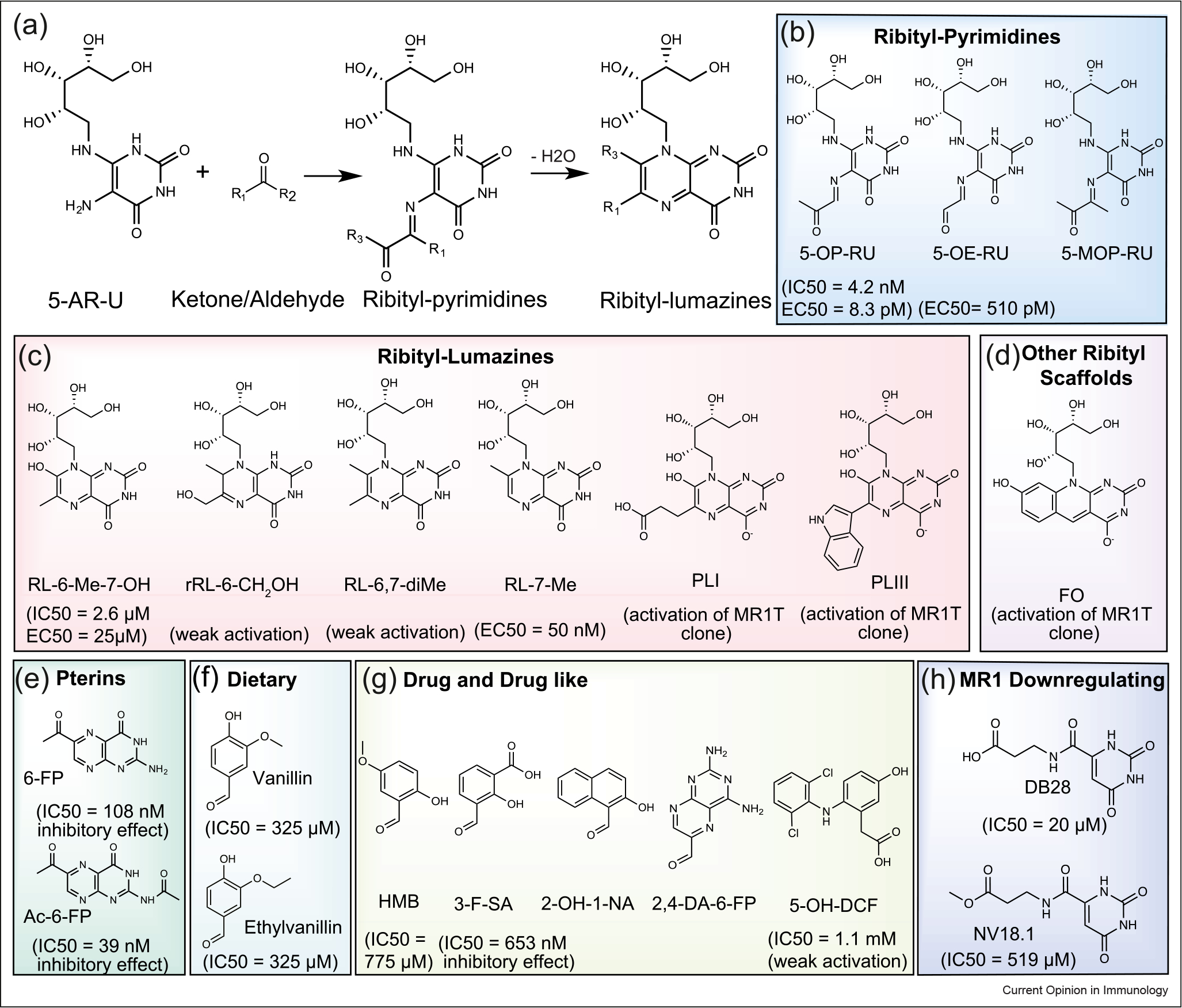
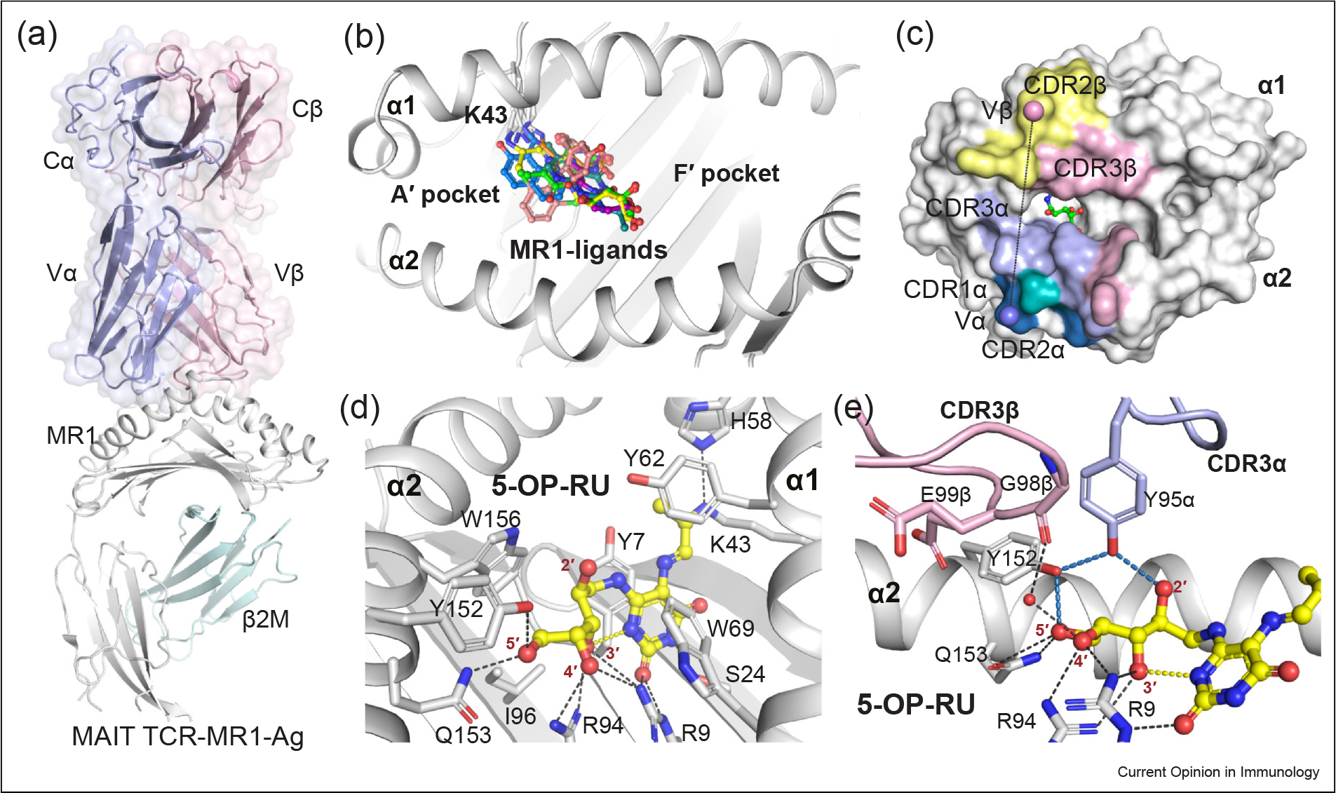
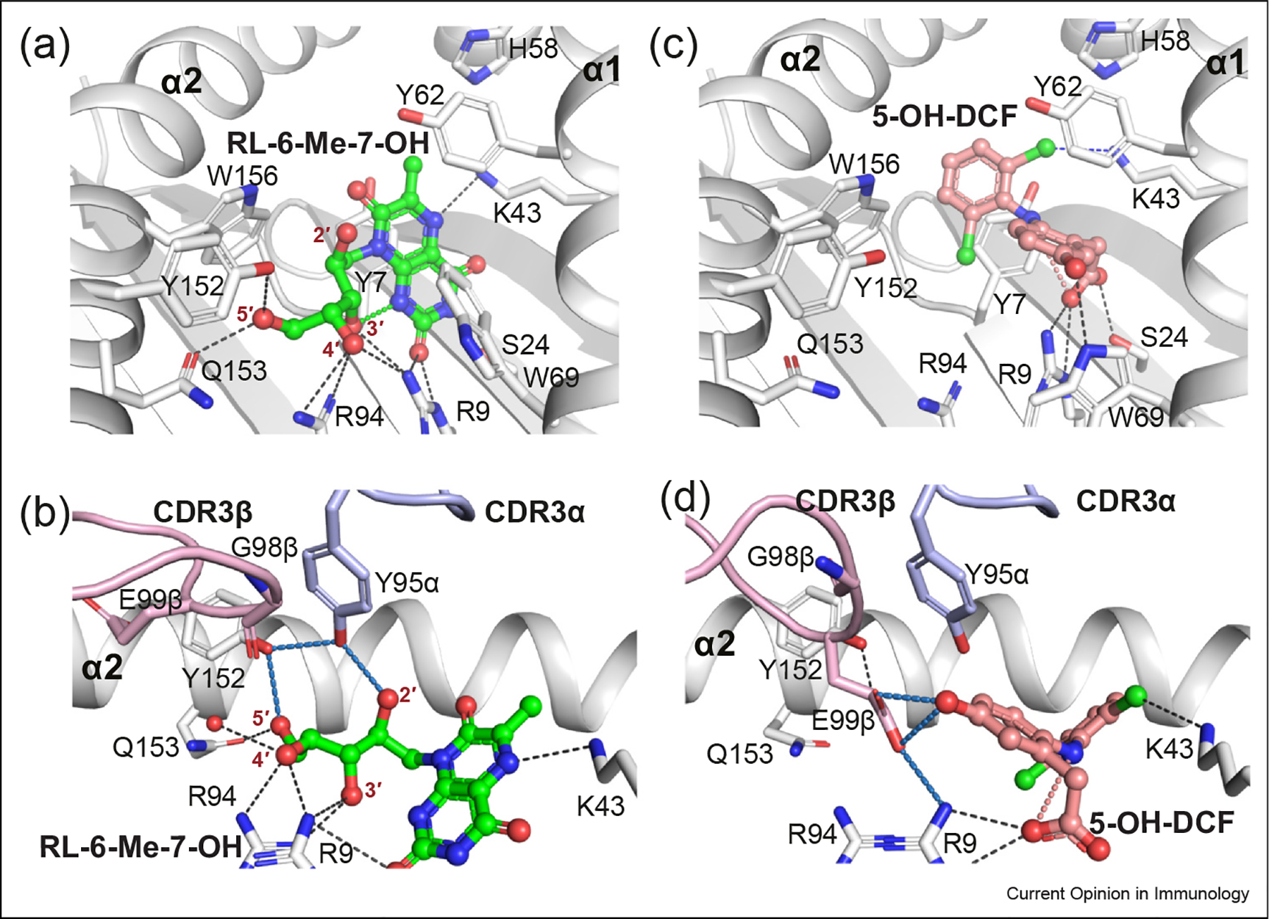
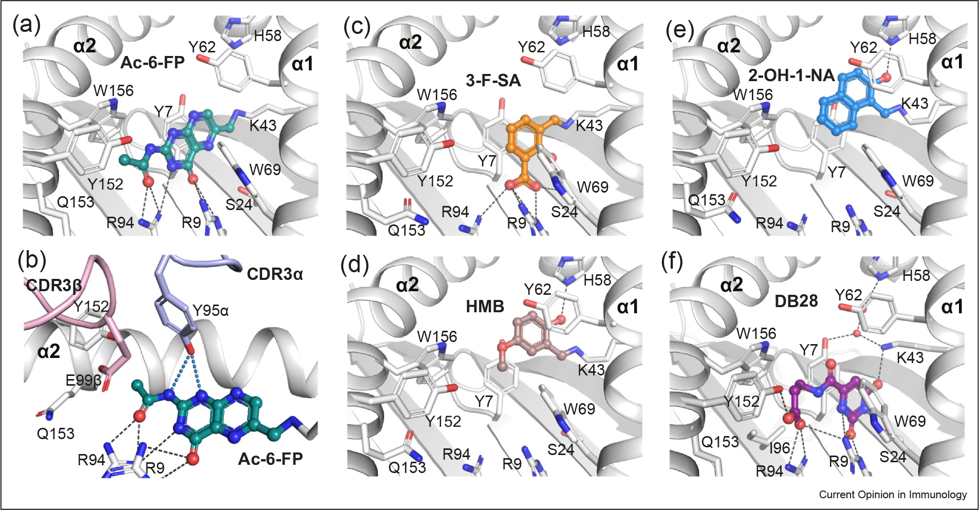
Metabolite-based T-cell immunity is emerging as a major player in antimicrobial immunity, autoimmunity, and cancer. Here, small-molecule metabolites were identified to be captured and presented by the major histocompatibility complex class-I-related molecule (MR1) to T cells, namely mucosal-associated invariant T cells (MAIT) and diverse MR1-restricted T cells. Both MR1 and MAIT are evolutionarily conserved in many mammals, suggesting important roles in host immunity. Rational chemical modifications of these naturally occurring metabolites, termed altered metabolite ligands (AMLs), have advanced our understanding of the molecular correlates of MAIT T cell receptor (TCR)-MR1 recognition. This review provides a generalized framework for metabolite recognition and modulation of MAIT cells.

Keywords: Altered Metabolites Ligands; MAIT immunity; MR1; MR1-restricted T; Metabolite antigens.


Conflict of interest statement

Declaration of Competing Interest J.M., D.P.F., and J.R. are inventors on patents describing MR1 ligands and MR1-tetramer reagents.

Figures

Figure 1
Figure 1
Broad range of MR1 Ags. (a) Condensation reactions of bacterially produced riboflavin intermediate 5-A-RU with reactive ketones/aldehydes producing pyrimidine-based Ags, which subsequently dehydrate to ribityllumazine compounds if not captured by MR1. (b–h) Chemical structures of different categories of MR1-binding Ags. (b–d) Riboflavin-based Ags. The MR1 ligand affinities (IC50) as measured by the fluorescence-based polarization assay and the activation potency (EC50) of MAIT cells are included. (b) Ribityl-pyrimidines: 5-OP-RU, 5-OE-RU, and 5-(1-methyl-2-oxopropylideneamino)-6-d-ribitylaminouracil (5-MOP-RU). (c) Ribityl-lumazines: RL-6-Me-7-OH, reduced 6-hydroxymethyl-8-d-ribityllumazine (rRL-6-CH2OH), 6,7-dimethyl-8-d-ribityllumazine (RL-6,7-diMe), and RL-7-Me, 6-(2-carboxyethyl)-7-hydroxy-8-ribityllumazine (PLI), and 6-(1H-indol-3-yl)-7-hydroxy-8-ribityllumazine (PLIII). (d) Other ribityl scaffolds: FO. (e) Folate-based metabolites, including 6-FP and Ac-6-FP. (f–h) Various classes of small-molecule scaffolds aside from vitamin-B derivatives, including (f) dietary compounds∷ Vallin, and Ethylvanillin; (g) drug-related compounds: HMB, 3-F-SA, 2-OH-1-NA, 2,4-DA-6-FP, and DCF; and (h) MR1 downregulated compounds: DB28 and NV18.1.
Figure 2
Figure 2
MAIT TCR recognition of MR1–pyrimidine-based metabolites. (a) Cartoon representation of the crystal structure of the ternary complex of the typical MAIT TRAV1–2+ A-F7 TCR–MR1–5-OP-RU (PDB: 6PUC). The constant (C) and variable (V) domains of the TCRα and TCRβ chains are shown as light-blue and light-pink surfaces, respectively. The MR1 and β2-microglobulin (β2M) molecules are shown as white and pale-cyan cartoons, respectively. (b) Surface representation of the MR1–Ag- binding A′ and F′ pockets that are formed between MR1 α1 and α2-helices. All recognized MR1 ligands (represented as colored sticks), to date, dock in the A′ pocket. (c) The footprint of the MAIT A-F7 TCR on the surface of MR1–5-OP-RU. The atomic footprint is colored according to the TCR segment making contact via its complementarity-determining region (CDR) loops colored as follows: CDR1α, teal; CDR2α, sky-blue; CDR3α, light-blue; frameworks of α-chain, dark-green; CDR1β, roseberry; CDR2β, pink; CDR3β, yellow-orange; and frameworks of β-chain, dark-gray. (d) Interactions of 5-OP-RU (yellow sticks) within the MR1-binding groove. MR1 α-helices and β-sheets are presented as white cartoon. (e) Interactions between the CDR3α and CDR3β loops of the MAIT A-F7 TCR and MR1–5-OP-RU. The relevant intramolecular H-bonds of 5-OP-RU are colored yellow. The MR1 pocket H-bonds are colored black, the TCR-related H-bonds are colored sky-blue. The CDR3α and CDR3β loop are colored as light-blue and light-pink, respectively. Waters are shown as red spheres. The structural illustrations in Figures 2, 3 and 4 were prepared using PyMOL Molecular Graphics System, Version 2.0, Schrodinger.
Figure 3
Figure 3
MR1 presentation of noncovalent MAIT agonists. The RL-6-Me-7-OH (a–b) and 5-OH-DCF (c–d) metabolites are shown as colored sticks in the MR1-binding pocket (upper panels) and their interactions with A-F7 MAIT TCR (bottom panels) (PDB: 4L4V and 5U72, respectively). RL-6-Me-7-OH and 5-OH-DCF ligands and their intramolecular H-bonds are colored green and salmon, respectively. Halogen bonds are colored in blue. H-bonds, CDR loops are colored as in Figure 2.
Figure 4
Figure 4
MR1 presentation of small-molecule MAIT nonstimulating ligands. (a–b) Capture of Ac-6-FP ligand (deep-teal sticks) within the MR1-binding cleft (a) and its interaction with the CDR loops of MAIT TCR (b). (c–f) Docking of diverse chemical identities aside from vitamin-B derivatives within the MR1 groove: (c) 3-F-SA (orange), (d) HMB (brown), (e) 2-OH-1-NA (marine), and (f) DB28 (purple). Coloring as in Figure 3 2. Crystal structures of MAIT–MR1 ligands: Ac-6-FP (PDB: 4PJ5), 3-F-SA (PDB: 5U6Q), HMB (PDB: 5U2V), 2-OH-1-NA (PDB: 5U16), DB28 (PDB: 6PVC), and NV18.1 (PDB: 6PVD), used in figure.

References

    1. Tilloy F, Treiner E, Park S-H, Garcia C, Lemonnier F, de la Salle H, Bendelac A, Bonneville M, Lantz O: An invariant T cell receptor α chain defines a novel TAP-independent major histocompatibility complex class Ib–restricted α/β T cell subpopulation in mammals. J Exp Med 1999, 189:1907–1921. - PMC - PubMed
    1. Treiner E, Duban L, Bahram S, Radosavljevic M, Wanner V, Tilloy F, Affaticati P, Gilfillan S, Lantz O: Selection of evolutionarily conserved mucosal-associated invariant T cells by MR1. Nature 2003, 422:164–169. - PubMed
    1. Godfrey DI, Koay HF, McCluskey J, Gherardin NA: The biology and functional importance of MAIT cells. Nat Immunol 2019, 20:1110–1128. - PubMed
    1. Eckle SB, Birkinshaw RW, Kostenko L, Corbett AJ, McWilliam HE, Reantragoon R, Chen Z, Gherardin NA, Beddoe T, Liu L, et al.: A molecular basis underpinning the T cell receptor heterogeneity of mucosal-associated invariant T cells. J Exp Med 2014, 211:1585–1600. - PMC - PubMed
    1. Gherardin NA, Keller AN, Woolley RE, Le Nours J, Ritchie DS, Neeson PJ, Birkinshaw RW, Eckle SB, Waddington JN, Liu L, et al.: Diversity of T cells restricted by the MHC class I-related molecule MR1 facilitates differential antigen recognition. Immunity 2016, 44:32–45. - PubMed

Publication types

MeSH terms

Substances

LinkOut - more resources