. 2023 Jun 6;42(1):142.
doi: 10.1186/s13046-023-02720-2.

Ferroptosis inducers enhanced cuproptosis induced by copper ionophores in primary liver cancer

Affiliations

Ferroptosis inducers enhanced cuproptosis induced by copper ionophores in primary liver cancer

Weikai Wang et al. J Exp Clin Cancer Res. .

Abstract

Introduction: Cuproptosis and ferroptosis are the two newly defined metal-related regulated cell death. However, the crosstalk between cuproptosis and ferroptosis is obscure.

Materials and methods: We analyzed the effect of ferroptosis inducers on copper ionophores-induced cell death through CCK-8 assay. Cuproptosis was studied using immunofluorescence and protein soluble-insoluble fraction isolation. GSH assay, qRT-PCR and western blot were adopted to explore the machinery of ferroptosis inducers enhanced cuproptosis. And mouse xenograft model was built to detect the synergy effect of elesclomol-Cu and sorafenib in vivo.

Results: Herein we found that ferroptosis inducers sorafenib and erastin could enhance cuproptosis in primary liver cancer cells by increasing copper dependent lipoylated protein aggregation. Mechanically, sorafenib and erastin upregulated protein lipoylation via suppressing mitochondrial matrix-related proteases mediated ferredoxin 1 (FDX1) protein degradation, and reduced intracellular copper chelator glutathione (GSH) synthesis through inhibiting cystine importing.

Discussion/conclusion: Our findings proposed that combination of ferroptosis inducers and copper ionophores to co-targeting ferroptosis and cuproptosis could be a novel therapeutic strategy for primary liver cancer.

Keywords: Copper ionophores; Cuproptosis; Ferredoxin 1 (FDX1); Ferroptosis; Lipoylation.


Conflict of interest statement

There is no conflict of interest to declare.

Figures

Fig. 1
Fig. 1
Ferroptosis inducers enhanced copper ionophores-induced cell death dependent on copper in liver cancer cells A and C. Cell viability of MHCC-97 H (A) or QBC939 (C) cells after Elesclomol (ES) 48 h treatment with DMSO, 10µM sorafenib (Sora) or 10µM Sora + 10µM TTM was measured with CCK-8 assay B and D. Cell viability of MHCC-97 H (B) or QBC939 (D) cells after Elesclomol (ES) 48 h treatment with DMSO, 10µM erastin (Era) or 10µM Era + 10µM TTM was measured with CCK-8 assay E and G. Cell viability of MHCC-97 H (E) or QBC939 (G) cells after diethyldithiocarbamate (DDC) 48 h treatment with DMSO, 10µM Sora or 10µM Sora + 10µM TTM was measured with CCK-8 assay F and H. Cell viability of MHCC-97 H (F) or QBC939 (H) cells after DDC 48 h treatment with DMSO, 10µM Era or 10µM Era + 10µM TTM was measured with CCK-8 assay. For A to G, media were supplemented with 1µM CuCl2
Fig. 2
Fig. 2
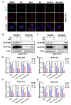
Ferroptosis inducers promoted cuproptosis in liver cancer cells A. MHCC-97 H cells were treated with indicated drugs for 24 h (DMSO, 10µM erastin (Era), 10µM sorafenib (Sora), 10nM elesclomol (ES), 10µM Era + 10nM ES, 10µM Sora + 10nM ES), DLAT protein aggregation was analyzed by immunofluorescence (IF) (green, DLAT; red, Mitotracker; blue, DAPI). White scale bars on full tiled images are 5μm B and C. The distribution of DLAT protein in soluble or insoluble fraction after treatment with indicated drugs for 24 h (DMSO, 10nM ES, 10µM Sora (or 10µM Era) + 10nM ES) was detected by western blotting in MHCC-97 H cells D and E. MHCC-97 H cells were transiently transfected with siFDX1, siNC was used as negative control, then the transfected cells were treated with sorafenib (Sora) or erastin (Era) and Elesclomol (ES) for another 48 h before cell viability was measured with CCK-8 assay F and G. MHCC-97 H cells were transiently transfected with siLIAS, siNC was used as negative control, then the transfected cells were treated with sorafenib (Sora) or erastin (Era) and Elesclomol (ES) for another 48 h before cell viability was measured with CCK-8 assay. For A to G, media were supplemented with 1µM CuCl2
Fig. 3
Fig. 3
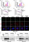
Ferroptosis inducers promoted cuproptosis through depleting intracellular GSH A and B. Relative GSH level in MHCC-97 H (A) or QBC939 (B) cells after treatment with DMSO, 10µM erastin (Era), 10µM sorafenib (Sora), 10nM elesclomol (ES), 10µM Era + 10nM ES or 10µM Sora + 10nM ES. C and D. Cell viability of MHCC-97 H (C) or QBC939 (D) cells after ES 48 h treatment with DMSO, 10µM Sora, 10mM GSH or 10µM Sora + 10mM GSH was measured with CCK-8 assay E. MHCC-97 H cells were treated with indicated drugs for 24 h (DMSO, 10nM ES, 10µM Era + 10nM ES, 10µM Sora + 10nM ES, 10µM Era + 10nM ES + 10mM GSH, 10µM Sora + 10nM ES + 10mM GSH), DLAT protein aggregation was analyzed by immunofluorescence (green, DLAT; red, Mitotracker; blue, DAPI). White scale bars on full tiled images are 5μm F and G. The effect of GSH (10mM) on elesclomol (ES 10nM) and Sora (10µM) or Era (10µM)-induced DLAT protein aggregation was analyzed by western blotting in MHCC-97 H cells. For A to G, media were supplemented with 1µM CuCl2
Fig. 4
Fig. 4
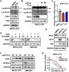
Ferroptosis inducers promoted protein lipoylation through suppressing FDX1 protein mitochondria proteases dependent degradation A. DLAT and DLST protein lipoylation (lip-DLAT and lip-DLST), FDX1 and LIAS protein level in MHCC-97 H cells treated with 10µM sorafenib (Sora) for 24 h were blotted with indicated antibodies, GAPDH was used as the loading control B. lip-DLAT, lip-DLST, FDX1 protein level in MHCC-97 H cells with FDX1 knocking down and Sora (10µM) treatment for 24 h were blotted with indicated antibodies C. FDX1 mRNA expression in MHCC-97 H cells treated with Sora or Era 24 h was determined by real-time RT-PCR. D. The effect of sorafenib on FDX1 protein stability in MHCC-97 H (left) or QBC939 (right) cells with Cycloheximide (CHX, 100 µg/ml) and indicated concentration of Sora 12 h treatment was analyzed by immunoblotting E. FDX1 protein level in MHCC-97 H cells with DMSO, 10µM MG132 or 100nM Baf-A1 treatment for 12 h was analyzed by immunoblotting. ATF4 and p62 were used as positive controls of MG132 and Baf-A1 respectively F. The effect of AFG3L2 knockdown on FDX1 expression was analyzed by western blotting after 48 h of siRNA transfection in MHCC-97 H (left) or QBC939 (right) cells G. Cell viability of MHCC-97 H cells with Elesclomol-Cu after AFG3L2 knockdown in MHCC-97 H cells was measured with CCK-8 assay
Fig. 5
Fig. 5
Ferroptosis inducers promoted cuproptosis in liver cancer in vivo A-F. Nude mice xenograft model (n = 6 per group) was generated by subcutaneous inoculation of MHCC-97 H (A-C) or QBC939 (D-F) cells. Mice were then treated with indicated drugs. Tumor pictures (A and D), tumor growth curve (B and E) and tumor weight (C and F) were summarized and shown respectively G. Frozen tissue sections from each group of MHCC-97 H xenograft model were labeled with DLAT (green) and DAPI (blue). White scale bars on full tiled images are 5μm
Fig. 6
Fig. 6
Schematic diagram of ferroptosis inducers enhanced cuproptosis induced by copper ionophores in primary liver cancer Ferroptosis inducers (FINs) sorafenib (Sora) and erastin (Era) promoted copper ionophores (CINs)-induced cuproptosis through stabilizing FDX1 protein and depleting intracellular GSH level. Mechanically, FINs stabilized FDX1 protein by suppressing mitochondrial proteases (AFG3L2 etc.) mediated FDX1 protein turnover. The stabilized FDX1 enhanced the protein lipoylation process and promoted the transfer of reduced copper ion, Cu1+. In addition, FINs could also inhibit the import of cystine through suppressing the catalytic subunit (SLC7A11) of system Xc, resulting in the decrease of GSH level, which could elevate the concentration of liable copper ion. Overall, these factors together augmented the aggregation of lipoylated proteins, and thus promoted cuproptosis in liver cancer cells. The diagram was created from BioRender.com

References

    1. Rumgay H, Arnold M, Ferlay J, Lesi O, Cabasag CJ, Vignat J et al. Global burden of primary liver cancer in 2020 and predictions to 2040. J Hepatol. 2022. - PMC - PubMed
    1. Sung H, Ferlay J, Siegel RL, Laversanne M, Soerjomataram I, Jemal A, et al. Global Cancer Statistics 2020: GLOBOCAN estimates of incidence and Mortality Worldwide for 36 cancers in 185 countries. CA Cancer J Clin. 2021;71(3):209–49. doi: 10.3322/caac.21660. - DOI - PubMed
    1. Dubyak GR. Ion homeostasis, channels, and transporters: an update on cellular mechanisms. Adv Physiol Educ. 2004;28(1–4):143–54. doi: 10.1152/advan.00046.2004. - DOI - PubMed
    1. Denoyer D, Masaldan S, La Fontaine S, Cater MA. Targeting copper in cancer therapy: ‘copper that Cancer’. Metallomics. 2015;7(11):1459–76. doi: 10.1039/C5MT00149H. - DOI - PubMed
    1. Wang Y, Yu L, Ding J, Chen Y. Iron Metabolism in Cancer. Int J Mol Sci. 2018;20(1). - PMC - PubMed