. 2023 Dec;28(1):2218684.
doi: 10.1080/13510002.2023.2218684.

Liraglutide ameliorates oxidized LDL-induced endothelial dysfunction by GLP-1R-dependent downregulation of LOX-1-mediated oxidative stress and inflammation

Affiliations

Liraglutide ameliorates oxidized LDL-induced endothelial dysfunction by GLP-1R-dependent downregulation of LOX-1-mediated oxidative stress and inflammation

Wu Ying et al. Redox Rep. 2023 Dec.

Abstract

Objective: To investigate the effects of glucagon-like peptide 1 receptor (GLP-1R) agonist liraglutide on endothelial dysfunction in LDL receptor-deficient (LDLR-KO) mice and ox-LDL-challenged human umbilical vein endothelial cells (HUVECs) and its possible mechanism.

Methods: LDLR-KO mice were randomly treated with normal saline, liraglutide, or liraglutide plus a GLP-1R antagonist exendin-9 for four weeks. In parallel, HUVECs were cultured with ox-LDL alone or combined with liraglutide, in the presence or absence of lectin-like ox-LDL receptor-1(LOX-1) overexpression or GLP-1R knockdown. Endothelial-dependent relaxation and LOX-1 protein expression of thoracic aorta, circulating levels of oxidative and inflammatory markers in mice, and cell survival, reactive oxygen species production, and expression of adhesion molecules and signal regulators in ox-LDL cultured endothelial cells were measured.

Results: liraglutide effectively enhanced acetylcholine-induced vasodilation, reduced LOX-1 expression in aortas, and decreased circulatory oxidative and inflammatory levels in LDLR-KO mice, which were abolished by cotreatment with exendin-9. HUVECs exposed to ox-LDL exhibited reduced cell viability, increased reactive oxygen species production and apoptosis, and elevated protein expression of ICAM-1, VCAM-1, LOX-1, NOX4, and NF-κB, which were markedly ameliorated by liraglutide treatment. The protective effects of liraglutide against ox-LDL-induced cell injury were abrogated in HUVECs overexpressing LOX-1 or silencing GLP-1R.

Conclusions: Liraglutide improved oxidized LDL-induced endothelial dysfunction via GLP-1R-dependent downregulation of LOX-1-mediated oxidative stress and inflammation.

Keywords: Liraglutide; endothelial dysfunction; inflammation; lectin-like ox-LDL receptor-1; oxidative stress; oxidized low-density lipoprotein.


Conflict of interest statement

No potential conflict of interest was reported by the author(s).

Figures

Figure 1.
Figure 1.
Effects of liraglutide on the acetylcholine-induced relaxation of the aorta from LDLR-KO mice and the role of exendin-9. After 1 h of incubation, the aortic rings were precontracted with norepinephrine (10−5 M), and then the rings were exposed to a cumulative concentration of acetylcholine (10−9–10−5 M) to test the endothelial-dependent vasodilation. LIR indicates liraglutide. EXE indicates exendin-9. Results are mean ± SE, n = 4 in each group, *P < 0.05 compared to wild-type at the same concentration, #P < 0.05 compared to LDLR-KO at the same concentration.
Figure 2.
Figure 2.
Effects of liraglutide on the circulatory levels of ox-LDL, oxidative and inflammatory markers, and NO bioavailability in LDLR-KO mice and the role of exendin-9. LIR indicates liraglutide. EXE indicates exendin-9. Results are mean ± SE, n = 10 in each group, *P < 0.05 compared to wild-type mice, #P < 0.05 compared to LDLR-KO mice, &P < 0.05 compared to liraglutide-treated LDLR-KO mice.
Figure 3.
Figure 3.
Effect of liraglutide on the LOX-1 expression in the thoracic aorta from LDLR-KO mice and the role of exendin-9. LIR indicates liraglutide. EXE indicates exendin-9. The results of three independent experiments were expressed as mean ± SE. *P < 0.05 compared to wild-type mice, #P < 0.05 compared to LDLR-KO mice, &P < 0.05 compared to liraglutide-treated LDLR-KO mice.
Figure 4.
Figure 4.
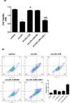
Effect of liraglutide on ox-LDL-induced LOX-1-mediated cytotoxicity. (A) cells were treated with ox-LDL 0, 10, 20, 40, or 80 µg/ml, respectively, for 24 h and then intracellular ROS levels were determined by flow cytometry. The results of three independent experiments were expressed as mean ± SE. *P < 0.05 compared to control, #P < 0.05 compared to ox-LDL 10 µg/ml. (B) Cells were treated with liraglutide 10–1000 nM for 24 h and then cell viability was measured by CCK-8 assay. The results of three independent experiments were expressed as a percentage relative to the control. *P < 0.05 compared to control. (C) Cells were transfected with pcDNA3.1 null control or pcDNA3.1-LOX-1, respectively, and LOX-1 protein expression was determined by immunoblotting. The results of three independent experiments were expressed as a fold of control. *P < 0.05 compared to control. (D) Cells transfected with pcDNA3.1 null control or pcDNA3.1-LOX-1 were treated with 20 µg/mL ox-LDL alone or combined with 1000 nM liraglutide and cell viability was determined by CCK-8 assay. The results of three independent experiments were expressed as a percentage relative to the control. *P < 0.05 compared to control, #P < 0.05 compared to ox-LDL group, &P < 0.05 compared to ox-LDL + liraglutide group. (E) Cells transfected with pcDNA3.1 null control or pcDNA3.1-LOX-1 were treated with ox-LDL alone or combined with liraglutide 1000 nM and cell apoptosis was determined by flow cytometry. The results of three independent experiments were expressed as mean ± SE. *P < 0.05 compared to control, #P < 0.05 compared to ox-LDL group, &P < 0.05 compared to ox-LDL + liraglutide group.
Figure 4.
Figure 4.
Effect of liraglutide on ox-LDL-induced LOX-1-mediated cytotoxicity. (A) cells were treated with ox-LDL 0, 10, 20, 40, or 80 µg/ml, respectively, for 24 h and then intracellular ROS levels were determined by flow cytometry. The results of three independent experiments were expressed as mean ± SE. *P < 0.05 compared to control, #P < 0.05 compared to ox-LDL 10 µg/ml. (B) Cells were treated with liraglutide 10–1000 nM for 24 h and then cell viability was measured by CCK-8 assay. The results of three independent experiments were expressed as a percentage relative to the control. *P < 0.05 compared to control. (C) Cells were transfected with pcDNA3.1 null control or pcDNA3.1-LOX-1, respectively, and LOX-1 protein expression was determined by immunoblotting. The results of three independent experiments were expressed as a fold of control. *P < 0.05 compared to control. (D) Cells transfected with pcDNA3.1 null control or pcDNA3.1-LOX-1 were treated with 20 µg/mL ox-LDL alone or combined with 1000 nM liraglutide and cell viability was determined by CCK-8 assay. The results of three independent experiments were expressed as a percentage relative to the control. *P < 0.05 compared to control, #P < 0.05 compared to ox-LDL group, &P < 0.05 compared to ox-LDL + liraglutide group. (E) Cells transfected with pcDNA3.1 null control or pcDNA3.1-LOX-1 were treated with ox-LDL alone or combined with liraglutide 1000 nM and cell apoptosis was determined by flow cytometry. The results of three independent experiments were expressed as mean ± SE. *P < 0.05 compared to control, #P < 0.05 compared to ox-LDL group, &P < 0.05 compared to ox-LDL + liraglutide group.
Figure 5.
Figure 5.
Effect of liraglutide on ox-LDL-induced LOX-1-mediated ROS generation. Cells transfected with pcDNA3.1 null control or pcDNA3.1-LOX-1 were treated with 20 µg/mL ox-LDL alone or combined with 1000 nM liraglutide and intracellular ROS levels were determined by flow cytometry. The results of three independent experiments were expressed as mean ± SE. *P < 0.05 compared to control, #P < 0.05 compared to ox-LDL group, &P < 0.05 compared to ox-LDL + liraglutide group.
Figure 6.
Figure 6.
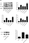
Effect of liraglutide on ox-LDL-induced LOX-1-mediated upregulation of ICAM-1 and VCAM-1. Cells transfected with pcDNA3.1 null control or pcDNA3.1-LOX-1 were treated with 20 µg/mL ox-LDL alone or combined with 1000 nM liraglutide and ICAM-1 and VCAM-1 level was determined by ELISA assay. The results of three independent experiments were expressed as mean ± SE. *P < 0.05 compared to control, #P < 0.05 compared to ox-LDL group, &P < 0.05 compared to ox-LDL + liraglutide group.
Figure 7.
Figure 7.
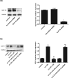
Effect of liraglutide on ox-LDL-induced activation of LOX-1/NOX4/NF-κB pathway and the role of GLP-R. (A) Cells transfected with pcDNA3.1 null control or pcDNA3.1-LOX-1 were treated with ox-LDL alone or combined with liraglutide 1000 nM and protein expression levels of LOX-1, NOX4, and NF-κB p65 were determined by immunoblotting. The results of three independent experiments were expressed as mean ± SE. *P < 0.05 compared to control, #P < 0.05 compared to ox-LDL group, &P < 0.05 compared to ox-LDL + liraglutide group. (B) Cells were pretreated with the NOX-4 inhibitor (GKT137831, 10 μmol/l) for 1 h and then exposed to 20 µg/ml ox-LDL for 24 h. The protein expression of NF-κB p65 was determined by immunoblotting. The results of three independent experiments were expressed as mean ± SE. *P < 0.05 compared to control, #P < 0.05 compared to ox-LDL group. (C) Cells were transfected with scrambled siRNA or GLP-1R siRNA, respectively, and GLP-1R protein expression was determined by immunoblotting. The results of three independent experiments were expressed as a fold of control. *P < 0.05 compared to control. (D) Cells transfected with scrambled siRNA or GLP-1R siRNA were treated with ox-LDL alone or combined with liraglutide, and then the protein expression of LOX-1 was determined by immunoblotting. The results of three independent experiments were expressed as mean ± SE. *P < 0.05 compared to control, #P < 0.05 compared to ox-LDL group, &P < 0.05 compared to ox-LDL + liraglutide group.
Figure 7.
Figure 7.
Effect of liraglutide on ox-LDL-induced activation of LOX-1/NOX4/NF-κB pathway and the role of GLP-R. (A) Cells transfected with pcDNA3.1 null control or pcDNA3.1-LOX-1 were treated with ox-LDL alone or combined with liraglutide 1000 nM and protein expression levels of LOX-1, NOX4, and NF-κB p65 were determined by immunoblotting. The results of three independent experiments were expressed as mean ± SE. *P < 0.05 compared to control, #P < 0.05 compared to ox-LDL group, &P < 0.05 compared to ox-LDL + liraglutide group. (B) Cells were pretreated with the NOX-4 inhibitor (GKT137831, 10 μmol/l) for 1 h and then exposed to 20 µg/ml ox-LDL for 24 h. The protein expression of NF-κB p65 was determined by immunoblotting. The results of three independent experiments were expressed as mean ± SE. *P < 0.05 compared to control, #P < 0.05 compared to ox-LDL group. (C) Cells were transfected with scrambled siRNA or GLP-1R siRNA, respectively, and GLP-1R protein expression was determined by immunoblotting. The results of three independent experiments were expressed as a fold of control. *P < 0.05 compared to control. (D) Cells transfected with scrambled siRNA or GLP-1R siRNA were treated with ox-LDL alone or combined with liraglutide, and then the protein expression of LOX-1 was determined by immunoblotting. The results of three independent experiments were expressed as mean ± SE. *P < 0.05 compared to control, #P < 0.05 compared to ox-LDL group, &P < 0.05 compared to ox-LDL + liraglutide group.
Figure 8.
Figure 8.
Schematic diagram illustrating the proposed signaling pathway involved in the protective effect of liraglutide against ox-LDL-associated endothelial dysfunction.

References

    1. Herrington W, Lacey B, Sherliker P, et al. . Epidemiology of atherosclerosis and the potential to reduce the global burden of atherothrombotic disease. Circ Res 2016;118(4):535–546. DOI:10.1161/CIRCRESAHA.115.307611 - DOI - PubMed
    1. Gimbrone MA, García-Cardeña G.. Endothelial cell dysfunction and the pathobiology of atherosclerosis. Circ Res 2016;118(4):620–636. - PMC - PubMed
    1. Xu S, Ilyas I, Little PJ, et al. . Endothelial dysfunction in atherosclerotic cardiovascular diseases and beyond: from mechanism to pharmacotherapies. Pharmacol Rev 2021;73(3):924–967. - PubMed
    1. Murdaca G, Colombo BM, Cagnati P, et al. . Endothelial dysfunction in rheumatic autoimmune diseases. Atherosclerosis. 2012;224(2):309–317. - PubMed
    1. Colombo BM, Murdaca G, Caiti M, et al. . Intima-media thickness: a marker of accelerated atherosclerosis in women with systemic lupus erythematosus. Ann N Y Acad Sci 2007;1108:121–126. DOI:10.1196/annals.1422.014 - DOI - PubMed

MeSH terms