

Identification of In Vitro Inhibitors of Monkeypox Replication
- PMID: 37278625
- PMCID: PMC10434227
- DOI: 10.1128/spectrum.04745-22
Identification of In Vitro Inhibitors of Monkeypox Replication
Erratum in
-
Correction for Chiem et al., "Identification of In Vitro Inhibitors of Monkeypox Replication".Microbiol Spectr. 2024 Jan 11;12(1):e0362123. doi: 10.1128/spectrum.03621-23. Epub 2023 Dec 6. Microbiol Spectr. 2024. PMID: 38054706 Free PMC article. No abstract available.
Abstract
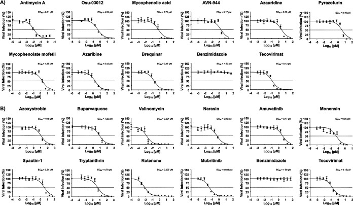
Monkeypox virus (MPXV) infections in humans have historically been restricted to regions of endemicity in Africa. However, in 2022, an alarming number of MPXV cases were reported globally, with evidence of person-to-person transmission. Because of this, the World Health Organization (WHO) declared the MPXV outbreak a public health emergency of international concern. The supply of MPXV vaccines is limited, and only two antivirals, tecovirimat and brincidofovir, approved by the U.S. Food and Drug Administration (FDA) for the treatment of smallpox, are currently available for the treatment of MPXV infection. Here, we evaluated 19 compounds previously shown to inhibit different RNA viruses for their ability to inhibit orthopoxvirus infections. We first used recombinant vaccinia virus (rVACV) expressing fluorescence (mScarlet or green fluorescent protein [GFP]) and luciferase (Nluc) reporter genes to identify compounds with antiorthopoxvirus activity. Seven compounds from the ReFRAME library (antimycin A, mycophenolic acid, AVN-944, pyrazofurin, mycophenolate mofetil, azaribine, and brequinar) and six compounds from the NPC library (buparvaquone, valinomycin, narasin, monensin, rotenone, and mubritinib) showed inhibitory activity against rVACV. Notably, the anti-VACV activity of some of the compounds in the ReFRAME library (antimycin A, mycophenolic acid, AVN-944, mycophenolate mofetil, and brequinar) and all the compounds from the NPC library (buparvaquone, valinomycin, narasin, monensin, rotenone, and mubritinib) were confirmed with MPXV, demonstrating their inhibitory activity in vitro against two orthopoxviruses. IMPORTANCE Despite the eradication of smallpox, some orthopoxviruses remain important human pathogens, as exemplified by the recent 2022 monkeypox virus (MPXV) outbreak. Although smallpox vaccines are effective against MPXV, access to those vaccines is limited. In addition, current antiviral treatment against MPXV infections is limited to the use of the FDA-approved drugs tecovirimat and brincidofovir. Thus, there is an urgent need to identify novel antivirals for the treatment of MPXV infection and other potentially zoonotic orthopoxvirus infections. Here, we show that 13 compounds, derived from two different libraries, previously found to inhibit several RNA viruses, also inhibit VACV. Notably, 11 compounds also displayed inhibitory activity against MPXV.
Keywords: GFP; antivirals; luciferase; mScarlet; monkeypox; orthopoxvirus; poxvirus; vaccinia virus.
Conflict of interest statement
The authors declare no conflict of interest.
Figures


Similar articles
-
Antivirals against monkeypox infections.bioRxiv [Preprint]. 2023 Apr 19:2023.04.19.537483. doi: 10.1101/2023.04.19.537483. bioRxiv. 2023. PMID: 37131608 Free PMC article. Preprint.
-
Identification of IMP Dehydrogenase as a Potential Target for Anti-Mpox Virus Agents.Microbiol Spectr. 2023 Aug 17;11(4):e0056623. doi: 10.1128/spectrum.00566-23. Epub 2023 Jul 6. Microbiol Spectr. 2023. PMID: 37409948 Free PMC article.
-
Treatment efficacy of cidofovir and brincidofovir against clade II Monkeypox virus isolates.Antiviral Res. 2024 Nov;231:105995. doi: 10.1016/j.antiviral.2024.105995. Epub 2024 Sep 5. Antiviral Res. 2024. PMID: 39243894
-
Monkeypox, a Literature Review: What Is New and Where Does This concerning Virus Come From?Viruses. 2022 Aug 27;14(9):1894. doi: 10.3390/v14091894. Viruses. 2022. PMID: 36146705 Free PMC article. Review.
-
Molecular Virology of Orthopoxviruses with Special Reference to Monkeypox Virus.Adv Exp Med Biol. 2024;1451:111-124. doi: 10.1007/978-3-031-57165-7_7. Adv Exp Med Biol. 2024. PMID: 38801574 Review.
Cited by
-
Inhibition Effects and Mechanisms of Marine Compound Mycophenolic Acid Methyl Ester against Influenza A Virus.Mar Drugs. 2024 Apr 23;22(5):190. doi: 10.3390/md22050190. Mar Drugs. 2024. PMID: 38786581 Free PMC article.
-
Novel replication-competent reporter-expressing Rift Valley fever viruses for molecular studies.J Virol. 2025 Jan 31;99(1):e0178224. doi: 10.1128/jvi.01782-24. Epub 2024 Dec 12. J Virol. 2025. PMID: 39665546 Free PMC article.
-
Antivirals against Monkeypox (Mpox) in Humans: An Updated Narrative Review.Life (Basel). 2023 Sep 26;13(10):1969. doi: 10.3390/life13101969. Life (Basel). 2023. PMID: 37895350 Free PMC article. Review.
-
Repurposing Drugs for Synergistic Combination Therapies to Counteract Monkeypox Virus Tecovirimat Resistance.Viruses. 2025 Jan 13;17(1):92. doi: 10.3390/v17010092. Viruses. 2025. PMID: 39861882 Free PMC article.
References
-
- WHO. 2022. Multi-country monkeypox outbreak in non-endemic countries. https://www.who.int/europe/news/item/23-07-2022-who-director-general-dec....
-
- WHO. 2022. WHO Director-General declares the ongoing monkeypox outbreak apublic health emergency of international concern.
-
- Kraemer MUG, Tegally H, Pigott DM, Dasgupta A, Sheldon J, Wilkinson E, Schultheiss M, Han A, Oglia M, Marks S, Kanner J, O’Brien K, Dandamudi S, Rader B, Sewalk K, Bento AI, Scarpino SV, de Oliveira T, Bogoch II, Katz R, Brownstein JS. 2022. Tracking the 2022 monkeypox outbreak with epidemiological data in real-time. Lancet Infect Dis 22:941–942. doi:10.1016/S1473-3099(22)00359-0. - DOI - PMC - PubMed
Publication types
MeSH terms
Substances
Grants and funding
- R01 AI141607/AI/NIAID NIH HHS/United States
- R21 AI173816/AI/NIAID NIH HHS/United States
- R01 AI142985/AI/NIAID NIH HHS/United States
- U19 AI171403/AI/NIAID NIH HHS/United States
- U19 AI171110/AI/NIAID NIH HHS/United States
- R43 AI165089/AI/NIAID NIH HHS/United States
- R01 AI161175/AI/NIAID NIH HHS/United States
- U19 AI171443/AI/NIAID NIH HHS/United States
- R01 AI145332/AI/NIAID NIH HHS/United States
- R01 AI161363/AI/NIAID NIH HHS/United States
- U19 AI135972/AI/NIAID NIH HHS/United States
- 75N93021C00014/AI/NIAID NIH HHS/United States
LinkOut - more resources
Full Text Sources
Medical
