. 2023 Jul 24;8(14):e167211.
doi: 10.1172/jci.insight.167211.

Alveolar repair following LPS-induced injury requires cell-ECM interactions

Affiliations

Alveolar repair following LPS-induced injury requires cell-ECM interactions

Jennifer Ms Sucre et al. JCI Insight. .

Abstract

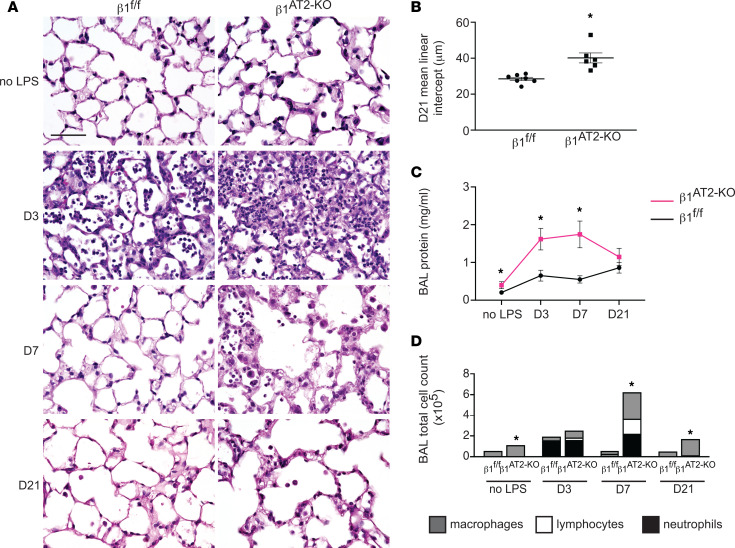
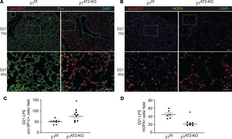
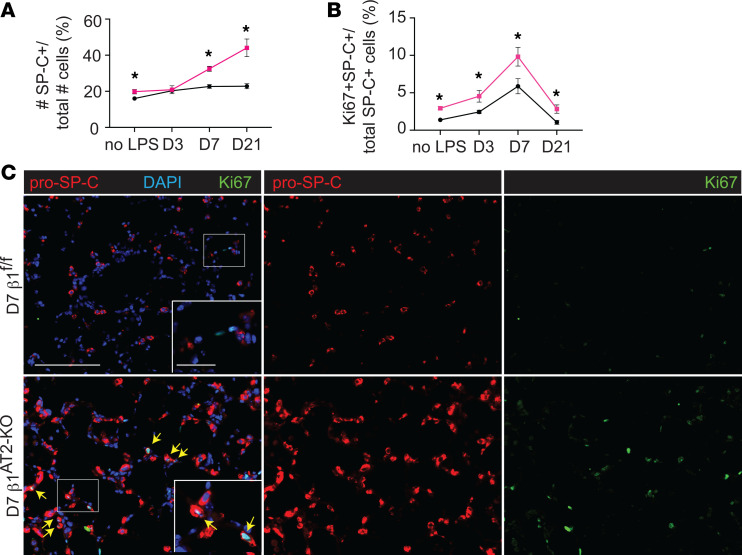
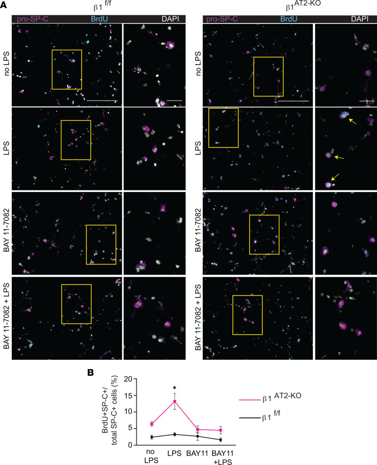
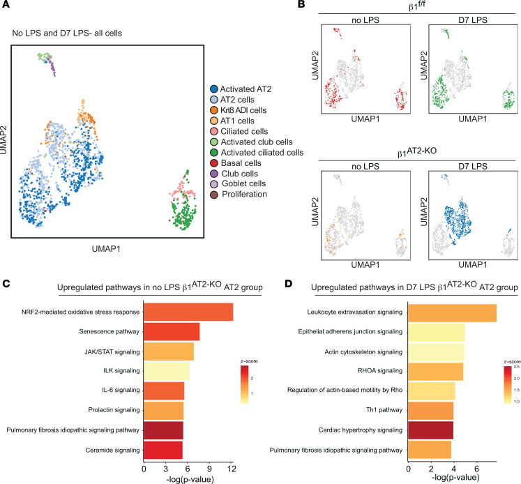
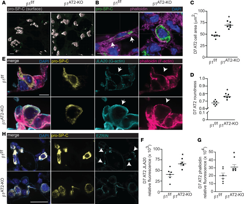
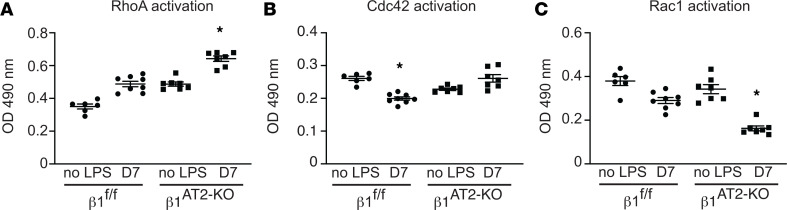
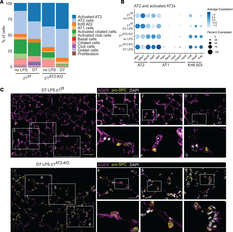
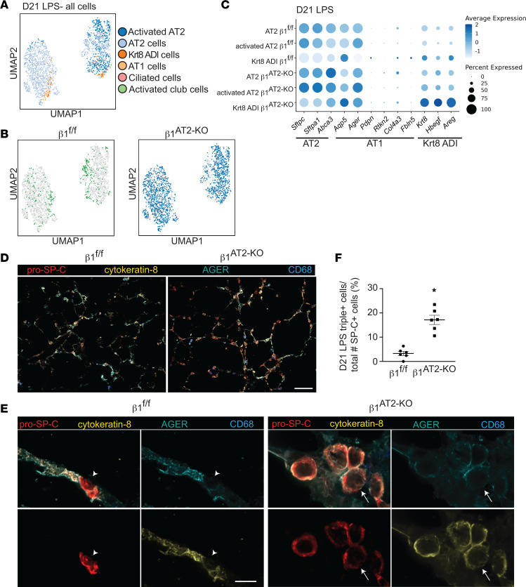
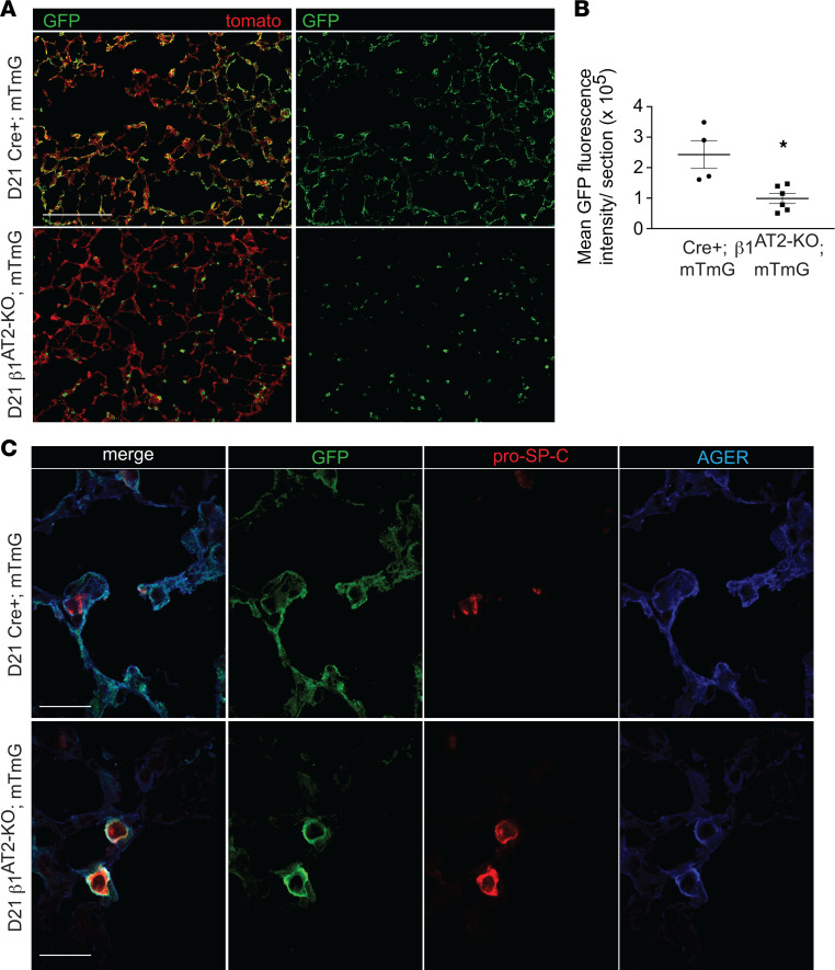
During alveolar repair, alveolar type 2 (AT2) epithelial cell progenitors rapidly proliferate and differentiate into flat AT1 epithelial cells. Failure of normal alveolar repair mechanisms can lead to loss of alveolar structure (emphysema) or development of fibrosis, depending on the type and severity of injury. To test if β1-containing integrins are required during repair following acute injury, we administered E. coli lipopolysaccharide (LPS) by intratracheal injection to mice with a postdevelopmental deletion of β1 integrin in AT2 cells. While control mice recovered from LPS injury without structural abnormalities, β1-deficient mice had more severe inflammation and developed emphysema. In addition, recovering alveoli were repopulated with an abundance of rounded epithelial cells coexpressing AT2 epithelial, AT1 epithelial, and mixed intermediate cell state markers, with few mature type 1 cells. AT2 cells deficient in β1 showed persistently increased proliferation after injury, which was blocked by inhibiting NF-κB activation in these cells. Lineage tracing experiments revealed that β1-deficient AT2 cells failed to differentiate into mature AT1 epithelial cells. Together, these findings demonstrate that functional alveolar repair after injury with terminal alveolar epithelial differentiation requires β1-containing integrins.

Keywords: Cell Biology; Integrins; Pulmonology; Respiration.


Conflict of interest statement

Conflict of interest: The authors have declared that no conflict of interest exists.

Figures

Figure 1
Figure 1. Deletion of β1 integrin in AT2 cells results in increased inflammation, abnormal repair, and decreased survival after LPS-induced injury.
(A) Representative images of lung histology demonstrate increased edema at 3 and 7 days postinjury in β1AT2-KO lungs compared with β1fl/fl lungs, as well as persistent inflammation and emphysematous remodeling in β1AT2-KO lungs by 21 days post-LPS. D3, D7, and D21 refer to 3, 7, and 21 days after LPS injury, respectively. (B) Mean linear intercept quantified emphysematous alveolar remodeling at 21 days after LPS, 28.5 ± 0.9 μm in β1fl/fl lungs versus 40.2 ± 2.8 μm in β1AT2-KO lungs (n = 6–7 mice/group, P = 0.0014 by 2-tailed t test). (C) Bicinchoninic acid (BCA) protein assay quantified increased bronchoalveolar lavage (BAL) fluid protein in uninjured β1AT2-KO lungs and at 3 and 7 days post-LPS injury in β1AT2-KO lungs compared with β1fl/fl lungs at the same time points (n = 6–14 mice/group, 2-tailed t test comparing genotypes at each time point, P = 0.0485 for uninjured mice; P = 0.0036 at D3; P = 0.005 at D7; P = 0.2628 at D21). (D) BAL cell counts are significantly increased in β1AT2-KO lungs compared with β1fl/fl littermates in uninjured mice and at 7 and 21 days post-LPS. Peak inflammation is present at 7 days in β1AT2-KO lungs, 55,663 ± 3,306 cells/mL in β1fl/fl BAL fluid versus 624,000 ± 118,753 cells/mL in β1AT2-KO BAL (n = 6–26 mice/group, 2-tailed t test comparing genotypes at each time point, P = 0.0002 for uninjured mice; P = 0.0730 at D3; P = 0.0007 at D7; P < 0.0001 at D21). Total numbers of BAL fluid macrophages are significantly increased in uninjured β1AT2-KO mice and at D7 and D21; lymphocytes and neutrophils are significantly increased in β1AT2-KO BAL at D7 only. Scale bar = 50 μm for A. * P < 0.05.
Figure 2
Figure 2. Lungs deficient in β1have increased AT2 cell number and decreased AT1 cells at 21 days after LPS injury.
(A) Representative low-power (original magnification, 10×) and inset high-power images (original magnification, 40×, denoted by yellow boxes) of β1fl/fl and β1AT2-KO lungs 21 days after LPS (D21), immunostained for AT2 marker pro–SP-C (red) and AT1 marker T1α (green). (B) Representative images of D21 β1fl/fl and β1AT2-KO lungs immunostained for pro–SP-C (red) with the AT1 marker Hopx (green), insets as in A. (C) Quantification of the number of pro–SP-C+ AT2 cells per field (n = 8 mice/group, 10 original magnification, 40× sections/mouse; P = 0.0086). (D) Hopx+ cells per field (n = 6 mice/group, 6 original magnification, 20×, sections/mouse; P = 0.0003). * P < 0.05. Scale bar = 100 μm for 10× in A and B; scale bar = 50 μm for 40× in A and B. Two-tailed t test was used to compare genotypes in C and D.
Figure 3
Figure 3. β1AT2-KO mice have increased AT2 proliferation during alveolar repair.
(A) Quantification of pro–SP-C+ cells per total cells (n = 6–8 mice/group; 5 sections per mouse; P = 0.0247 for uninjured (no LPS) mice; P = 0.8220 at D3; P = 0.0001 at D7; P = 0.0009 at D21). (B) Quantification of proliferating AT2 cells by percentage of total pro–SP-C+ AT2s (n = 6–8 mice/group, 10 sections/mouse; P = 0.0003 for uninjured mice; P = 0.0311 at D3; P = 0.0310 at D7; P = 0.0128 at D21). (C) Immunodetection of the proliferation marker Ki-67 (green) and pro–SP-C (red) shows peak AT2 proliferation in β1AT2-KO lungs at day 7 (arrows) as merged or single-channel panels. Two-tailed t test was used to compare genotypes at each time point for A and B. * P < 0.05. Scale bar = 200 μm for low-power image in C, 50 μm for inset in C.
Figure 4
Figure 4. AT2 proliferation is NF-κB dependent in LPS-treated β1AT2-KO lungs.
(A) Representative images of BrdU-incorporated precision-cut lung slices (PCLS) treated with LPS and/or NF-κB inhibitor BAY 11-7082 for 48 hours. Slices were immunostained for BrdU (cyan) and pro–SP-C (magenta) with DAPI nuclear marker (white). (B) Quantification of proliferating AT2 cells by percentage of total AT2 cells (BrdU+pro–SP-C+ over total number of SP-C+ cells) by condition as indicated (n = 6–8 mice/group, 1 slice per mouse per condition, imaged and quantified 10 original magnification, 40× sections/mouse per condition, data from 5 separate experiments; P = 0.0010, F value = 6.3 for treatment variation; P < 0.0001, F value = 26.1 for genotype variation). Two-way ANOVA was used to compare treatment conditions and genotype in B. * P < 0.05. Scale bar = 100 μm for low-power images in A, 50 μm for inset in A.
Figure 5
Figure 5. Overabundant AT2 cells are transcriptionally distinct during repair in β1AT2-KO mice.
(A) Uniform manifold approximation and projection (UMAP) of all epithelial cells from β1fl/fl and β1AT2-KO lungs with/without LPS clustered by label transfer from Strunz et al. (11). (B) Individual epithelial populations by group reveal transcriptionally distinct AT2s and activated AT2s in day 7 LPS-treated β1AT2-KO lungs. (C) Ingenuity Pathway Analysis (QIAGEN) on combined AT2 groups from uninjured β1fl/fl and β1AT2-KO lungs demonstrates upregulation of oxidative stress, senescence, and inflammatory pathways in β1AT2-KO lungs compared with β1fl/fl lungs. (D) Ingenuity Pathway Analysis shows upregulation of actin cytoskeleton signaling pathways in β1AT2-KO AT2 cells compared with β1fl/fl AT2 cells at 7 days after LPS treatment.
Figure 6
Figure 6. During alveolar repair, β1 integrin regulates actin localization and RhoA GTPase activation.
(A) Surface rendering high-power images of pro–SP-C–immunostained, thick, frozen sections from day 7 (D7) LPS-treated β1fl/fl and β1AT2-KO lungs. (B) High-power images of thick, frozen sections from D7 LPS-treated β1fl/fl and β1AT2-KO lungs immunostained for pro–SP-C (green) with phalloidin F-actin probe (magenta); arrows indicate areas of actin-rich lateral protrusions. (C) Area of pro–SP-C+CD68 AT2 cells from D7 LPS-treated β1fl/fl and β1AT2-KO mice (46.8 ± 2.0 μm2 in β1fl/fl lungs compared with 69.6 ± 2.8 μm2 in β1AT2-KO lungs, n = 6 mice/group, ≥40 cells measured/mouse imaged from 5 sections, 2-tailed t test, P < 0.0001). (D) Roundness score calculated from pro–SP-C+CD68 cells from D7 LPS-treated β1fl/fl and β1AT2-KO mice (38–60 cells measured/mouse from 5 sections, n = 6 mice/group, 2-tailed t test comparing genotypes, P = 0.0009). (E) High-power images of frozen sections prepared at D7 after LPS β1fl/fl and β1AT2-KO lungs immunostained for pro–SP-C (gold) with JLA20 (cyan) and phalloidin (magenta) probes applied to detect G-actin and F-actin, respectively. Membrane localization of G-actin denoted by arrows and F-actin by arrowheads. (F and G) Quantification of JLA20 (F) and phalloidin (G) expression in pro–SP-C+ AT2 cells in β1fl/fl and β1AT2-KO lungs D7 after LPS (n = 5–6 mice/group, 10 sections/mouse, 2-tailed t test with P = 0.0088 for JLA20 and P = 0.0482 for phalloidin). (H) Representative high-power images from D7 LPS-treated β1fl/fl and β1AT2-KO lungs immunostained for ezrin (cyan) with AT2 cells identified by RNA in situ hybridization for Sftpc (gold). Arrows indicate ezrin expression localized to lateral extensions in β1fl/fl AT2 cells, whereas diffuse, nonfocal ezrin expression along the cell membrane is seen in β1AT2-KO AT2 cells. * P < 0.05. Scale bar = 5 μm for A, B, and E; scale bar = 25 μm for panels in H.
Figure 7
Figure 7. During repair, β1 integrin regulates GTPase activation in AT2 cells.
(AC) GTPase activation assay performed on AT2 cell lysates collected from uninjured and D7 LPS-treated β1fl/fl and β1AT2-KO lungs (n = 6–8 mice/group for each assay; in A RhoA 1-way ANOVA * P < 0.0001, F value 53.42, df = 3; in B Cdc42 1-way ANOVA * P < 0.001, F value 17.46, df = 3; in C Rac1 1-way ANOVA * P < 0.0001, F value 31.09, df = 3).
Figure 8
Figure 8. Postinjury β1-deficient AT2s exhibit an AT2-AT1 mixed epithelial transcriptomic phenotype.
(A) Stacked bar graph of epithelial proportions demonstrates an expansion of the AT2 and activated AT2 populations in day 7 (D7) LPS-treated β1AT2-KO lungs. (B) Marker gene expression by genotype and treatment group in AT2/activated AT2 cluster, in which higher expression is represented with a darker color and the size of the dot reflects the proportion of cells expressing that marker. (C) Representative low-power images from day 7 LPS-treated lung sections immunostained for the AT1 marker AGER (purple) and AT2 marker pro–SP-C (gold) demonstrate overall decreased AGER in β1AT2-KO lungs. Three insets per low-power field show colocalization of AGER with pro–SP-C+ AT2 cells in D7 β1AT2-KO lungs. Scale bar = 100 μm for low-power image in C, 50 μm for middle inset, and 10 μm for high-power inset.
Figure 9
Figure 9. AT2s of mixed transcriptomic phenotype persist, proliferate, and maintain an enlarged, rounded cell shape in late alveolar repair.
(A) UMAP of all epithelial cells from β1fl/fl and β1AT2-KO lungs 21 days after LPS treatment clustered by label transfer from Strunz et al. (11). (B) Individual epithelial populations by group reveal transcriptionally abundant AT2s and activated AT2s in day 21 LPS-treated β1AT2-KO lungs. (C) Hallmark gene expression by genotype in AT2, activated AT2, and Krt8 ADI clusters, in which higher expression is represented with a darker color and the size of the dot reflects the proportion of cells expressing that marker. (D) Representative low-power images of β1fl/fl and β1AT2-KO lungs 21 days after LPS co-immunostained for pro–SP-C (red), cytokeratin 8 (gold), AGER (cyan), and CD68 (blue). β1AT2-KO lungs are notable for enlarged airspaces and increased numbers of round, large pro–SP-C+ AT2 cells, which are distinct from alveolar macrophages. (E) High-power images of lung sections immunostained for pro–SP-C, cytokeratin 8, AGER, and CD68, as above, demonstrate round, large pro–SP-C+ AT2 cells that colocalize with cytokeratin 8, with a subset also triple positive (pro–SP-C+cytokeratin 8+AGER+). Arrow denotes occasional triple-positive cells in β1AT2-KO lungs, and arrowhead marks AGER AT2 cells in β1fl/fl lungs. (F) Quantification of pro–SP-C+cytokeratin 8+AGER+ triple-positive cells as a percentage of total pro–SP-C+ cells in day 21 LPS-treated lungs (n = 6 mice/group, 10 original magnification, 60×, sections/mouse, P < 0.0001 by 2-tailed t test). * P < 0.05. Scale bar = 100 μm in D, 10 μm in E.
Figure 10
Figure 10. β1AT2-KO mice fail to repopulate alveolus with cells of AT1 morphology.
(A) Low-power images of Cre+ mTmG control mice demonstrate Cre-recombinase reporter–labeled GFP+ cells of both AT1 and AT2 cell shape repopulate the injured alveolus 21 days after LPS. GFP-labeled cells in β1AT2-KO mTmG mice retain only AT2 morphology and fail to attain an AT1 cell shape at 21 days. (B) Quantification of mean GFP fluorescence intensity shows decreased area of lung repopulated by GFP-labeled cells at 21 days post-LPS in β1AT2-KO mTmG mice (n = 4 Cre+ mTmG and 6 β1AT2-KO mTmG mice; 10 original magnification, 20×, sections/mouse, P = 0.0087 by 2-tailed t test). (C) High-power images of Cre+ mTmG and β1AT2-KO mTmG lung sections immunostained for pro–SP-C (red) and AGER (blue); tomato omitted in imaging. Large, round GFP+-labeled cells acquire both AT2 (pro–SP-C+) and AT1 (AGER+) markers in β1AT2-KO mTmG lungs. GFP+-labeled cells possess either AT2 or AT1 markers at 21 days post-LPS in Cre+ mTmG lungs. * P < 0.05. Scale bar = 200 μm in A and 25 μm in C.

References

    1. Mizikova I, Morty RE. The extracellular matrix in bronchopulmonary dysplasia: target and source. Front Med (Lausanne) 2015;2:91. - PMC - PubMed
    1. Sokocevic D, et al. The effect of age and emphysematous and fibrotic injury on the re-cellularization of de-cellularized lungs. Biomaterials. 2013;34(13):3256–3269. doi: 10.1016/j.biomaterials.2013.01.028. - DOI - PMC - PubMed
    1. Liang Q, et al. Effect of SIS3 on extracellular matrix remodeling and repair in a lipopolysaccharide-induced ARDS rat model. J Immunol Res. 2020;2020:6644687. - PMC - PubMed
    1. Roychaudhuri R, et al. ADAM9 is a novel product of polymorphonuclear neutrophils: regulation of expression and contributions to extracellular matrix protein degradation during acute lung injury. J Immunol. 2014;193(5):2469–2482. doi: 10.4049/jimmunol.1303370. - DOI - PMC - PubMed
    1. Humphrey JD, et al. Mechanotransduction and extracellular matrix homeostasis. Nat Rev Mol Cell Biol. 2014;15(12):802–812. doi: 10.1038/nrm3896. - DOI - PMC - PubMed

Publication types