. 2023 Jul 24;8(14):e169855.
doi: 10.1172/jci.insight.169855.

Platelets exacerbate cardiovascular inflammation in a murine model of Kawasaki disease vasculitis

Affiliations

Platelets exacerbate cardiovascular inflammation in a murine model of Kawasaki disease vasculitis

Begüm Kocatürk et al. JCI Insight. .

Abstract

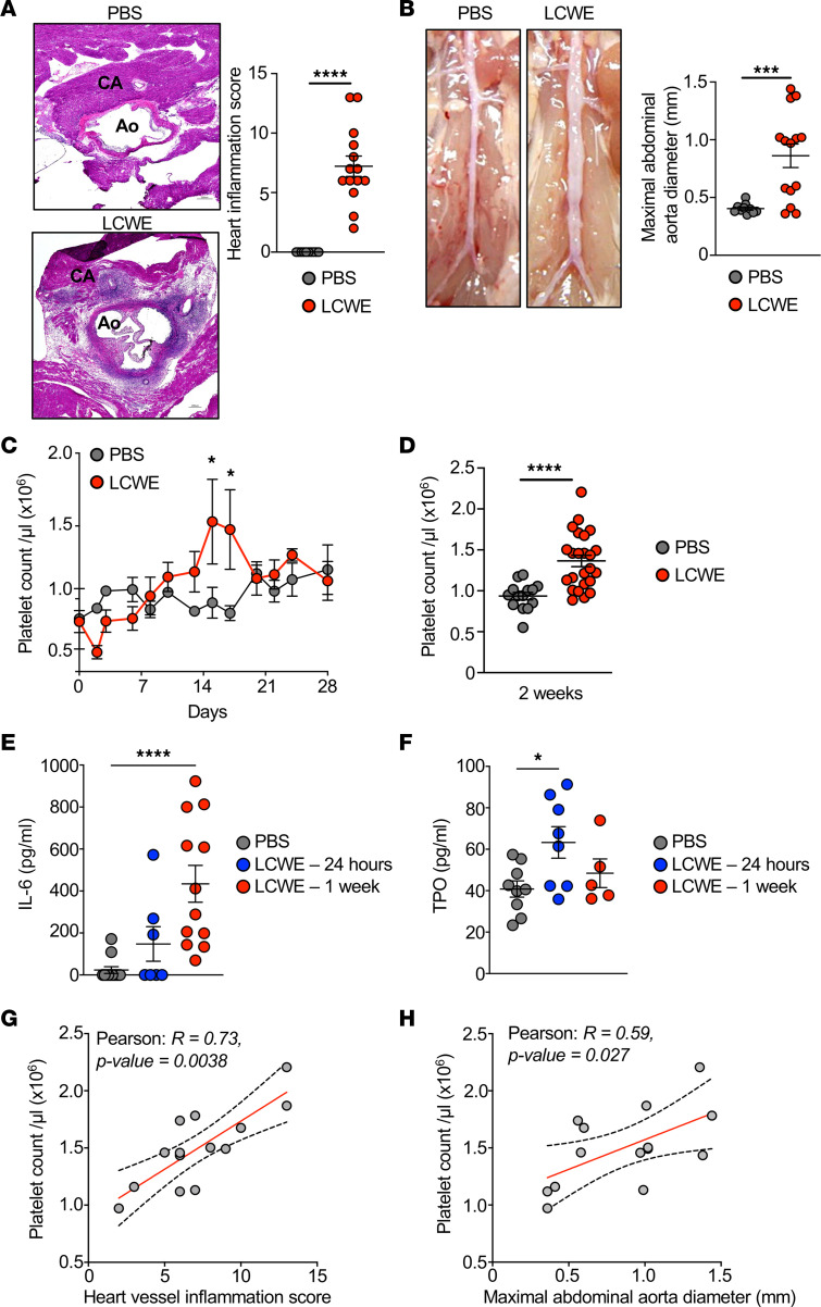
Kawasaki disease (KD) is the leading cause of acquired heart disease among children. Increased platelet counts and activation are observed during the course of KD, and elevated platelet counts are associated with higher risks of developing intravenous immunoglobulin resistance and coronary artery aneurysms. However, the role of platelets in KD pathogenesis remains unclear. Here, we analyzed transcriptomics data generated from the whole blood of patients with KD and discovered changes in the expression of platelet-related genes during acute KD. In the Lactobacillus casei cell wall extract (LCWE) murine model of KD vasculitis, LCWE injection increased platelet counts and the formation of monocyte-platelet aggregates (MPAs), upregulated the concentration of soluble P-selectin, and increased circulating thrombopoietin and interleukin 6 (IL-6). Furthermore, platelet counts correlated with the severity of cardiovascular inflammation. Genetic depletion of platelets (Mpl-/- mice) or treatment with an anti-CD42b antibody significantly reduced LCWE-induced cardiovascular lesions. Furthermore, in the mouse model, platelets promoted vascular inflammation via the formation of MPAs, which likely amplified IL-1B production. Altogether, our results indicate that platelet activation exacerbates the development of cardiovascular lesions in a murine model of KD vasculitis. These findings enhance our understanding of KD vasculitis pathogenesis and highlight MPAs, which are known to enhance IL-1B production, as a potential therapeutic target for this disorder.

Keywords: Cardiovascular disease; Inflammation; Platelets; Vascular Biology; Vasculitis.


Figures

Figure 1
Figure 1. Change in the expression of platelet signature genes during acute KD.
(A) List of the 40 genes either expressed by platelets or associated with platelet activity included in the platelet signature genes. (B and C) Heatmaps showing platelet signature genes that are differentially expressed (at least 1.5-fold change [FC] in either direction and with an adjusted P value < 0.05) in whole blood from patients with acute KD compared with HCs (B: GSE68004, n = 76 KD patients and n = 37 HCs, and C: GSE73461, n = 77 KD patients and n = 55 HCs). (D) List of 19 genes from the platelet signature genes that were commonly upregulated during acute KD in B and C. (E) Expression of common platelet signature differentially expressed genes (DEGs) (D) in whole blood of patients with acute KD compared with convalescent IVIG-treated KD patients (GSE63881; n = 146 acute KD patients and n = 145 convalescent KD patients). Genes were selected based on an adjusted P value of less than 0.05. (B, C, and E) Blue–red color gradient: Low to high expression relative to the mean of each row. Each column represents 1 patient of the defined groups. Differential expression was analyzed with GEO2R. HC, healthy control; KD, Kawasaki disease.
Figure 2
Figure 2. Platelet count correlates with the severity of LCWE-induced KD vasculitis.
(A) H&E-stained heart tissue sections and heart vessel inflammation score from PBS- and LCWE-injected mice at 2 weeks after LCWE injection (n = 10 to 14 mice/group). Scale bar: 200 μm. (B) Representative pictures of the abdominal aorta area and maximal abdominal aorta diameter of PBS- and LCWE-injected mice at 2 weeks after LCWE injection (n = 10 to 14 mice/group). (C) Four-week time-course analysis of platelet counts in the blood of PBS- and LCWE-injected mice (n = 4 to 5 mice/group). (D) Blood platelet count of PBS- and LCWE-injected mice at 2 weeks after injection (n = 14 to 24 mice/group). (E and F) IL-6 and TPO levels in the serum of PBS- and LCWE-injected mice (n = 8 to 12 mice/group) at 24 hours and 1 week after LCWE injection. (G and H) Correlation of blood platelet counts with heart inflammation score (G) and maximal abdominal aorta diameter (H). Each symbol represents 1 mouse. Results presented as mean ± SEM. *P < 0.05, ***P < 0.001, ****P < 0.0001 obtained by unpaired 2-tailed Student’s t test with Welch’s correction (A, B, and D), 2-way ANOVA with Bonferroni’s multiple-comparison test (C), 1-way ANOVA with Tukey’s multiple-comparison test (E), Kruskal-Wallis with Dunn’s multiple-comparison test (F), or Pearson’ r correlation test (G and H). CA, coronary artery; Ao, Aorta; TPO, thrombopoietin.
Figure 3
Figure 3. Platelet depletion attenuates the severity of LCWE-induced KD vasculitis.
(A and B) Representative H&E-stained heart tissue sections (A) and heart vessel inflammation score (B) from LCWE-injected mice, or LCWE-injected mice that received either the platelet-depleting anti-CD42b antibody or IgG control at 1 week after LCWE injection (n = 7 to 8 mice/group). Scale bars: 200 μm. (CE) Representative pictures of the abdominal aorta area (C), maximal abdominal aorta diameter (D), and abdominal aorta area measurements (E) from LCWE-injected mice, or LCWE-injected mice that received either the platelet-depleting anti-CD42b or IgG control at 1 week after LCWE injection (n = 7 to 8 mice/group). (F) Levels of IL-1B in the serum of PBS- or LCWE-injected mice that received either IgG isotype control or the platelet-depleting anti-CD42b antibody on day 1 or day 7 after LCWE injection (n = 4 to 7 mice/group). (G) Representative H&E staining and immunostaining staining of CD41 (red) in serial heart sections of WT mice injected with PBS, LCWE, or LCWE-injected mice treated with the platelet-depleting anti-CD42b. (H) CD41+ cell counts in the coronary artery of WT mice injected with PBS, LCWE, or LCWE-injected mice treated with the platelet-depleting anti-CD42b. Scale bars: 200 μm (H&E) and 100 μm (immunofluorescence). Each symbol represents 1 mouse. Results presented as mean ± SEM. *P < 0.05; **P < 0.01; ***P < 0.001; ****P < 0.0001 obtained by 1-way ANOVA with Tukey’s multiple-comparison test (B and DG).
Figure 4
Figure 4. Decreased development of LCWE-induced cardiovascular lesions in thrombocytopenic Mpl–/– mice.
(A and B) Representative H&E-stained heart tissue sections (A) and heart inflammation score (B) of LCWE-injected WT and Mpl–/– mice 1 week after LCWE injection (n = 12/group). Scale bars: 200 μm. (C and D) Representative pictures of the abdominal aorta area (C) and maximal abdominal aorta diameter and abdominal aorta area measurements (D) from WT and Mpl–/– mice (n = 15–17/group) at 1 week after LCWE injection. (E) Heatmap illustrating the expression of S100a8 and S100a9 (at least 1.5 FC with an adjusted P value < 0.05) in abdominal aorta tissues of PBS- and LCWE-injected mice (n = 5/groups; GSE141072). Blue–red color gradient: Low to high expression relative to the mean of each column. Each row represents 1 mouse of the defined groups. (F) Calprotectin levels in the serum of PBS- and LCWE-injected WT and Mpl–/– mice, at 1 week after injection (n = 4 to 12/group). Each symbol represents 1 mouse. Results presented as mean ± SEM pooled from 2–3 independent experiments. *P < 0.05; ****P < 0.0001 obtained by unpaired 2-tailed Student’s t test with Welch’s correction (B), unpaired 2-tailed Student’s t test (D), or 2-way ANOVA with Tukey’s multiple-comparison test (F).
Figure 5
Figure 5. Increased frequencies of circulating monocyte-platelet aggregates (MPAs) during LCWE-induced KD vasculitis.
(A) Serum levels of soluble P-selectin in PBS- or LCWE-injected WT and Mpl–/– mice (n = 6–7/group). (B and C) Representative flow cytometry plots (B) and frequencies (C) of CD61+CD11b+CD115+ MPAs in whole blood of WT and Mpl–/– mice 3 days after LCWE injection (n = 5–7/group). (D) Serum IL-1B concentration in PBS-and LCWE-injected WT and Mpl–/– mice at baseline and 3 days after injection (n = 6–7/group). Each symbol represents 1 mouse. Results presented as mean ± SEM. *P < 0.05; **P < 0.01; ***P < 0.001; ****P < 0.0001 obtained by 2-way ANOVA with Bonferroni’s multiple-comparison test (A and D) or 2-tailed Mann-Whitney test (C).

Comment in

References

    1. McCrindle BW, et al. Diagnosis, treatment, and long-term management of Kawasaki disease: a scientific statement for health professionals from the American Heart Association. Circulation. 2017;135(17):e927–e999. doi: 10.1161/CIR.0000000000000484. - DOI - PubMed
    1. Soni PR, et al. A comprehensive update on Kawasaki disease vasculitis and myocarditis. Curr Rheumatol Rep. 2020;22(2):6. doi: 10.1007/s11926-020-0882-1. - DOI - PubMed
    1. Rowley AH. Is Kawasaki disease an infectious disorder? Int J Rheum Dis. 2018;21(1):20–25. doi: 10.1111/1756-185X.13213. - DOI - PMC - PubMed
    1. Skochko SM, et al. Kawasaki disease outcomes and response to therapy in a multiethnic community: a 10-year experience. J Pediatr. 2018;203:408–415. doi: 10.1016/j.jpeds.2018.07.090. - DOI - PubMed
    1. Maury CP, et al. Circulating interleukin-1 beta in patients with Kawasaki disease. N Engl J Med. 1988;319(25):1670–1671. doi: 10.1056/NEJM198812223192515. - DOI - PubMed

Publication types