. 2023 Jun 13;120(24):e2220867120.
doi: 10.1073/pnas.2220867120. Epub 2023 Jun 6.

ZBTB20 is essential for cochlear maturation and hearing in mice

Affiliations

ZBTB20 is essential for cochlear maturation and hearing in mice

Zhifang Xie et al. Proc Natl Acad Sci U S A. .

Abstract

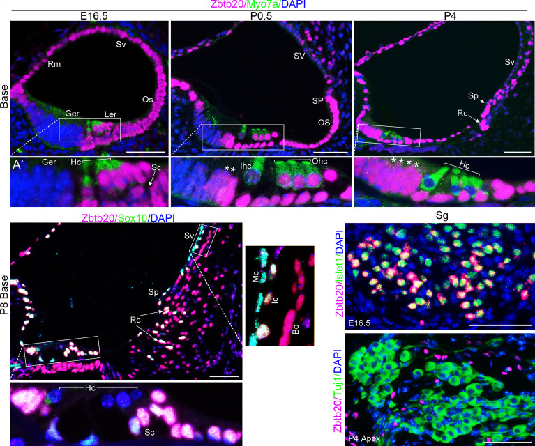
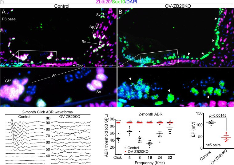
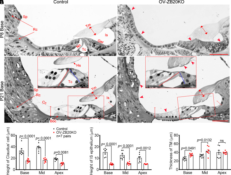
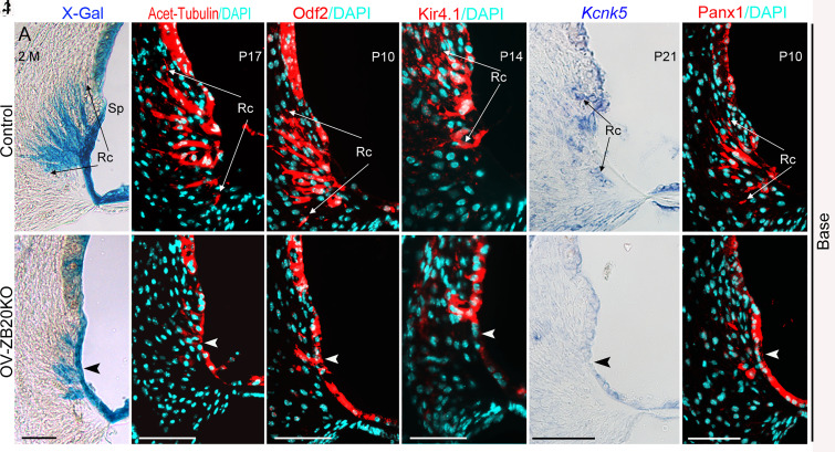
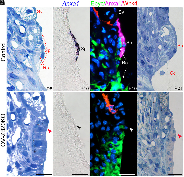
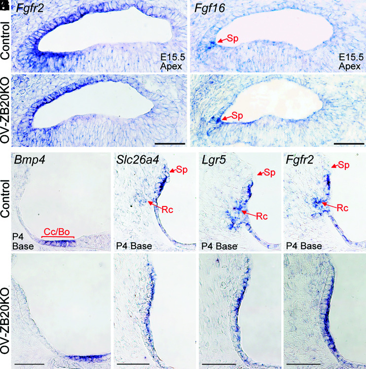
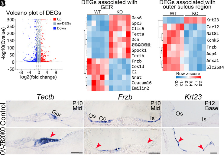
The mammalian cochlear epithelium undergoes substantial remodeling and maturation before the onset of hearing. However, very little is known about the transcriptional network governing cochlear late-stage maturation and particularly the differentiation of its lateral nonsensory region. Here, we establish ZBTB20 as an essential transcription factor required for cochlear terminal differentiation and maturation and hearing. ZBTB20 is abundantly expressed in the developing and mature cochlear nonsensory epithelial cells, with transient expression in immature hair cells and spiral ganglion neurons. Otocyst-specific deletion of Zbtb20 causes profound deafness with reduced endolymph potential in mice. The subtypes of cochlear epithelial cells are normally generated, but their postnatal development is arrested in the absence of ZBTB20, as manifested by an immature appearance of the organ of Corti, malformation of tectorial membrane (TM), a flattened spiral prominence (SP), and a lack of identifiable Boettcher cells. Furthermore, these defects are related with a failure in the terminal differentiation of the nonsensory epithelium covering the outer border Claudius cells, outer sulcus root cells, and SP epithelial cells. Transcriptome analysis shows that ZBTB20 regulates genes encoding for TM proteins in the greater epithelial ridge, and those preferentially expressed in root cells and SP epithelium. Our results point to ZBTB20 as an essential regulator for postnatal cochlear maturation and particularly for the terminal differentiation of cochlear lateral nonsensory domain.

Keywords: auditory; cell differentiation; cochlea; hearing; transcription factor.


Conflict of interest statement

The authors declare no competing interest.

Figures

Fig. 1.
Fig. 1.
ZBTB20 is differentially expressed in cochlear epithelial cells. (AD) Double immunofluorescence staining showing spatial and temporal expression pattern of ZBTB20 (magenta) in the cochlea. (A) ZBTB20 was expressed strongly in Myosin7a-positive (green) hair cells (Hc) and the adjacent supporting cells (Sc), outer sulcus (Os), stria vascularis (Sv), Reissner’s membrane (Rm), but undetectable in the greater epithelial ridge (Ger) at E16.5. (B and C) ZBTB20 staining intensity becomes weaker in Myosin7a-positive inner hair cells (Ihc) at P0.5 (B) and undetectable in all HCs at P4 (C), while becomes stronger in the lateral and throughout GER at P0.5 and P4, respectively (indicated with *). Note the developing root cells (Rc) and spiral prominence epithelium (Sp) showing strong signals for ZBTB20. Idc, interdental cells, Ohc, outer hair cells. (D) At P8, ZBTB20 was strongly expressed by cochlear Sox10-positive (green) nonsensory epithelial cells including supporting cells (Sc) and root cells (Rc, indicated by arrowheads), with the exception of Sox10-positive SV marginal cells (Mc), which showed very weak staining. Ic and Bc indicate SV intermediate cell and basal cell, respectively; Ler, lesser epithelial ridge. (A’, B’, C’, D’, and D’’) are magnified views of indicated boxed areas in (AD). (E and F) Double immunofluorescence staining showing that ZBTB20 (magenta) was expressed by a subset of Islet-positive (green) spiral ganglion (Sg) neurons at E16.5 but undetectable in Tuj1-positive (green) SG neurons at P4. Nuclei were stained with DAPI (blue). n = 4 mice for each developmental stage. (Scale bars in all panels, 50 µm.) See also SI Appendix, Figs. S2 and S3.
Fig. 2.
Fig. 2.
Conditional knockout of Zbtb20 causes endolymph potential reduction and deafness. (A and B) Immunofluorescence staining confirming that ZBTB20 protein (magenta) was detected in Sox10-positive (green) supporting cells (Sc), greater epithelial ridge (Ger), root cells (Rc), and spiral prominence epithelial cells (Sp) from control (A) but not from OV-ZB20KO mice (B) at P8. Arrowheads indicate negative staining in OV-ZB20KO. (A’) and (B’) are magnified views of indicated boxed regions in (A) and (B), respectively. Nuclei were stained with DAPI (blue). (Scale bars: 50 µm.) n = 4 mice/group. Hc, hair cell; Sv, stria vascularis; Tbc, tympanic border cell. (C) Representative click-evoked ABR waveforms for control and OV-ZB20KO mice at 2 months (M) of age. Broadband click stimuli were applied at sound pressure levels (SPL) indicated in decibels (dB). (D) Averaged thresholds of click- and pure tone–evoked ABRs of control and OV-ZB20KO mice at 2 M. A two-way ANOVA followed by post-hoc pair-wise tests (Bonferroni method) was used to compare differences in ABR threshold at multiple frequencies between the two groups. **P < 0.01; ****P < 0.0001. (E) Values of endolymph potential (EP) recorded from the basal cochlear turns from control and OV-ZB20KO mice at 2 M. All values are represented as mean ±SEM. EP values were compared by using a paired Student’s t test (two tailed). Abbreviations are consistent across figures in this manuscript. See also SI Appendix, Figs. S4 and S5.
Fig. 3.
Fig. 3.
Targeted Zbtb20 ablation permanently arrests postnatal cochlear development. (A and B) Representative semi-thin sections from control and OV-ZB20KO mice at P8, showing the defects (indicated by arrowheads) in OV-ZB20KO cochleae, including the closed spaces of Nuel (Nu) and tunnel of Corti (Tc), and delayed root cell (Rc) development. In addition, the spiral prominence (Sp) appeared flattened, the greater epithelial ridge (Ger) showed initial sign of regression leading to a limited opening of the inner sulcus (Is), and the tectorial membrane (Tm, indicated by the line with arrowheads in both ends) was thickened and abnormally attached to the underlying Corti’s organ. (C and D) Representative sections showing the defects in OV-ZB20KO cochleae at P21, including a partially collapsed tunnel of Corti (Tc) with a deformed inner pillar cell (the red and blue lines indicating the length of the outer and the inner pillar cells, respectively), a thicker tectorial membrane (Tm) with deformation at its lower surface medial to the Hensen’s stripe (Hs), an immature outer border region with no identifiable Boettcher cells (Boc), an immature outer sulcus epithelia with very limited root cells (Rc), and a flattened spiral prominence (Sp). The insets are magnified images of boxed regions in each image. (EG) Histomorphometry on semi-thin cochlear sections showing significantly decreased height of Claudius cells (Cc) (E) and inner sulcus (IS) epithelium (F) and increased TM thickness (G) in OV-ZB20KO at P21. n = 7 mice/group. All values are represented as mean ±SEM. A two-way ANOVA followed by post-hoc pair-wise tests (Bonferroni method) was used to compare differences between the two groups. ns, not significant. See also SI Appendix, Figs. S6–S9.
Fig. 4.
Fig. 4.
Deletion of Zbtb20 causes severe defects in root cell development. (A and B) X-gal staining (blue) showing root cell processes (Rc) within the spiral ligament from control (FoxG1-Cre) and OV-ZB20KO mice crossed with ROSA26-lacZ reporter mice at 2 months (M). The arrowhead indicates very limited root cell (Rc) processes in the mutant mice. (CF) Immunofluorescence staining showing acetylated tubulin (red) and Odf2 (red) expression in the control and mutant lateral wall at indicated postnatal days and cochlear locations. (GL) Kir4.1 protein (red), Kcnk5 mRNA (blue), and Panx1 protein (red) were detected by either immunofluorescence staining or ISH at indicated postnatal days. Arrowheads in (D, F, H, J, and L) highlight alterations in the expression patterns of indicated molecules in the mutant root cells. Nuclei were stained with DAPI (turqoise). Sp, spiral prominence. (Scale bars, 50 µm.) n = 4 animals/group for each marker. See also SI Appendix, Figs. S10–S12.
Fig. 5.
Fig. 5.
Ablation of Zbtb20 disrupts postnatal development of spiral prominence. (A and B) Representative semi-thin toluidine blue–stained cochlear sections at P8 showing a delay in the differentiation of spiral prominence (Sp) epithelial cells and root cells (Rc) in OV-ZB20KO mice compared with those in control mice at P8. Sv: Stria vascularis. (C and D) In situ hybridization showing that Anxa1 mRNA was detected in WT control but not OV-ZB20KO spiral prominence epithelium at P10. (E and F) Representative immunostained cochlear sections showing an absence of Anxa1 expression (magenta) in OV-ZB20KO spiral prominence epithelium at P10. SV was demarcated by Wnk4-positive (red) basal cells. Root cells were stained with anti-Epyc (green). Nuclei were stained with DAPI (Blue). Of note, Anxa1-positive Sp epithelial cells covered the underlying Epyc-positive root cells (Rc) in WT control. (G and H) Representative semi-thin cochlear sections showing a flattened spiral prominence in OV-ZB20KO mice at P21 while WT counterparts exhibiting a mature morphology with squamoid epithelial cells directly connected with Claudius cells (Cc, indicated by black dotted line) covering the underlying root cells. [Scale bars: 20 µm in (A and B and EH), 50 µm in (C and D).] n = 4 mice/group. See also SI Appendix, Fig. S13.
Fig. 6.
Fig. 6.
Zbtb20 deletion does not affect the lineage commitment of cochlear lateral epithelium. (AF) In situ hybridization showing comparable expression patterns of Fgfr2, Fgf16, and Bmp4 mRNA in OV-ZB20KO cochleae and their control cochleae at indicated ages. Sp, spiral prominence; Cc, Claudius cells; Bo, Bottcher cells. (GL) In situ hybridization showing that expression domains of Slc26a4, Lgr5, and Fgfr2 mRNA were confined in the outer sulcus epithelium lining the scala media in OV-ZB20KO at P4, though lacking an extension of those domains within the spiral ligament (corresponding to the developing root cells, Rc) observed in WT controls. (Scale bars: 50 µm.) n = 4 mice/group.
Fig. 7.
Fig. 7.
ZBTB20 regulates gene expression for cochlear maturation. (A) Volcano plot showing the significantly differentially expressed genes (DEGs) detected in cochlear tissues from P10 OV-ZB20KO compared to WT controls by RNA-seq analysis (n = 5 pairs, fold change ≧ 2, Q value ≦ 0.05). (B and C) Heatmap of the DEGs associated with GER (B) or outer sulcus region (C) in OV-ZB20KO mice vs. WT controls. (D) Validation of DEGs by in situ hybridization analysis. (Scale bars: 50 µm.) n = 3 mice/group. Ger, greater epithelial ridge; Os, outer sulcus; Cc, Claudius cells; Is, inner sulcus. See also SI Appendix, Fig. S14 and Dataset S1.

Similar articles

Cited by

References

    1. Walters B. J., Zuo J., Postnatal development, maturation and aging in the mouse cochlea and their effects on hair cell regeneration. Hear. Res. 297, 68–83 (2013). - PMC - PubMed
    1. Lim D. J., Anniko M., Developmental morphology of the mouse inner ear. A scanning electron microscopic observation. Acta Otolaryngol. Suppl. 422, 1–69 (1985). - PubMed
    1. Roth B., Bruns V., Postnatal development of the rat organ of Corti. I. General morphology, basilar membrane, tectorial membrane and border cells. Anat. Embryol. (Berl) 185, 559–569 (1992). - PubMed
    1. Jagger D. J., Nevill G., Forge A., The membrane properties of cochlear root cells are consistent with roles in potassium recirculation and spatial buffering. J. Assoc. Res. Otolaryngol. 11, 435–448 (2010). - PMC - PubMed
    1. Jagger D. J., Forge A., The enigmatic root cell–emerging roles contributing to fluid homeostasis within the cochlear outer sulcus. Hear. Res. 303, 1–11 (2013). - PubMed

Publication types

Substances