. 2023 Jun 13;120(24):e2304874120.
doi: 10.1073/pnas.2304874120. Epub 2023 Jun 6.

A specialized integrin-binding motif enables proTGF-β2 activation by integrin αVβ6 but not αVβ8

Affiliations

A specialized integrin-binding motif enables proTGF-β2 activation by integrin αVβ6 but not αVβ8

Viet Q Le et al. Proc Natl Acad Sci U S A. .

Abstract

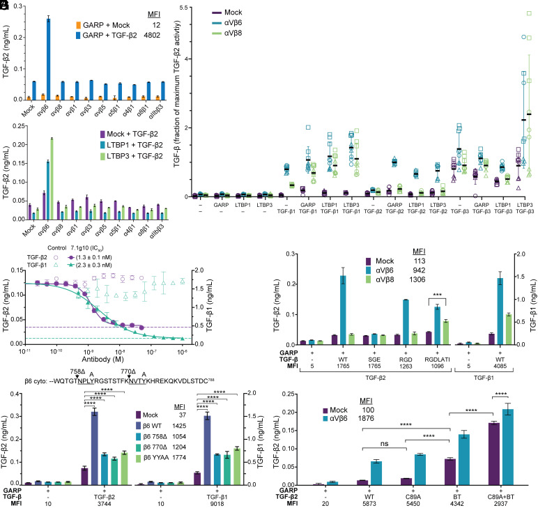
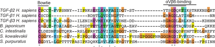
Activation of latent transforming growth factor (TGF)-β2 is incompletely understood. Unlike TGF-β1 and β3, the TGF-β2 prodomain lacks a seven-residue RGDLXX (L/I) integrin-recognition motif and is thought not to be activated by integrins. Here, we report the surprising finding that TGF-β2 contains a related but divergent 13-residue integrin-recognition motif (YTSGDQKTIKSTR) that specializes it for activation by integrin αVβ6 but not αVβ8. Both classes of motifs compete for the same binding site in αVβ6. Multiple changes in the longer motif underlie its specificity. ProTGF-β2 structures define interesting differences from proTGF-β1 and the structural context for activation by αVβ6. Some integrin-independent activation is also seen for proTGF-β2 and even more so for proTGF-β3. Our findings have important implications for therapeutics to αVβ6 in clinical trials for fibrosis, in which inhibition of TGF-β2 activation has not been anticipated.

Keywords: X-ray crystallography; integrins; transforming growth factor beta; TGFb2.


Conflict of interest statement

T.A.S. is a stockholder of Morphic Therapeutic and is a founder and board member of Morphic Therapeutic.

Figures

Fig. 1.
Fig. 1.
Integrin αVβ6–mediated activation of TGF-β2. (AG) CAGA luciferase reporter coculture assays characterizing integrin-mediated activation of TGF-β standardized with purified TGF-β growth factor. Median fluorescence intensities (MFI) for cell surface integrins or FLAG-tagged TGF-βs measured by FACS are reported in keys or below graphs in panels (A and EG). (A and B) Integrin dependence of activation of TGF-β2 coexpressed with GARP (A) or alone or with LTBP1 or LTBP3 (B). (C) Effect of milieu anchor co-expression on αVβ6 and αVβ8-mediated activation of TGF-β1, TGF-β2 and TGF-β3. TGF-β activation over three independent experiments is shown as the fraction of the amount of TGF-β2 released by αVβ6 transfectants from GARP/TGF-β2 transfectants in each experiment. (D) Inhibition of αVβ6-mediated activation of TGF-β2 and TGF-β1 by the αVβ6 function-blocking antibody 7.1g10 or mouse IgG X63 as isotype control. IC50 values are from fits to a four parameter dose response curve (solid lines). Dashed lines show the levels of αVβ6-independent activation. (E) Effect of replacements in the SGDQKTI sequence in TGF-β2 on αVβ6-mediated activation. (F) Effect of truncations and mutations that eliminate talin or kindlin binding sites in the integrin β6 cytoplasmic domain on TGF-β2 activation. A segment of the integrin β6 cytoplasmic domain sequence is shown with the talin and kindlin binding sites underlined. The positions of Tyr-to-Ala mutations (A) and truncations (∆) are indicated above the sequence. (G) Effect of Cys-to-Ala mutations in TGF-β2 inter-prodomain disulfides on activation. BT, bowtie triple Cys to Ala mutation. Data show mean ± SD of three biological replicates from representative experiments. Overall results from three such independent experiments are shown in SI Appendix, Fig. S2. P-values were determined using the Tukey multiple comparisons test following a two-way ANOVA (ns: P > 0.05, *P < 0.05, **P < 0.01, ***P < 0.001, ****P < 0.0001).
Fig. 2.
Fig. 2.
ProTGF-β2 structure. (A) Sequence alignment of TGF-β2 and β1 with deuterium uptake after 60 s keyed to the color scale (19). Sequences of disordered regions are italicized. Numbering is based on the immature protein, and dots represent decadal positions in TGF-β2. Structural elements are labeled and shown as lines for TGF-β2 (Upper) and TGF-β1 (Lower). Assoc, association region; F, fastener; BT, bowtie; arrowhead, proconvertase cleavage site. (B and C) Crystal structures of the proTGF-β2/Nb18 complex (B) and proTGF-β1 with one monomer complexed with αVβ6 (PDB ID:5ffo) (C). Nanobodies and prodomains are shown in ribbon cartoon and growth factor domains are shown as solvent-accessible surfaces. Cysteine sidechains are shown in yellow stick. Termini that flank disordered regions are shown as Cα spheres. Integrin binding loops that are disordered in TGF-β2 are shown as orange dashes and that rearrange in TGF-β1 are depicted in orange. Other disordered regions are shown as dashes in the same color as their domains. (D) Electron density for the fastener element of the TGF-β2 Nb18 complex shown in stick with the 2Fo–Fc map contoured at 1σ in mesh. (E and F) The three bowtie disulfides of the TGF-β2/Nb9 complex (E) and the two bowtie disulfides of TGF-β1 (5ffo) (F). One TGF-β monomer is colored blue, the other is colored green, and disulfides are shown as yellow sticks. (G) Comparison of integrin-binding loops from both Nb complex structures (green and blue) to the αVβ6-bound TGF-β1 monomer (orange) and the unbound TGF-β1 monomer (pink) after superposition on arm domains. Residues T256 and G276 in TGF-β2 are equivalent in sequence alignments to residues F239 and M253 in TGF-β1 and are shown as spheres.
Fig. 3.
Fig. 3.
Defining the integrin αVβ6 binding site in TGF-β2. (AD) Affinity measurements with fluorescence polarization (FP). TGF-β1 and TGF-β2 proteins and peptides were used to compete binding of 10 nM FITC-TGF-β3 GRGDLGRLK peptide to 20 nM αVβ6 ectodomain. Data in panels (A, C, and D) are mean ± SE of three independent experiments [five for the 13-mer peptide in panels (C and D)] each performed in duplicate and were fitted to a variable slope (four-parameter) dose–response curve. (A) Comparison of intact TGF-β1 and TGF-β2 to peptides. (B) Summary table of KD’s measured in this study. Fold change comparisons are enclosed in brackets and are relative to proTGF-β1, proTGF-β2, or the TGF-β2 13-mer peptide as indicated. (C and D) TGF-β2 13-mer peptides with Ala mutations. Panels (C and D) display the same data for the wild-type 13-mer. (E) Inhibition of αVβ6-mediated activation of TGF-β2 by the K265A 13-mer TGF-β2 peptide compared to the 9-mer TGF-β1 peptide and the αVβ6 antibody 7.1g10. Data were fit to a four-parameter dose–response curve to calculate IC50 values. (F) Effect of mutations in the integrin αVβ6 binding motif in TGF-β2 on activation. Expi293 GARP/TGF-β2 cotransfectants were cocultured with mock or αVβ6 Expi293 transfectants and CAGA-reporter cells. Data are mean SD of three technical replicates from a representative experiment. Overall data from three independent experiments are reported in SI Appendix, Fig. S5E. To control for mutants with lower expression, WT GARP/TGF-β2 cotransfection with 1/4 amount of plasmid was included. MFI for cell surface integrins and FLAG-tagged TGF-β2 constructs are reported in the key and below the graph, respectively. (G) Correlation of TGF-β2 mutant peptide affinity (B) with the effect of the corresponding mutation in TGF-β2/GARP complexes on integrin αVβ6-dependent activation (activation with αVβ6 transfectants–activation with mock transfectants in F).
Fig. 4.
Fig. 4.
Bowtie and bowtie tail sequences of TGF-βs. Full-length TGF-β sequences were aligned with MAFFT (37); the portion between the β7 and β10 strands (Fig. 2A) is shown. The three human TGF-βs (Chordata, Vertebrata; all from RefSeq) were aligned with the sole TGF-β's from Branchiostoma japonicum (Chordata, Cephalochordata; UniProt: F6M2M4), Ciona intestinalis (Chordata, Tunicata; UniProt: Q4H2P5), Saccoglossus kowalevskii (Hemichordata; UniProt: A0A0U2L5S7), and Strongylocentrotus purpuratus (Echinodermata; UniProt: A0A7M7RFG3). The αVβ6-binding motifs are indicated for TGF-β2 (Top line) and for TGF-β1 and TGF-β3 (Lower line).

References

    1. Sanford L. P., et al. , TGFβ2 knockout mice have multiple developmental defects that are non-overlapping with other TGFβ knockout phenotypes. Development 124, 2659–2670 (1997). - PMC - PubMed
    1. Doetschman T., et al. , Transforming growth factor β signaling in adult cardiovascular diseases and repair. Cell Tissue Res. 347, 203–223 (2012). - PMC - PubMed
    1. Morikawa M., Derynck R., Miyazono K., TGF-β and the TGF-β family: Context-dependent roles in cell and tissue physiology. Cold Spring Harb. Perspect. Biol. 8, a021873 (2016). - PMC - PubMed
    1. Sun T., et al. , TGFβ2 and TGFβ3 isoforms drive fibrotic disease pathogenesis. Sci. Transl. Med. 13, eabe0407 (2021). - PubMed
    1. Hinck A. P., Mueller T. D., Springer T. A., Structural biology and evolution of the TGF-β family. Cold Spring Harb. Perspect. Biol. 8, a022103 (2016). - PMC - PubMed

Publication types

MeSH terms

LinkOut - more resources