. 2023 Aug 1;44(29):2763-2783.
doi: 10.1093/eurheartj/ehad262.

Cationic proteins from eosinophils bind bone morphogenetic protein receptors promoting vascular calcification and atherogenesis

Affiliations

Cationic proteins from eosinophils bind bone morphogenetic protein receptors promoting vascular calcification and atherogenesis

Zhaojie Meng et al. Eur Heart J. .

Abstract

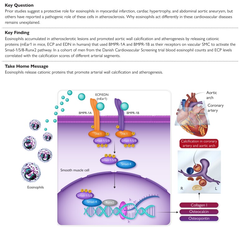
Aims: Blood eosinophil count and eosinophil cationic protein (ECP) concentration are risk factors of cardiovascular diseases. This study tested whether and how eosinophils and ECP contribute to vascular calcification and atherogenesis.

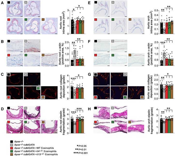
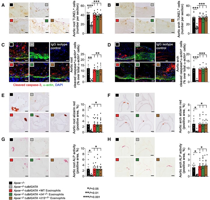
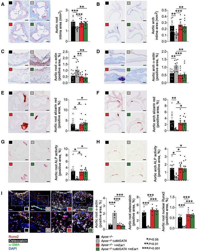
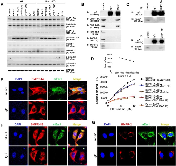
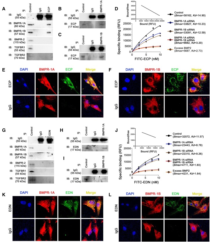
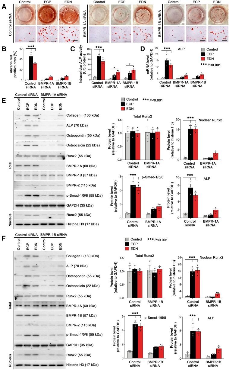
Methods and results: Immunostaining revealed eosinophil accumulation in human and mouse atherosclerotic lesions. Eosinophil deficiency in ΔdblGATA mice slowed atherogenesis with increased lesion smooth muscle cell (SMC) content and reduced calcification. This protection in ΔdblGATA mice was muted when mice received donor eosinophils from wild-type (WT), Il4-/-, and Il13-/- mice or mouse eosinophil-associated-ribonuclease-1 (mEar1), a murine homologue of ECP. Eosinophils or mEar1 but not interleukin (IL) 4 or IL13 increased the calcification of SMC from WT mice but not those from Runt-related transcription factor-2 (Runx2) knockout mice. Immunoblot analyses showed that eosinophils and mEar1 activated Smad-1/5/8 but did not affect Smad-2/3 activation or expression of bone morphogenetic protein receptors (BMPR-1A/1B/2) or transforming growth factor (TGF)-β receptors (TGFBR1/2) in SMC from WT and Runx2 knockout mice. Immunoprecipitation showed that mEar1 formed immune complexes with BMPR-1A/1B but not TGFBR1/2. Immunofluorescence double-staining, ligand binding, and Scatchard plot analysis demonstrated that mEar1 bound to BMPR-1A and BMPR-1B with similar affinity. Likewise, human ECP and eosinophil-derived neurotoxin (EDN) also bound to BMPR-1A/1B on human vascular SMC and promoted SMC osteogenic differentiation. In a cohort of 5864 men from the Danish Cardiovascular Screening trial and its subpopulation of 394 participants, blood eosinophil counts and ECP levels correlated with the calcification scores of different arterial segments from coronary arteries to iliac arteries.

Conclusion: Eosinophils release cationic proteins that can promote SMC calcification and atherogenesis using the BMPR-1A/1B-Smad-1/5/8-Runx2 signalling pathway.

Keywords: Bone morphogenetic protein receptors; Calcification; Eosinophil; Eosinophil cationic protein; Smooth muscle cell.


Conflict of interest statement

Conflict of interest All authors declare no conflict of interest for this contribution.

Figures

Structured Graphical Abstract
Eosinophils release cationic proteins ECP and EDN in humans (or their orthologue mEar1 in mice) that use BMPR-1A and BMPR-1B as their receptors on vascular SMC. Their ligation of these receptors activates downstream Smad-1/5/8 and Runx2 that induce the expression of osteogenic proteins as a mechanism that promotes arterial wall calcification and atherogenesis. ECP, eosinophilic cationic protein; EDN, eosinophil-derived neurotoxin; mEar1, mouse eosinophil-associated-ribonuclease-1; BMPR, bone morphogenetic protein receptor.
Figure 1
Figure 1
Eosinophils accelerate atherogenesis in apoe−/− mice. Apoe−/− and Apoe−/−ΔdblGATA mice were fed with an atherosclerotic diet for 12 weeks. Apoe−/−ΔdblGATA mice were also reconstituted with eosinophils from wild-type, Il4−/−, or Il13−/− mice every 2 weeks. Aortic root intima area, scale: 500 μm (A), α-actin+ smooth muscle cell contents in arterial wall, scale: 200 μm (B), Sirius red collagen staining in grade, scale: 200 μm (C), and elastin fragmentation in grade, scale: 200 μm (D). Aortic arch intima area, scale: 500 μm (E), α-actin+ smooth muscle cell contents in arterial wall, scale: 200 μm (F), Sirius red collagen staining in grade, scale: 200 μm (G), and elastin fragmentation in grade, scale: 200 μm (H). The results are expressed as mean ± standard error of mean of 9–10 mice per group.
Figure 2
Figure 2
Eosinophils promote arterial calcification and lesion apoptosis in apoe−/− mice. Apoe−/− and Apoe−/−ΔdblGATA mice were fed with an atherosclerotic diet for 12 weeks. Apoe−/−ΔdblGATA mice were reconstituted with eosinophils from WT, Il4−/−, or Il13−/− mice every 2 weeks. (A) Aortic root and (B) arch TUNEL-positive apoptotic cell contents. Scale: 100 μm. (C) Root and (D) arch immunofluorescence staining for cleaved caspase-3 + α-actin+ smooth muscle cell. IgG isotypes were used as experimental controls. Scale: 50 μm. (E) Root and (F) arch Alizarin red-positive calcium deposition. Scale: 500 μm. (G) Root and (H) arch alkaline phosphatase activity-positive area. Scale: 500 μm. The results are expressed as mean ± standard error of mean of 9–10 mice per group.
Figure 3
Figure 3
Mouse eosinophil-associated-ribonuclease-1 exacerbates atherogenesis and arterial calcification in apoe−/− mice. Apoe−/− and Apoe−/− ΔdblGATA mice were fed with an atherosclerotic diet and administrated with recombinant mouse eosinophil-associated-ribonuclease-1 (5 μg/mouse/time) twice per week for 12 weeks. (A) Aortic root and (B) arch intima area. Scale: 500 μm. (C) Root and (D) arch α-actin+ smooth muscle cell contents. Scale: 200 μm. (E) Root and (F) arch Alizarin red-positive calcium deposition. Scale: 500 μm. (G) Root and (H) arch alkaline phosphatase activity-positive area. Scale: 500 μm. (I) Aortic root α-actin, osteocalcin, and nuclear Runt-related transcription factor-2 contents by immunofluorescence staining. Scale: 50 μm (top) and 25 μm (bottom). The results are expressed as mean ± standard error of mean of 10–11 (A–H) and 5 mice (I) per group.
Figure 4
Figure 4
Mouse eosinophil-associated-ribonuclease-1 aggravates vascular smooth muscle cell calcification via Runt-related transcription factor-2. Wild-type and Runt-related transcription factor-2 KO mouse aortic smooth muscle cell were exposed to osteogenic media with or without mouse eosinophil-associated-ribonuclease-1, interleukin-4, or interleukin-13 proteins, eosinophil lysates from wild-type, Il4−/− or Il13−/− mice, and wild-type eosinophil lysate plus anti-mouse eosinophil-associated-ribonuclease-1 antibody. (A) Representative images of stained dishes (top), photomicrographs (bottom, Scale: 200 μm). Quantification of (B) Alizarin red staining for mineralized calcium and (C) intracellular alkaline phosphatase activity. (D) Immunoblots of collagen I, alkaline phosphatase, osteocalcin, osteopontin, total and nuclear Runt-related transcription factor-2. (E) Immunofluorescence staining of α-actin, osteocalcin, and Runt-related transcription factor-2. Scale: 50 μm. The results are expressed as mean ± standard error of mean of three independent experiments.
Figure 5
Figure 5
Mouse eosinophil-associated-ribonuclease-1 uses bone morphogenetic protein receptors to activate the smad-1/5/8-Runt-related transcription factor-2 signalling pathway. (A) Wild-type and Runt-related transcription factor-2 KO mouse smooth muscle cell were exposed to osteogenic media with or without mouse eosinophil-associated-ribonuclease-1, interleukin-4, interleukin-13, eosinophil lysates from wild-type, Il4−/− or Il13−/− mice. Immunoblots of different bone morphogenetic protein receptors, TGFβRs, and phosphorylated Smad-2, Smad-3, and Smad-1/5/8. (B–G) Wild-type smooth muscle cell were exposed to osteogenic media for 14 days and stimulated with mouse eosinophil-associated-ribonuclease-1 for 30 min before collection. Immunoprecipitation with anti-mouse eosinophil-associated-ribonuclease-1 (B), anti-bone morphogenetic protein receptor-1A (C, top) and anti-bone morphogenetic protein receptor-1B (C, bottom) antibodies, followed by immunoblotting detection of different bone morphogenetic protein receptors, TGFβRs, or mouse eosinophil-associated-ribonuclease-1. (D) FITC-mouse eosinophil-associated-ribonuclease-1 (0∼10.0 nM) binding affinity and Scatchard plot on smooth muscle cell treated with or without bone morphogenetic protein receptor siRNA or excessive BMP2 (1000 ng/mL). Immunofluorescence double-staining of mouse eosinophil-associated-ribonuclease-1 with bone morphogenetic protein receptor-1A (E), bone morphogenetic protein receptor-1B (F) or bone morphogenetic protein receptor-2 (G).
Figure 6
Figure 6
Tamoxifen-induced depletion of bone morphogenetic protein receptor-1A and bone morphogenetic protein receptor-1B in aortic smooth muscle cell blocks mouse eosinophil-associated-ribonuclease-1-induced calcification. Aortic smooth muscle cell were isolated from BMPR-1Afl/fl/1Bfl/fl and Myh11CreER(T)BMPR-1Afl/fl/1Bfl/fl mice and treated with tamoxifen (0.5 µM) or vehicle (DMSO) for 24 h. Cells were then exposed to osteogenic media with or without mouse eosinophil-associated-ribonuclease-1 for 14 days. (A) Representative images of Alizarin red-stained dishes (top) and photomicrographs (bottom, scale: 200 um), and quantifications of Alizarin red staining for mineralized calcium and intracellular alkaline phosphatase activity. (B and C) RT-PCR detected the expression osteogenetic genes (B), bone morphogenetic protein receptor, Runt-related transcription factor-2, and TGF-β receptors (C). (D and E) Immunoblot analysis of bone morphogenetic protein receptor signalling molecules and osteogenetic proteins (D) and TGF-β receptors and signalling molecules (E). lRepresentative images are presented to the left (A, D, E). Data are mean ± standard error of mean from three independent experiments.
Figure 7
Figure 7
Binding of eosinophil cationic protein and eosinophil-derived neurotoxin on human smooth muscle cell bone morphogenetic protein receptors. Human smooth muscle cell were exposed to osteogenic media for 14 days and stimulated with recombinant eosinophil cationic protein or eosinophil-derived neurotoxin for 30 min before collection. Immunoprecipitation with anti-eosinophil cationic protein (A), anti-bone morphogenetic protein receptor-1A (B) and anti-bone morphogenetic protein receptor-1B (C) antibodies, followed by immunoblotting detection of different bone morphogenetic protein receptors, TGFβRs, and eosinophil cationic protein. (D) FITC-eosinophil cationic protein (0∼10.0 nM) binding affinity and Scatchard plot on smooth muscle cell treated with or without bone morphogenetic protein receptor siRNA or excessive BMP2 (1000 ng/mL). Immunofluorescence double-staining of eosinophil cationic protein and bone morphogenetic protein receptor-1A (E) or bone morphogenetic protein receptor-1B (F). Immunoprecipitation with anti-eosinophil-derived neurotoxin (G), anti-bone morphogenetic protein receptor-1A (H), and anti-bone morphogenetic protein receptor-1B (I) antibodies, followed by immunoblotting detection of different bone morphogenetic protein receptors, TGFBRs, and eosinophil-derived neurotoxin. (J) FITC-eosinophil-derived neurotoxin (0∼10.0 nM) binding affinity and Scatchard plot on smooth muscle cell treated with or without with bone morphogenetic protein receptor siRNA or excessive BMP2 (1000 ng/mL). Immunofluorescence double-staining of eosinophil-derived neurotoxin and bone morphogenetic protein receptor-1A (K) or bone morphogenetic protein receptor-1B (L).
Figure 8
Figure 8
Human bone morphogenetic protein receptor silencing ameliorates eosinophil cationic protein- and eosinophil-derived neurotoxin-induced human smooth muscle cell calcification. Human vascular smooth muscle cell were transfected with bone morphogenetic protein receptor-1A, bone morphogenetic protein receptor-1B, or control siRNA and then exposed to osteogenic media with or without recombinant eosinophil cationic protein or eosinophil-derived neurotoxin for 14 days. (A) Representative images of stained dishes (top) and photomicrographs (bottom. Scale: 200 μm.) along with quantification (B) of Alizarin red staining for mineralized calcium, (C) intracellular alkaline phosphatase activity, and (D) alkaline phosphatase gene expression. Immunoblot analysis of different bone morphogenetic protein receptor-Smad-Runt-related transcription factor-2 signalling proteins in human vascular smooth muscle cell treated with or without eosinophil cationic protein or eosinophil-derived neurotoxin after cells were treated with bone morphogenetic protein receptor-1A (E), bone morphogenetic protein receptor-1B (F), and control siRNAs. The results are expressed as mean ± standard error of mean from three to four independent experiments.

Comment in

References

    1. Hirasawa R, Shimizu R, Takahashi S, Osawa M, Takayanagi S, Kato Y, et al. Essential and instructive roles of GATA factors in eosinophil development. J Exp Med 2002;195:1379–1386. 10.1084/jem.20020170 - DOI - PMC - PubMed
    1. Collins PD, Marleau S, Griffiths-Johnson DA, Jose PJ, Williams TJ. Cooperation between interleukin-5 and the chemokine eotaxin to induce eosinophil accumulation in vivo. J Exp Med 1995;182:1169–1174. 10.1084/jem.182.4.1169 - DOI - PMC - PubMed
    1. Weller PF, Spencer LA. Functions of tissue-resident eosinophils. Nat Rev Immunol 2017;17:746–760. 10.1038/nri.2017.95 - DOI - PMC - PubMed
    1. Miller M, Sung KL, Muller WA, Cho JY, Roman M, Castaneda D, et al. Eosinophil tissue recruitment to sites of allergic inflammation in the lung is platelet endothelial cell adhesion molecule independent. J Immunol 2001;167:2292–2297. 10.4049/jimmunol.167.4.2292 - DOI - PubMed
    1. Lehrer RI, Szklarek D, Barton A, Ganz T, Hamann KJ, Gleich GJ. Antibacterial properties of eosinophil major basic protein and eosinophil cationic protein. J Immunol 1989;142:4428–4434. 10.4049/jimmunol.142.12.4428 - DOI - PubMed

Publication types