. 2023 Jul 11;56(7):1548-1560.e5.
doi: 10.1016/j.immuni.2023.05.006. Epub 2023 Jun 5.

Platelets and mast cells promote pathogenic eosinophil recruitment during invasive fungal infection via the 5-HIAA-GPR35 ligand-receptor system

Affiliations

Platelets and mast cells promote pathogenic eosinophil recruitment during invasive fungal infection via the 5-HIAA-GPR35 ligand-receptor system

Marco De Giovanni et al. Immunity. .

Abstract

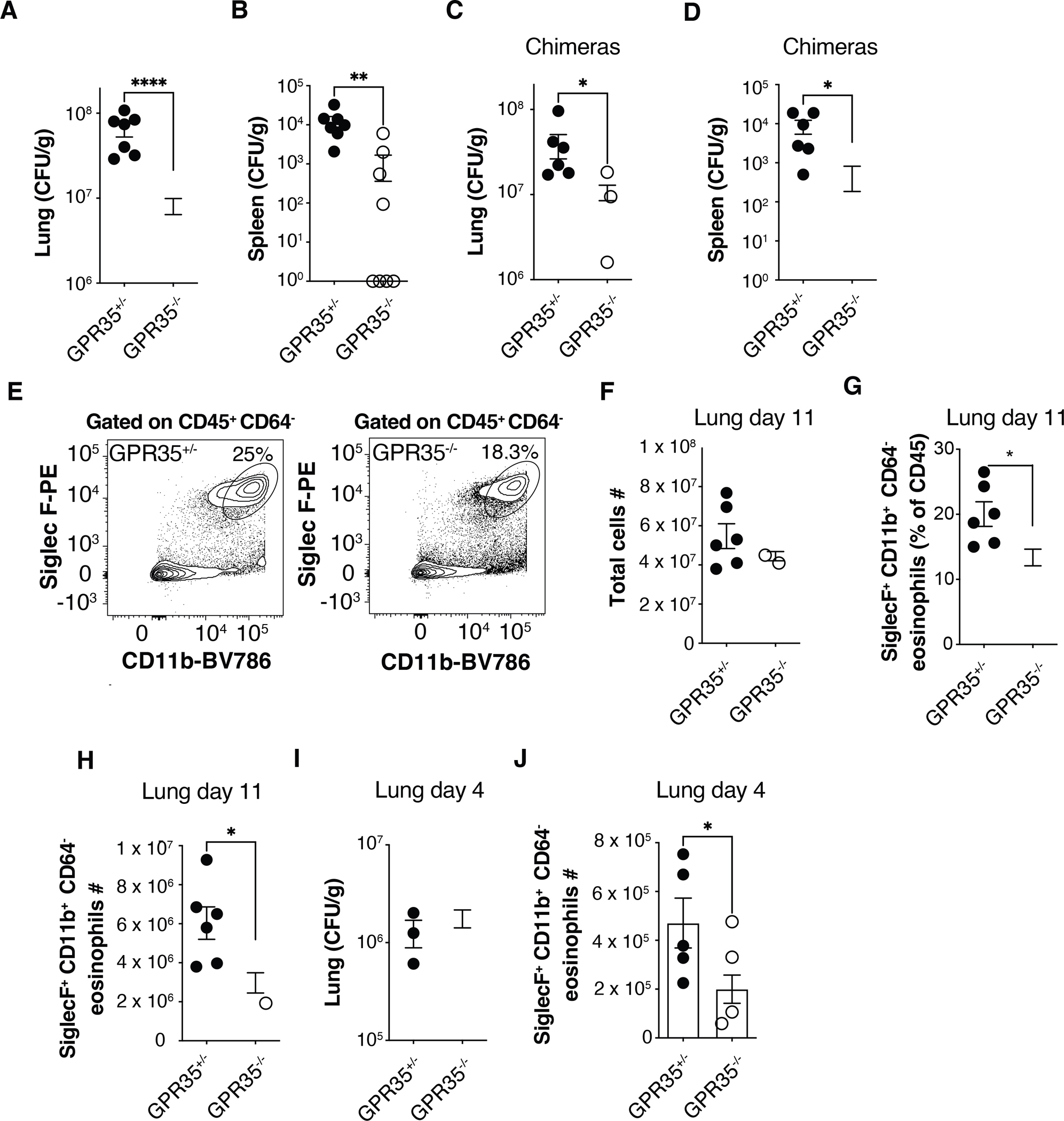
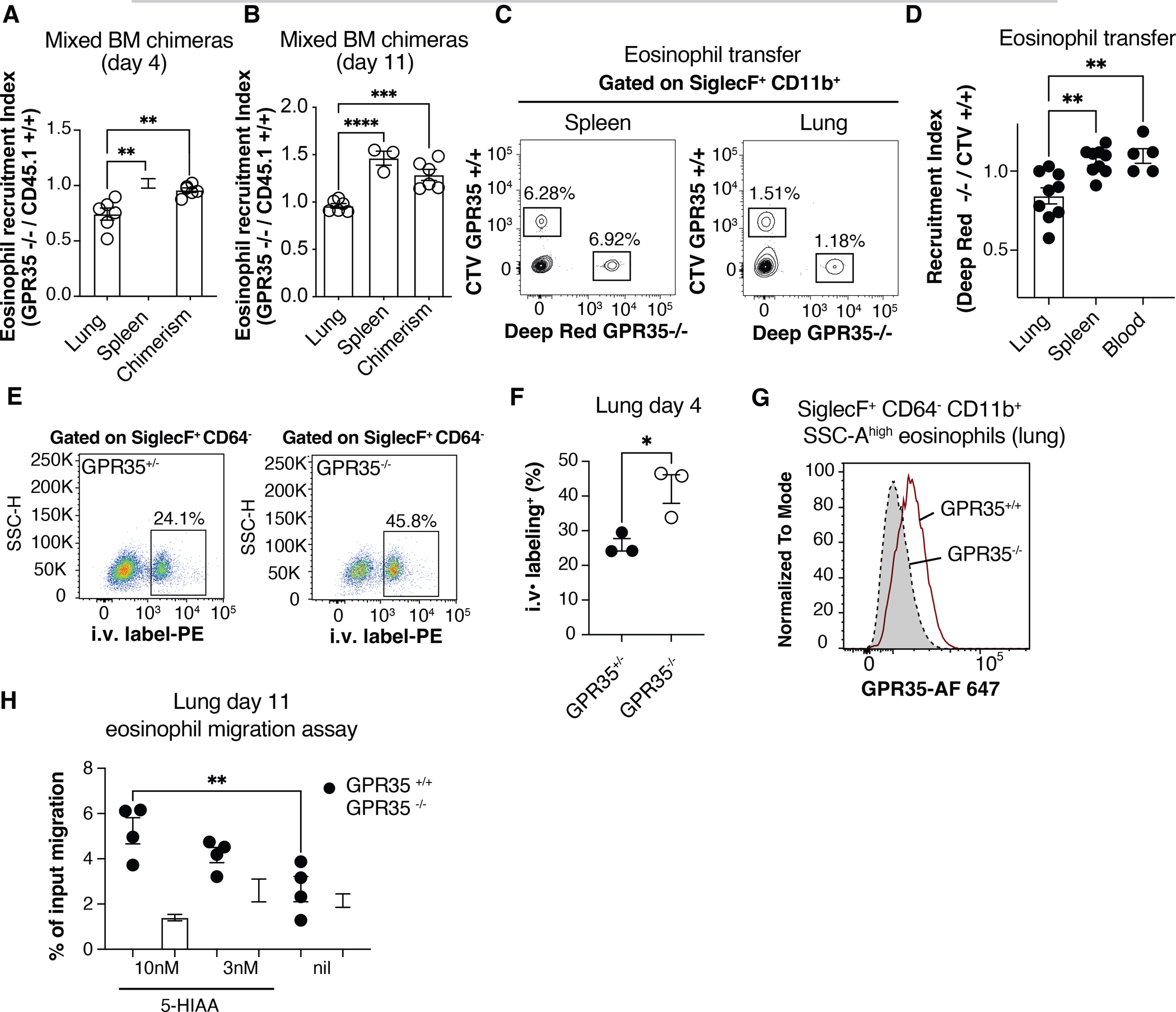
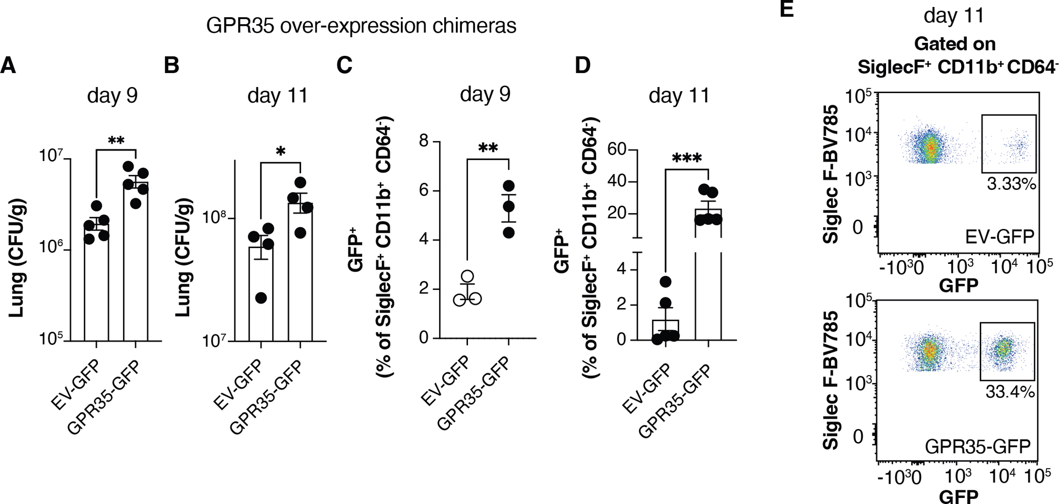
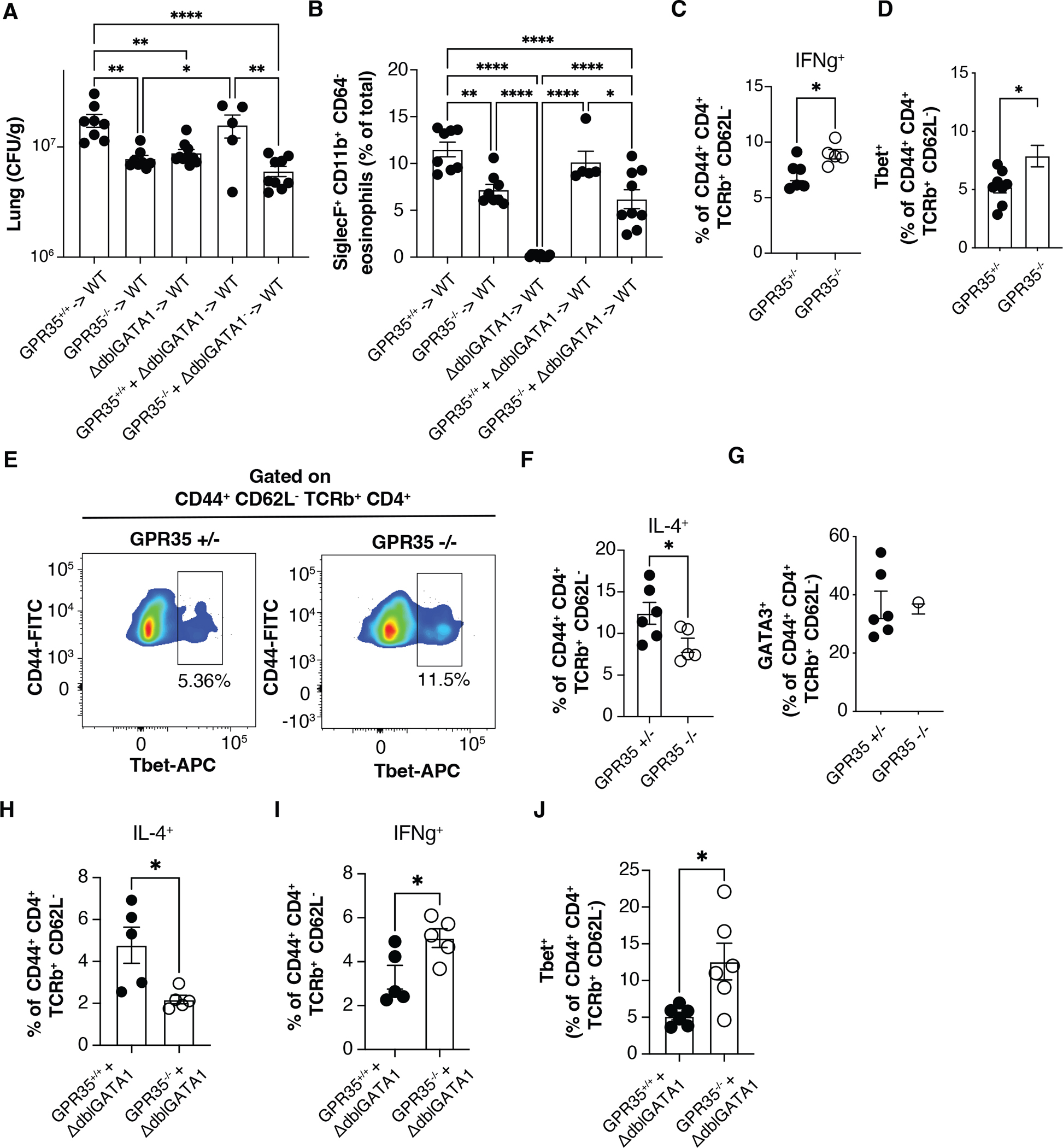
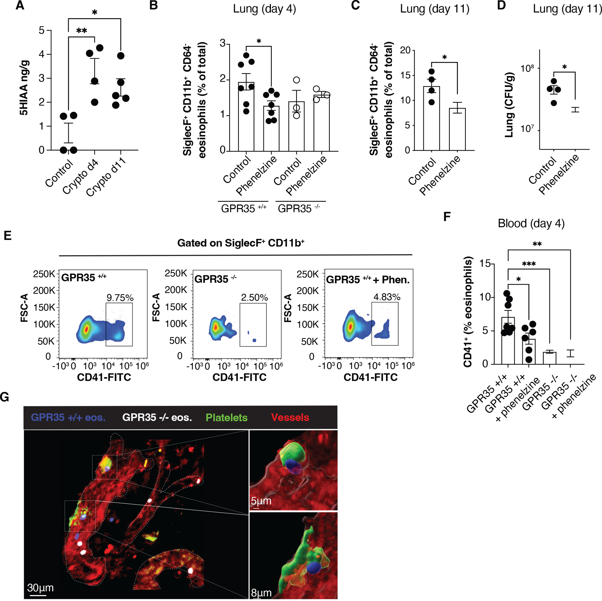
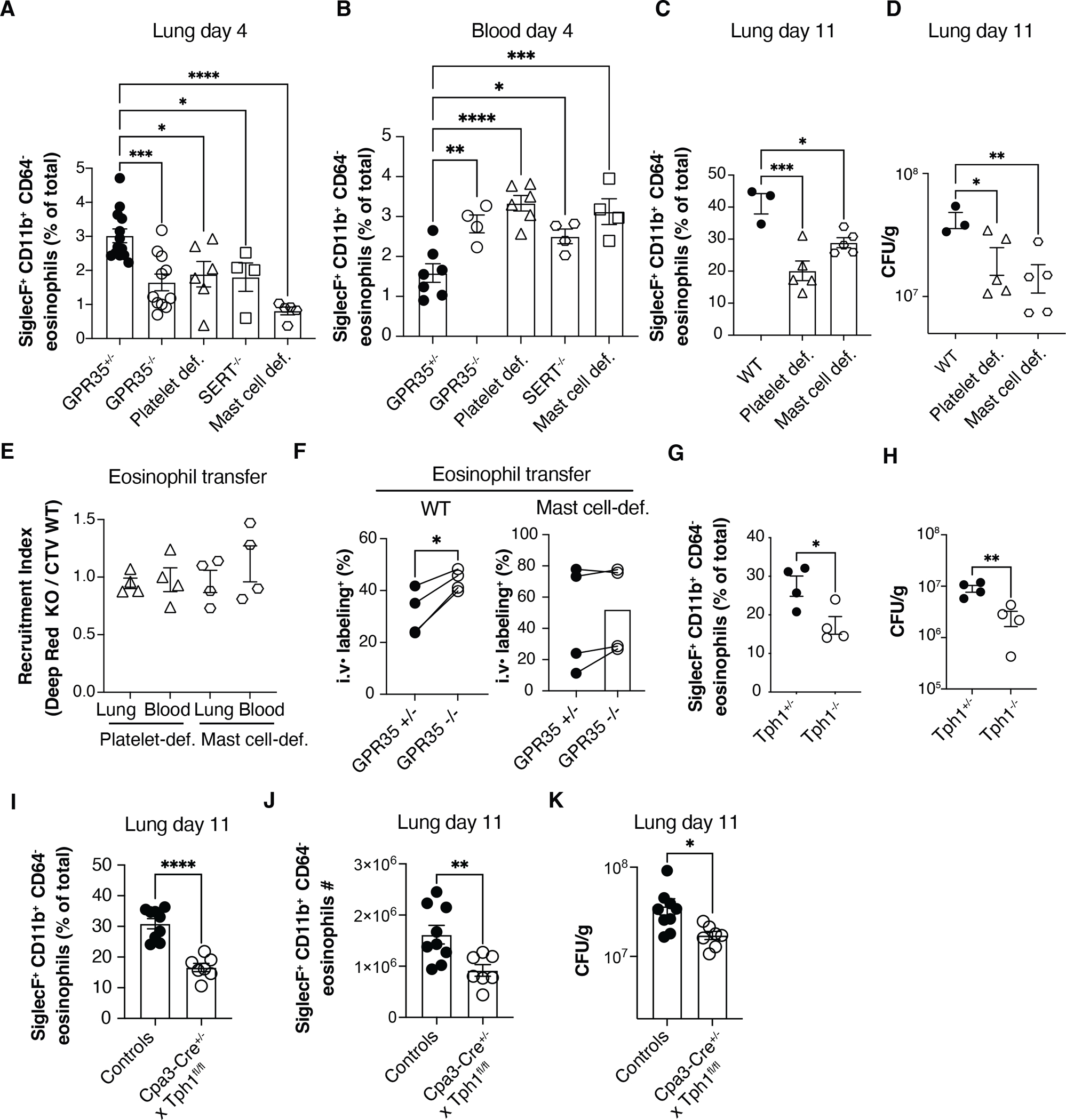
Cryptococcus neoformans is the leading cause of fungal meningitis and is characterized by pathogenic eosinophil accumulation in the context of type-2 inflammation. The chemoattractant receptor GPR35 is expressed by granulocytes and promotes their migration to the inflammatory mediator 5-hydroxyindoleacetic acid (5-HIAA), a serotonin metabolite. Given the inflammatory nature of cryptococcal infection, we examined the role of GPR35 in the circuitry underlying cell recruitment to the lung. GPR35 deficiency dampened eosinophil recruitment and fungal growth, whereas overexpression promoted eosinophil homing to airways and fungal replication. Activated platelets and mast cells were the sources of GPR35 ligand activity and pharmacological inhibition of serotonin conversion to 5-HIAA, or genetic deficiency in 5-HIAA production by platelets and mast cells resulted in more efficient clearance of Cryptococcus. Thus, the 5-HIAA-GPR35 axis is an eosinophil chemoattractant receptor system that modulates the clearance of a lethal fungal pathogen, with implications for the use of serotonin metabolism inhibitors in the treatment of fungal infections.

Keywords: 5-HIAA; Cryptococcus; GPR35; cell migration; eosinophils; lung; mast cells; platelets.


Conflict of interest statement

Declaration of interests The authors declare no competing interests.

Figures

Figure 1.
Figure 1.. GPR35 in BM-derived cells promotes increased Cryptococcus burden and eosinophil recruitment to the infected lung
(A-D) Quantification of C. neoformans CFUs in lung and spleen of GPR35+/− and GPR35−/− mice (A, B) or full chimeras (C, D) 11 days after intranasal infection. A, B, n=7–9; C, D, n=6. Data are pooled from 2 independent experiments. (E) Flow cytometry plots showing percentages of SiglecF+ CD11b+ eosinophils in GPR35+/− (left) and GPR35−/− (right) out of CD45+ CD64 cells in the lung. (F-H) Quantification of total cell numbers (F), SiglecF+ CD11b+ CD64 eosinophil percentages (out of CD45+ cells, G) and numbers (H) in the lung of GPR35+/− and GPR35−/− full chimeras 11 days after intranasal infection. n=6. Data are pooled from two independent experiments. (I, J) Quantification of C. neoformans CFUs (I) and SiglecF+ CD11b+ CD64 eosinophil numbers (J) in lungs of GPR35+/− and GPR35−/− mice 4 days after intranasal infection. F-H, n=6; I, n=3–5; J=5–7. Data are pooled from two independent experiments. * p<0.05; ** p<0.005; **** p<0.0001. Data are presented as mean ± SEM. See also Figure S1.
Figure 2.
Figure 2.. Intrinsic requirement for GPR35 in eosinophils
(A, B) Quantification of eosinophil recruitment index in lung and spleen of C. neoformans infected CD45.2+ GPR35−/− + CD45.1+ GPR35+/+ mixed chimeras, 4 (A) or 11(B) days after intranasal infection. The chimerism was calculated as the CD45.2+ / CD45.1+ ratio within blood B220+ cells at the time of tissue isolation. n=3–6. Data are pooled from 2 independent experiments. (C) Flow cytometry plots showing transferred CTV+ GPR35+/+ and Deep Red+ GPR35−/− BM-derived eosinophils in spleen and lung of mice infected 5 days before with C. neoformans, 24 hours after cell injection. (D) Quantification of transferred eosinophils determined as in C, shown as recruitment index in lung, spleen and blood of infected mice, 24 hr after cell transfer. n=5–9. Data are pooled from three independent experiments. (E, F) Flow cytometry plots (E) and quantification (F) of 2 min intravascular labeled (i.v. CD45-PE+) SiglecF+ CD64 eosinophils in lungs of GPR35+/− (left) and GPR35−/− (right) mice 4 days after C. neoformans infection. n=3. Data are representative of two independent experiments. (G) Flow cytometry plot showing GPR35 expression in GPR35+/+ and GPR35−/− SiglecF+ CD11b+ CD64 eosinophils in the lungs 11 days after intranasal infection. Data are representative of at least 2 independent experiments. (H) Transwell migration assay to 5-HIAA (nM) of GPR35+/+ and GPR35−/− eosinophils isolated from C. neoformans infected lung 11 days after infection. Nil indicates no added chemoattractant. n=3–4. Data are representative of 3 independent experiments. * p<0.05; ** p<0.005; *** p<0.0005; **** p<0.0001. Data are presented as mean ± SEM. See also Figure S1.
Figure 3.
Figure 3.. GPR35 over-expression augments Cryptococcus infection and eosinophil recruitment
(A-D) Quantification of C. neoformans CFUs (A, B) and GFP+ eosinophil percentages (C, D) in empty vector (EV)-GFP or GPR35-GFP overexpressing chimeras, 9 (A, C) or 11 (B, D) days after intranasal infection. A, B, D, n=4–5; C, n=3. E. Flow cytometry plots showing GFP+ percentages within SiglecF+ CD11b+ CD64 eosinophils quantified in D. Data are pooled (A) or representative (B-D) of two independent experiments. * p<0.05; ** p<0.005; *** p<0.0005. Data are presented as mean ± SEM. See also Figure S2.
Figure 4.
Figure 4.. GPR35 expressing eosinophils sustain Cryptococcus infection
(A, B) Quantification of C. neoformans CFUs (A) and SiglecF+ CD11b+ CD64 eosinophil percentages (B) in the lung of the indicated full and mixed BM chimeras 11 days after intranasal infection. n=5–10. Data are pooled from two independent experiments. (C, D) Quantification of ex-vivo stimulated IFNγ+ cells (C) and Tbet+ Th1 cell percentages (D) out of CD4+ CD44+ TCRβ+ CD62L in the lung of GPR35+/+ and GPR35−/− full chimeras 11 days after intranasal infection. C, n=5–6; d, n=8–9. Data are pooled from 3 (C) or 2 (D) independent experiments. (E) Flow cytometry plot showing Tbet+ Th1 percentages in GPR35+/− (left) and GPR35−/− (right) infected mice quantified in D. (F, G) Quantification of ex-vivo stimulated IL-4+ cell percentages (F) and GATA3+ Th2 cell percentages (G) out of CD4+ CD44+ TCRβ+ CD62L cells in the lung of GPR35+/+ and GPR35−/− mice 11 days after intranasal infection. n=5–6. (H-J) Quantification of ex-vivo stimulated IL-4+ (H), IFNγ+ (I) and Tbet+ (J) cells out of CD4+ CD44+ TCRβ+ CD62L cells in the lung of GPR35−/− + ΔdblGATA and control mixed BM chimeras, 11 days after intranasal infection. n=5–6. Data are pooled from two independent experiments. * p<0.05; ** p<0.005; **** p<0.0001. Data are presented as mean ± SEM. See also Figures S3 and S4.
Figure 5.
Figure 5.. 5-HIAA is required for GPR35-mediated eosinophil recruitment to the infected lung
(A) Quantification by ELISA of 5-HIAA concentrations in the lung of uninfected mice or C. neoformans infected mice (4 or 11 days after infection). n=4–5. Data are pooled from two independent experiments. (B-D) Quantification of SiglecF+ CD11b+ CD64 eosinophil percentages (B, C) and C. neoformans CFUs (D) in the lung of GPR35+/+ and GPR35−/− mice not treated or treated with phenelzine, 4 (B) or 11 (C, D) days after intranasal infection. B, n=3–7; C, D, n=4. Data are pooled from two independent experiments. e, f. Flow cytometry plots (E) and quantification (F) of CD41+ SiglecF+ CD64 eosinophils in the blood of not treated or phenelzine-treated GPR35+/+ and GPR35−/− mice, 4 days after intranasal infection. n=3–7. Data are pooled from two independent experiments. (G) Multiphoton micrograph of C. neoformans infected lung from platelet-reporter mice (Pf4-Cre x mTmG mice), 4 days after intranasal infection. Transferred BM-derived GPR35+/+ (CTV+, blue) and GPR35−/− (Deep Red+, white) eosinophils, and endogenous platelets (green) and vessels (red) are shown. Data are representative of at least 2 independent experiments. * p<0.05; ** p<0.005; *** p<0.0005. Data are presented as mean ± SEM.
Figure 6.
Figure 6.. Platelets and mast cells are required for GPR35-mediated eosinophil recruitment to the infected lung
(A, B) Quantification of SiglecF+ CD11b+ CD64 eosinophil percentages in lungs (A) and blood (B) of C. neoformans infected GPR35+/+, GPR35−/−, platelet-deficient (cmlp−/−), SERT−/− and mast cell-deficient (KitW/v) mice, 4 days after intranasal injection. A, n=4–13; B, n=4–7. (C, D) Quantification of SiglecF+ CD11b+ CD64 eosinophil percentages (C) and Cryptococcus CFUs (D) in the lung of WT, platelet-deficient or mast cell-deficient mice, 11 days after intranasal infection. n=3–5. Data are pooled from two independent experiments. (E, F) Quantification of transferred BM-derived CTV+ GPR35+/+ and Deep Red+ GPR35−/− eosinophil recruitment index (E) and intravascular labeling (F), in the lung and blood of C. neoformans infected (day 5) WT, platelet-deficient and/or mast cell-deficient mice, 24 hours after cell transfer. n=4. (G, H) Quantification of SiglecF+ CD11b+ CD64 eosinophil percentages (G) and C. neoformans CFUs (H) in the lung of Tph1+/− and Tph1−/− full BM chimeras, 11 days after intranasal infection. n=4. (I-K) Quantification of SiglecF+ CD11b+ CD64 eosinophil percentages (I), absolute numbers (J) or Cryptococcus CFUs in the lung (K) of Cpa3-Cre+ x Tph1fl/fl mice and littermate controls (Cpa3-Cre+ Tph1fl/wt and Cpa3-Cre x Tph1fl/fl) 11 days after intranasal infection. n=7–9. * p<0.05; ** p<0.005; *** p<0.0005, **** p<0.0001. Data are pooled from at least 2 independent experiments. Data are presented as mean ± SEM.

References

    1. Brown GD, Denning DW, Gow NA, Levitz SM, Netea MG, and White TC (2012). Hidden killers: human fungal infections. Sci Transl Med 4, 165rv113. 10.1126/scitranslmed.3004404. - DOI - PubMed
    1. Saluja R, Metz M, and Maurer M (2012). Role and relevance of mast cells in fungal infections. Front Immunol 3, 146. 10.3389/fimmu.2012.00146. - DOI - PMC - PubMed
    1. Mok AC, Mody CH, and Li SS (2021). Immune Cell Degranulation in Fungal Host Defence. J Fungi (Basel) 7. 10.3390/jof7060484. - DOI - PMC - PubMed
    1. Iyer KR, Revie NM, Fu C, Robbins N, and Cowen LE (2021). Treatment strategies for cryptococcal infection: challenges, advances and future outlook. Nat Rev Microbiol 19, 454–466. 10.1038/s41579-021-00511-0. - DOI - PMC - PubMed
    1. Lopes JP, Stylianou M, Backman E, Holmberg S, Ekoff M, Nilsson G, and Urban CF (2019). Cryptococcus neoformans Induces MCP-1 Release and Delays the Death of Human Mast Cells. Front Cell Infect Microbiol 9, 289. 10.3389/fcimb.2019.00289. - DOI - PMC - PubMed

Publication types