. 2023 Jun;26(6):1032-1041.
doi: 10.1038/s41593-023-01316-5. Epub 2023 Jun 5.

Psychedelics promote plasticity by directly binding to BDNF receptor TrkB

Affiliations

Psychedelics promote plasticity by directly binding to BDNF receptor TrkB

Rafael Moliner et al. Nat Neurosci. 2023 Jun.

Abstract

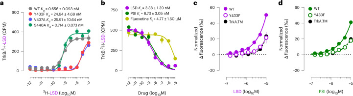
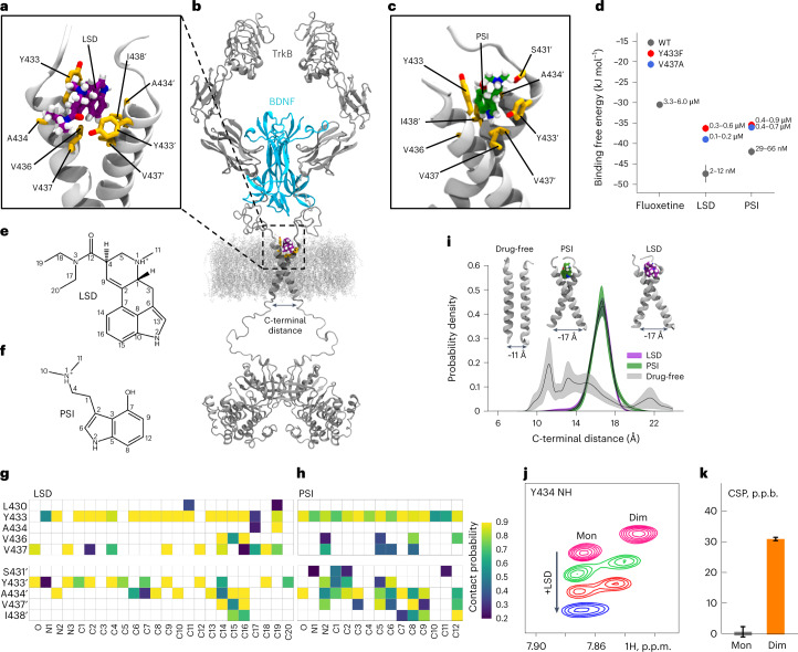
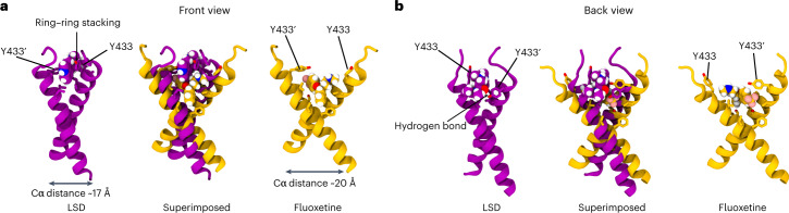
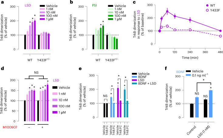
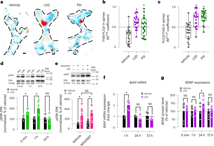
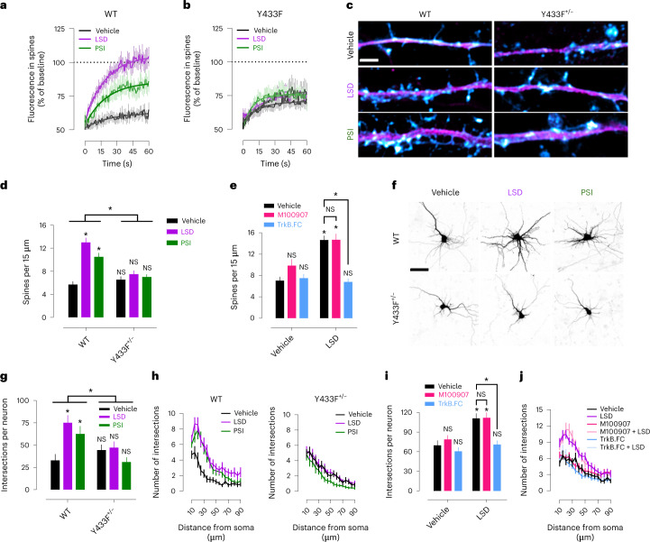
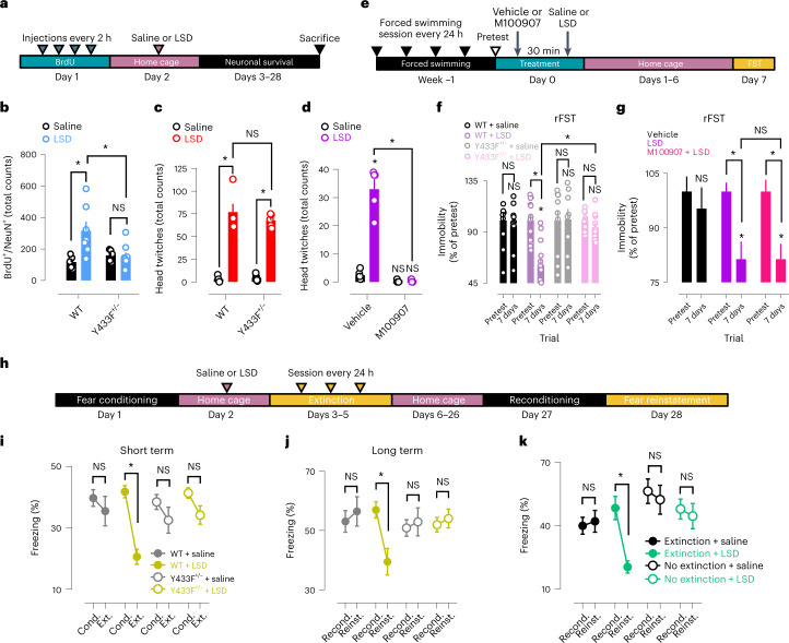
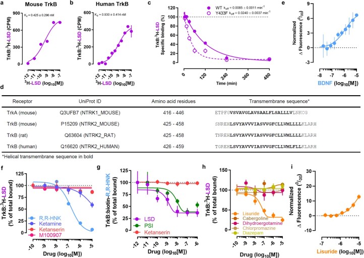
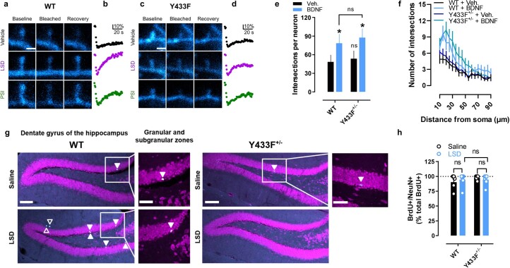
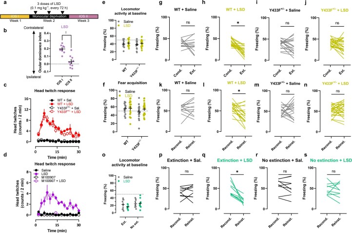
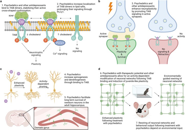
Psychedelics produce fast and persistent antidepressant effects and induce neuroplasticity resembling the effects of clinically approved antidepressants. We recently reported that pharmacologically diverse antidepressants, including fluoxetine and ketamine, act by binding to TrkB, the receptor for BDNF. Here we show that lysergic acid diethylamide (LSD) and psilocin directly bind to TrkB with affinities 1,000-fold higher than those for other antidepressants, and that psychedelics and antidepressants bind to distinct but partially overlapping sites within the transmembrane domain of TrkB dimers. The effects of psychedelics on neurotrophic signaling, plasticity and antidepressant-like behavior in mice depend on TrkB binding and promotion of endogenous BDNF signaling but are independent of serotonin 2A receptor (5-HT2A) activation, whereas LSD-induced head twitching is dependent on 5-HT2A and independent of TrkB binding. Our data confirm TrkB as a common primary target for antidepressants and suggest that high-affinity TrkB positive allosteric modulators lacking 5-HT2A activity may retain the antidepressant potential of psychedelics without hallucinogenic effects.


Conflict of interest statement

R.M., M.G., C.B., G.E., I.V., P.C.C. and E.C. are inventors in a patent application filed by the University of Helsinki that is related to these findings (GB2210278.4). E.C. has received speaker fees from Janssen Cilag. The other authors declare no competing interests.

Figures

Fig. 1
Fig. 1. Psychedelics bind to TrkB.
a, LSD binds with high affinity to WT TrkB, but binding is impaired by selective mutations in its binding pocket (Y433F and V437A). b, PSI and fluoxetine compete with LSD for TrkB binding. c,d, MST confirmed the interaction of LSD (c), and PSI (d), with TrkB, which is impaired by Y433F and TrkA.TM. CPM, counts per minute; M, molar. Data shown as mean ± s.e.m. Detailed statistics reported in Supplementary Table 1.
Fig. 2
Fig. 2. Characterization of the psychedelics binding site in the TrkB TMD.
ac, Representative MD snapshots showing the binding pocket for LSD (purple) (a) and PSI (green) (c) in the extracellular-facing crevice of the TrkB TMD dimer (gray). Side chains (yellow) of relevant binding site residues are displayed. A structural model of full-length TrkB dimer (gray) embedded in a lipid membrane is shown with bound BDNF (blue) and LSD (purple) (b). d, In silico binding free energy estimations for fluoxetine, LSD and PSI. Each free energy estimate (ΔG, circles) and its statistical error (SE, bars) were estimated from a separate set of FEP simulation (n = 1). Dissociation constants are given as a range with upper and lower bounds converted from ΔG − SE and ΔG + SE, respectively. e,f, Chemical structures of LSD (e) and PSI (f) with atom numbers annotated. g,h, Contact probability between binding pocket residues and LSD (g) or PSI (h). Residues of the second chain in the dimer are indicated with an apostrophe. i, Distributions of TMD dimer C-terminal distance show that LSD and PSI stabilize the cross-shaped conformation of TrkB favorable for receptor activation in a 40 mol% CHOL membrane. Lines represent the mean distribution, and bands represent the standard errors (n = 10 independent simulations). TMD conformations corresponding to indicated C-terminal distances and drug-bound states are shown in the inset. j, Expansions of overlaid 1H,15N-HSQC spectra of TrkB TMD in DMPC/DHPC bicelles showing the LSD-induced CSPs of resonance corresponding to Y433 amide group in the monomeric (Mon) and dimeric (Dim) states. p.p.m., parts per million. k, CSPs of the Y433 amide group in Mon and Dim states induced by the addition of LSD. p.p.b., parts per billion (10 p.p.b. = 0.01 p.p.m.). Data shown as mean ± s.e.m. Detailed statistics reported in Supplementary Table 1.
Fig. 3
Fig. 3. Different TrkB binding modes of LSD and fluoxetine.
a,b, Representative snapshots of atomistic MD simulations showing the front (a) and back (b) views of the binding pockets for LSD (purple) and fluoxetine (yellow) in the extracellular-facing crevice of TrkB TMD dimers. Side chains of relevant binding site residues are displayed. Superimposed structures of TrkB optimally bound to LSD or fluoxetine reveal that, while some residues involved in binding are shared (Y433 and V437), the binding modes are different. Fluoxetine binds at a site deeper within the dimer, locking the TMD dimers in a more open cross-shaped conformation (distance between the center of mass L451–L453 Cα atoms of each monomer ~20 Å). In contrast, LSD binds closer to the N-terminus of the TrkB TMD and establishes more stable interactions with the dimer: a hydrogen bond between the oxygen atom of the diethylamide group of LSD and the Y433 residue of one monomer, and pi-stacking of the aromatic backbone of the drug with the Y433 residue of the second monomer, locking the TMD dimer in a tighter cross-shaped conformation (L451–L453 Cα distance ~17 Å) compared with fluoxetine. Drugs are shown in van der Waals representation. Oxygen, nitrogen and hydrogen atoms are shown in red, blue and white, respectively.
Fig. 4
Fig. 4. Psychedelics promote TrkB dimerization.
a,b, LSD (a) and PSI (b) promote TrkB dimerization, but these effects are lost in Y433F+/−. c, Timeline of LSD-induced TrkB dimerization. d, M100907 does not prevent LSD-induced TrkB dimerization. e, TrkB receptor bodies (TrkB.FC) that hijack extracellular BDNF abolish the effects of both BDNF and LSD on dimerization. f, LSD potentiates the effects of low BDNF concentrations on TrkB dimerization. Data shown as mean ± s.e.m., *P < 0.05. NS, not significant. Detailed statistics reported in Supplementary Table 1.
Fig. 5
Fig. 5. Psychedelics recruit neurotrophic signaling through BDNF and TrkB.
a, Representative single-molecule localization microscopy maps of dendritic spines after treatment with vehicle, LSD and PSI. TrkB in light blue, PLCγ1 in yellow, TrkB:PLCγ1 in dark blue, PLCγ1:TrkB in red. Scale bar, 200 nm. b,c, Psychedelics increase colocalization of TrkB and PLCγ1 in dendritic spines. d, Timeline of LSD-induced increase in pERK. e, M100907 pretreatment does not prevent LSD-induced increase in pERK. f,g, Timelines of Bdnf mRNA (f) and BDNF protein expression (g) after LSD treatment. MW, molecular weight. Data shown as mean ± s.e.m. or box-and-whisker plots (center line, median; box limits, upper and lower quartiles; whiskers, maximum values; points, averaged Manders colocalization coefficient per neuron) (b and c), *P < 0.05. NS, not significant. Detailed statistics reported in Supplementary Table 1.
Fig. 6
Fig. 6. Psychedelic-induced neuroplasticity depends on TrkB and BDNF, but not 5-HT2A activation.
a,b, LSD and PSI produce rapid recovery of GFP-tagged TrkB fluorescence after photobleaching, indicating increased TrkB trafficking into dendritic spines (a). This effect is lost in GFP-tagged Y433F TrkB-expressing neurons (b). c, Representative images of spinogenesis experiments. MAP2 in magenta, phalloidin in cyan. Scale bar, 2 µm. d, LSD and PSI induce robust spinogenesis in mature neuronal cultures derived from WT but not Y433F+/− mice after 24 h of LSD treatment. e, TrkB.FC but not M100907 prevents increase in spinogenesis produced by LSD. f, Representative images of dendritogenesis experiments. Scale bar, 50 µm. g,h, LSD and PSI enhance dendritic arbor complexity 72 h after treatment in neuronal cultures of WT but not Y433F+/− mice as measured by Sholl analysis of neurite intersections. i,j, TrkB.FC but not M100907 prevents increase in dendritic arbor complexity produced by LSD. Data shown as mean ± s.e.m., *P < 0.05. NS, not significant. Detailed statistics reported in Supplementary Table 1.
Fig. 7
Fig. 7. TrkB mediates plasticity-related and antidepressant-like effects of LSD on neuronal networks and behavior.
a, Timeline of newborn DGCs long-term survival experiments. b, LSD increases survival of newborn DGCs of WT but not Y433F+/− mice at 4 weeks after a single administration. c,d, Head twitches in response to LSD occur normally in Y433F+/− mice (c) but are blocked by M100907 (d). e, Timeline of rFST experiments. f,g, A single dose of LSD produces a sustained antidepressant-like effect in rFST that is lost in Y433F+/− mice (f) but is unaffected by M100907 pretreatment (g). h, Timeline of fear conditioning experiments. i,j, A single injection of LSD facilitates extinction training and reduces contextual freezing at 3 days (i) and also at 4 weeks (j) after administration in WT but not Y433F+/− mice. k, LSD requires combination with extinction training to reduce contextual freezing in the long term. Locomotor activities at baseline and single traces of fear conditioning experiments shown in Extended Data Fig. 7e–s. Cond., conditioning; Ext., extinction; Recond., reconditioning; Reinst., reinstatement. Data shown as mean ± s.e.m., *P < 0.05. NS, not significant. Detailed statistics reported in Supplementary Table 1.
Extended Data Fig. 1
Extended Data Fig. 1. Psychedelics bind to TrkB.
a, b, LSD binds with high affinity to mouse (a) and human (b) TrkB. c, LSD displays a slow dissociation constant from TrkB WT, but fast dissociation kinetics from Y433F. d, Protein sequences of TrkA and TrkB TMDs of different species. e, BDNF induces a shift in fluorescence of GFP-tagged TrkB in MST. f, Ketamine and R,R-HNK displace LSD from TrkB at high concentrations, but not ketanserin or M100907. g, Psychedelics, but not ketanserin, displace R,R-HNK binding to TrkB with nM concentrations. h, Lisuride displaces LSD from its TrkB binding site, but other LSD-related compounds (cabergoline, dihydroergotamine) fail to do so. Negative control compounds chlorpromazine and diazepam also fail to displace LSD. i, MST confirms lisuride binding to TrkB. Data shown as mean ± s.e.m, *P < 0.05. Detailed statistics reported in Supplementary Table 1.
Extended Data Fig. 2
Extended Data Fig. 2. NMR confirms LSD binding to TrkB TMD.
a, 1H,15N-TROSY-HSQC spectrum of TrkB TMD in DMPC/DHPC bicelles. Spectrum was recorded at lipid-to-protein ratio (LPR) 80. Assigned are the signals of backbone amide groups. For splitted signals M and D stand for monomeric and dimeric forms of TrkB TMD, respectively. b, Propensity of alpha-helical structure of TrkB TMD calculated by TALOS-N software based on the NMR chemical shifts. Rectangle shows the transmembrane helix. c, Appearance of the cross-peak corresponding to the G444 amide group in 1H,15N-HSQC spectrum of TrkB TMD at various LPRs. Two states are clearly visible, the population of states depends on the LPR. LPR 80 corresponds to the equal intensity of cross-peaks, corresponding to the dimer and monomer of the protein. d, Purification of TrkB TMD by size-exclusion chromatography. SDS-PAGE analysis of TrkB TMD purification. to, protein sample before purification; MW, molecular weight marker (kDa); 9–10 and 11–21, fractions. e, Size-exclusion chromatography profile. Fractions of purified protein used for following sample preparation shown boxed in red. f, SDS-PAGE analysis of final NMR sample of TrkB TMD. g, Overlay of 1H,15N-HSQC spectra of TrkB TMD in phospholipid DMPC/DHPC bicelles recorded in absence (LSD-) and presence of ligand (LSD+). h, CSPs of several amide groups of TrkB are plotted as a function of the LSD-to-protein ratio. i, CSPs of amide groups found for TrkB TMD upon LSD binding. The CSPs of V433, Y434 and V443 are the most pronounced upon LSD addition, indicating that the LSD binding interface is close to the N-terminus of the transmembrane helix. Transmembrane helix is indicated according to the NMR chemical shift analysis in TALOS-N software. The dashed line corresponds to the two standard deviations of a half-normal distribution, describing the CSPs. j, CSPD of amide groups calculated for monomeric and dimeric TrkB TMD. Red rectangles indicate the absence of data for a residue. Substantial differences in the effects are seen for S432-Y434 and V437-I439, consistent with LSD binding close to the N-terminus of the TrkB TMD.
Extended Data Fig. 3
Extended Data Fig. 3. Representative atomistic MD snapshots showing the binding pocket for LSD+ (purple) and PSI+ (green) in the extracellular-facing crevice of the wild-type TrkB TMD dimer (gray).
a-c, g-i, Drugs are shown in licorice representation and protein is shown in surface representation. d-f, j-l, Drugs and protein are shown in van der Waals representation. Y433 residues are shown with yellow carbon atoms. m, n, 2D diagrams of protein-ligand interaction for protonated LSD (m) and protonated PSI (n) in their binding pockets. Oxygen, nitrogen, and hydrogen atoms are shown in red, blue, and white, respectively.
Extended Data Fig. 4
Extended Data Fig. 4. Modes and energetics of psychedelic binding to TrkB.
a, Binding free energies (ΔG ± s.e.m.) and the ranges for the corresponding dissociation constants are given for the drugs (neutral and protonated forms) and the WT and mutant variants (Y433F, V437A) of TrkB TMD dimers. CAR: cabergoline; DHE: dihydroergotamine. b, Representative MD snapshot showing the binding pocket for neutral LSD (purple) in the extracellular-facing side of TMD dimer (gray). Side chains (yellow) of relevant binding site residues are displayed. c, d, Contact probability between the neutral LSD heavy atoms (c) and binding pocket residues (d). Residues of the second chain in the dimer are indicated with an apostrophe (‘). e, Distributions of TMD dimer C-terminal distance show that neutral LSD stabilizes the cross-shaped conformation of TrkB favorable for receptor activation in a 40 mol% CHOL membrane. Lines represent the mean distribution and bands represent the standard errors (n = 10 independent simulations). TMD conformations corresponding to indicated C-terminal distances and drug-bound states are shown in the inset. Representative MD snapshots are shown for TMD conformations corresponding to indicated C-terminal distances of drug-free and neutral LSD-bound states. f, Representative MD snapshot showing the binding pocket for lisuride+ (orange) in the extracellular-facing side of TrkB TMD dimer. g, h, Contact probability between lisuride+ heavy atoms (g) and binding pocket residues (h). i, Distributions of TMD dimer C-terminal distance show that lisuride+ stabilizes the cross-shaped conformation of TrkB favorable for receptor activation in a 40 mol% CHOL membrane. Lines represent the mean distribution and bands represent the standard errors (n = 10 independent simulations). TMD conformations corresponding to indicated C-terminal distances and drug-bound states are shown in the inset. Representative MD snapshots are shown for TMD conformations corresponding to indicated C-terminal distances of drug-free and lisuride+-bound states. Detailed statistics reported in Supplementary Table 1.
Extended Data Fig. 5
Extended Data Fig. 5. Psychedelics promote TrkB signaling.
a, Lisuride promotes TrkB dimerization in a concentration-dependent manner, but these effects are lost in Y433F+/− heterodimers. b, Ketanserin does not prevent LSD-induced TrkB dimerization. c, Ketanserin does not block the effects of BDNF on TrkB dimerization. d, No differences in TrkB protein expression between cells transfected to express TrkB WT homodimers or Y433F heterodimers, with or without BDNF and LSD treatments. e, LSD increases TrkB cell surface retention independently of 5-HT2A activation. f, LSD rapidly increases TrkB retention in the cell surface, which is sustained for at least 1 h. g, h, LSD (g) and PSI (h) increase TrkB interaction with lipid raft-restricted Fyn, but this effect is lost in Y433F+/− dimers. i-k, LSD increases TrkB phosphorylation (pY816) and TrkB:PLCγ1 interaction in rat cortical cultures (i, k), and pY816 in hippocampal cultures (j). l, A single injection of LSD increases pY816 in the PFC and hippocampus of mice. m, n, A single administration of LSD increases TrkB:PLCγ1 interaction in the PFC (m) and hippocampus (n), but these effects are impaired in Y433F+/− mice. o, p, Total TrkB expression in PFC (o) or hippocampus (p) of WT or Y433F+/− mice remain unaffected by a single dose of LSD. q, r, Immunoblot confirms that a single administration of LSD increases pY816 (q) and TrkB:PLCγ1 co-immunoprecipitation (r) in the PFC of mice. s, Representative single-molecule localization maps (top) or corresponding Voronoi diagrams (bottom) of dendritic spines after treatment with vehicle, LSD or PSI. TrkB in green, PLCγ1 in purple. Scale bar = 200 nm. t, u, SMLM demonstrates that psychedelics increase colocalization of TrkB and PLCγ1 in dendritic spines of hippocampal neurons. Box-and-whisker plots (center line, median; box limits, upper and lower quartiles; whiskers, 1.5 interquartile range) show the distribution of Manders colocalization coefficients for TrkB:PLCγ1 (MTrkB) (t) and PLCγ1:TrkB (MPLCγ1) (u) per spine. v, Timeline of LSD-induced increase in pmTOR. w, Ntrk2 mRNA expression shows no change after LSD treatment. Data shown as mean ± s.e.m., *P < 0.05. Detailed statistics reported in Supplementary Table 1.
Extended Data Fig. 6
Extended Data Fig. 6. Effects of psychedelics on plasticity depend on TrkB.
a-d, Psychedelics promote fast and robust localization of WT but not Y433F TrkB into dendritic spines of hippocampal neurons. Representative FRAP image series (a,c) and recovery traces (b,d) of data shown in Fig. 6a, b. Scale bar = 1 µm. e,f, BDNF increases dendritic arbor complexity in neuronal cultures of both WT and Y433F+/- mice, indicating that the Y433F heterozygous mutation does not alter TrkB function and response to BDNF. g, Representative images of data shown in Fig. 7b from DGCs survival experiments with WT and Y433F+/- animals treated with saline or LSD. BrdU (cyan), NeuN (magenta). Filled arrows indicate BrdU and NeuN nuclear colocalization. Scale bar = 100 µm. h, Percentage of mature DGCs (BrdU+/NeuN+) over total BrdU+ cells. Data shown as mean ± s.e.m., *P < 0.05. Detailed statistics reported in Supplementary Table 1.
Extended Data Fig. 7
Extended Data Fig. 7. LSD effects on plasticity at the network and behavioral levels depend on TrkB.
a, Timeline of ocular dominance plasticity experiment. b, LSD treatment combined with monocular deprivation induces an ocular dominance shift in the primary visual cortex of mice in favor of the opened eye. c, d, Time course of the head-twitch response immediately after LSD administration for data shown in Fig. 7c, d. e-n, Locomotor activity at baseline (e), fear acquisition response (f) and single traces of fear conditioning and extinction data shown in Fig. 7i, j after the administration of saline or LSD to WT and Y433F+/- mice and their effects in the short-term (g-j) and long-term (k-n). o-s, Locomotor activity at baseline (o) and single traces (p-s) of fear conditioning data shown in Fig. 7k on administration of saline or LSD in combination with or without extinction training. Data shown as mean ± s.e.m, *P < 0.05. Detailed statistics reported in Supplementary Table 1.
Extended Data Fig. 8
Working model. a, Psychedelics bind to the TMD of TrkB dimers, stabilizing their active cross-shaped conformation even at high-cholesterol concentrations characteristic of synaptic membranes, where dimers would normally adopt their inactive parallel conformation. Psychedelics rapidly facilitate BDNF signaling and induction of plasticity by promoting TrkB dimerization, cell surface retention and localization into lipid rafts. There, TrkB interacts with Src kinases like Fyn and recruits PLCγ1, ERK and mTOR neurotrophic signaling pathways. b, Psychedelics promote synaptic plasticity in an activity-dependent manner. Activity increases endogenous BDNF release and psychedelics act as positive allosteric modulators facilitating TrkB neurotrophic signaling. This results in dendritic spines being formed or reinforced, while inactive spines are retracted. c, Psychedelics increase spinogenesis and dendritogenesis following TrkB activation, increasing the pool of available synapses. Neuronal networks are rewired following activity-dependent synaptic pruning. Psychedelics also increase survival of DGCs in the hippocampus, which can be incorporated into the dentate gyrus local circuitry after maturation. d, As psychedelics readily penetrate the brain and TrkB is widely expressed across many brain areas and cell-types, the enhanced state of plasticity induced by psychedelics and other antidepressants that bind to TrkB may allow for a relatively widespread rewiring of neuronal networks (for example PFC, visual cortex, hippocampus). However, the enduring changes at the network and behavioral levels following psychedelic-induced plasticity depend on environmental input (for example extinction training, monocular deprivation). Thus, the therapeutic potential of psychedelics for treating psychiatric disorders may lie in their combination with environmental support, and not in their induction of plasticity alone. Network representations in this panel were modified from Castrén, E..

References

    1. Liu Q, et al. Changes in the global burden of depression from 1990 to 2017: findings from the Global Burden of Disease study. J. Psychiatr. Res. 2020;126:134–140. doi: 10.1016/j.jpsychires.2019.08.002. - DOI - PubMed
    1. Vollenweider FX, Preller KH. Psychedelic drugs: neurobiology and potential for treatment of psychiatric disorders. Nat. Rev. Neurosci. 2020;21:611–624. doi: 10.1038/s41583-020-0367-2. - DOI - PubMed
    1. Carhart-Harris R, et al. Trial of psilocybin versus escitalopram for depression. N. Engl. J. Med. 2021;384:1402–1411. doi: 10.1056/NEJMoa2032994. - DOI - PubMed
    1. Johnson M, Richards W, Griffiths R. Human hallucinogen research: guidelines for safety. J. Psychopharmacol. 2008;22:603–620. doi: 10.1177/0269881108093587. - DOI - PMC - PubMed
    1. Ford H, et al. Hallucinogenic persisting perception disorder: a case series and review of the literature. Front. Neurol. 2022;13:878609. doi: 10.3389/fneur.2022.878609. - DOI - PMC - PubMed

Publication types

MeSH terms