. 2023 Nov;30(11):3273-3284.
doi: 10.1007/s43032-023-01273-1. Epub 2023 Jun 6.

Activation of SGK1/ENaC Signaling Pathway Improves the Level of Decidualization in Unexplained Recurrent Spontaneous Abortion

Affiliations

Activation of SGK1/ENaC Signaling Pathway Improves the Level of Decidualization in Unexplained Recurrent Spontaneous Abortion

Xiaoqian Di et al. Reprod Sci. 2023 Nov.

Abstract

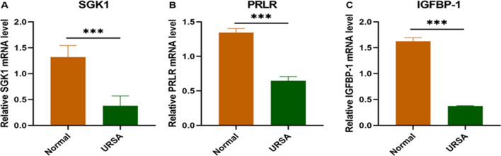
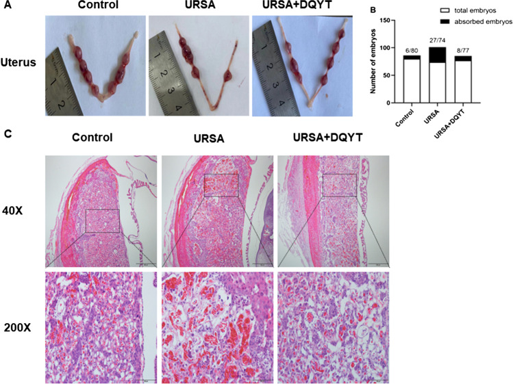
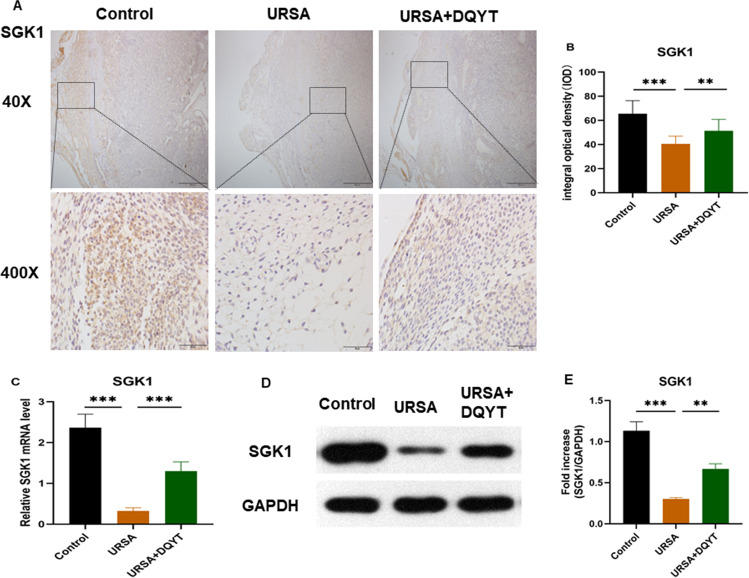
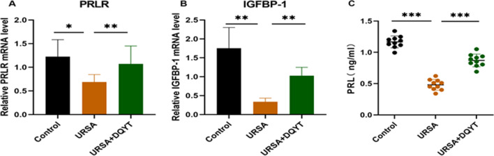
Recurrent spontaneous abortion (RSA) is one of the most common complications during pregnancy and seriously affects women's physical and mental health. About 50% of RSA cases are of unknown etiology. Our previous study found that the decidual tissue of patients with unexplained recurrent spontaneous abortion (URSA) had low expression levels of serum and glucocorticoid-induced protein kinase (SGK) 1. Endometrial decidualization is a key link in the early stage of pregnancy and is crucial to the development and maintenance of pregnancy. Decidualization is the proliferation and differentiation of endometrial stromal cells into deciduals, which involves a complex physiological process such as ovarian steroid hormones (estrogen, progesterone, prolactin, etc.), growth factors, and intercellular signaling. The binding of estrogen and its receptor stimulates the synthesis of endometrial deciduating markers prolactin (PRL) and insulin-like growth factor binding protein 1 (IGFBP-1), which mediates the occurrence of decidualization. Among them, SGK1/ENaC is a signaling pathway closely related to decidualization. The purpose of this study was to further investigate the expression of SGK1 and decidualization-related molecules in the decidual tissue of URSA patients and to explore the potential mechanism of SGK1's protective effect in URSA patients and in mouse models. Decidual tissue samples from 30 URSA patients and 30 women who actively terminated pregnancy were collected, and a URSA mouse model was established and treated with dydrogesterone. Expression levels of SGK1 and its signaling pathway-related proteins (p-Nedd4-2, 14-3-3 protein and ENaC-a), estrogen and progesterone receptors (ERβ, PR), and decidualization markers (PRLR, IGFBP-1) were assessed. Our study found that SGK1, p-Nedd4-2, 14-3-3 proteins, and ENaC-a expression levels were reduced in the decidual tissue, the SGK1/ENaC signaling pathway was inhibited, and the expression levels of the decidualization markers PRLR and IGFBP-1 were downregulated in the URSA group compared with the controls. Additionally, the concentrations of E2, P, and PRL in the serum of mice were decreased in the URSA group compared with the controls. However, SGK1/ENaC pathway-related proteins, estrogen and progesterone and their receptors, and decidualization-related molecules were upregulated by dydrogesterone. These data suggest that estrogen and progesterone can induce decidualization by activating the SGK1/ENaC signaling pathway; disruption of this pathway can lead to the development of URSA. Dydrogesterone can increase the expression level of SGK1 protein in decidual tissue.

Keywords: Decidualization; Estrogen and progesterone; SGK1/ENaC; URSA.


Conflict of interest statement

The authors declare no competing interests.

Figures

Fig. 1
Fig. 1
The expression of SGK1 and methylation-related molecule PRLR and IGFBP-1 in decidual tissue of URSA patients. A Expression of SGK1 mRNA in the URSA group and normal group by RT-PCR. B Expression of PRLR mRNA in the URSA group and normal group by RT-PCR. C Expression of IGFBP-1 mRNA in the URSA group and normal group by RT-PCR. All the results were representatives of three independent experiments and data were expressed as mean ± SD (n = 16/group). ***P < 0.001 compared to the control
Fig. 2
Fig. 2
Uterus anatomy and pathological changes of decidua in mice. A Left and right uterine horns from normal pregnant mice, aborted mice, and drug-treated mice. B The number of fetuses. C Decidua of the control group, URSA group, and URSA + DQYT group were stained by H&E staining. Scale bar represents 500 µm and 100 µm (n = 4/group)
Fig. 3
Fig. 3
The expression of SGK1 in mouse decidual tissue. A The expression of SGK1 in mouse decidual tissue was investigated by immunohistochemistry staining. Scale bar represents 500 µm and 50 µm (n = 4/group). B Quantitative assessment of the SGK1 positive staining area in decidua using Image-Pro Plus software. C Expression of SGK1 mRNA in the control group, URSA group, and URSA + DQYT group by RT-PCR. (n = 8/group). D The protein levels of SGK1 were identified by Western blot analysis. E The quantification data analysis of SGK1 in the mouse decidual tissues (n = 3/group). All the results were representatives of three independent experiments and data were expressed as mean ± SD. **P < 0.01, ***P < 0.001
Fig. 4
Fig. 4
The analysis of SGK1 signaling pathway in mouse decidua and serum. A Expression of protein 14–3-3 mRNA in the control group, URSA group, and URSA + DQYT group by RT-PCR (n = 8/group). B Expression of ENaC-a mRNA in the control group, URSA group, and URSA + DQYT group by RT-PCR. (n = 8/group). C The protein levels of p-Nedd4-2 were identified by Western blot analysis. D The quantification data analysis of p-Nedd4-2 in the mouse decidual tissues. (n = 3/group). E The serum E2 levels of mice among control group, URSA group, and URSA + DQYT group were analyzed by ELISA (n = 10/group). F The serum P levels of mice among control group, URSA group, and URSA + DQYT group were analyzed by ELISA (n = 10/group). G The expression of ERβ, PR, AND ENaC-a was detected by immunohistochemistry in decidua among control group, URSA group, and URSA + DQYT group. Scale bar represents 50 µm (n = 4/group). H Quantitative data analysis for ERβ. I Quantitative data analysis for PR. J Quantitative data analysis for ENaC-a. All the results were representatives of three independent experiments and data were expressed as mean ± SD. *P < 0.05, **P < 0.01, ***P < 0.001
Fig. 5
Fig. 5
The analysis of PRLR, IGFBP-1, and PRL in mouse decidua and serum. A Expression of PRLR mRNA in the control group, URSA group, and URSA + DQYT group by RT-PCR (n = 8/group). B Expression of protein IGFBP-1 mRNA in the control group, URSA group, and URSA + DQYT group by RT-PCR (n = 8/group). C The serum PRL levels of mice among control group, URSA group, and URSA + DQYT group were analyzed by ELISA (n = 10/group). All the results were representatives of three independent experiments and data were expressed as mean ± SD. *P < 0.05, **P < 0.01, ***P < 0.001
Fig. 6
Fig. 6
The summary of this study. Dydrogesterone promotes decidualization by increasing estrogen and progesterone levels and activating SGK1/ENaC signaling pathway

References

    1. RPL EGGo. Bender Atik R, Christiansen OB, Elson J, Kolte AM, Lewis S, et al. ESHRE guideline: recurrent pregnancy loss. Hum Reprod Open. 2018;2018(2):hoy004. doi: 10.1093/hropen/hoy004. - DOI - PMC - PubMed
    1. Practice Committee of the American Society for Reproductive Medicine. Definitions of infertility and recurrent pregnancy loss: a committee opinion. Fertil Steril. 2020;113(3):533–5. 10.1016/j.fertnstert.2019.11.025. - PubMed
    1. Kirshenbaum M, Orvieto R. Should we offer in vitro fertilization to couples with unexplained recurrent pregnancy loss? J Clin Med. 2019;8(11):2001. 10.3390/jcm8112001. - PMC - PubMed
    1. Gao Y, Wang PL. Increased CD56+ NK cells and enhanced Th1 responses in human unexplained recurrent spontaneous abortion. Genet Mol Res. 2015;14(4):18103–9. doi: 10.4238/2015.December.22.36. - DOI - PubMed
    1. Li M, Marin-Muller C, Bharadwaj U, Chow K-H, Yao Q, Chen C. MicroRNAs: control and loss of control in human physiology and disease. World J Surg. 2008;33(4):667–84. doi: 10.1007/s00268-008-9836-x. - DOI - PMC - PubMed