Case Reports
. 2023 Jun 7;21(1):172.
doi: 10.1186/s12957-023-03053-2.

Histological transformation to signet-ring cell carcinoma in a patient with clinically aggressive poorly differentiated adenocarcinoma of the ascending colon after response to chemotherapy plus cetuximab: a case report

Affiliations
Case Reports

Histological transformation to signet-ring cell carcinoma in a patient with clinically aggressive poorly differentiated adenocarcinoma of the ascending colon after response to chemotherapy plus cetuximab: a case report

Hideki Nagano et al. World J Surg Oncol. .

Abstract

Background: Alteration of chemosensitivity or tumor aggressiveness in response to chemotherapy has been reported, and liquid biopsy assessment during chemotherapy for colorectal cancers has confirmed the acquisition of mutations in various oncogenes. However, the occurrence of histological transformation seems to be extremely rare in colorectal cancers, and the few existing case reports of this transformation are from lung cancer and breast cancer. In this report, we describe the histological transformation of clinically aggressive scirrhous-type poorly differentiated adenocarcinoma of the ascending colon to signet-ring cell carcinoma in almost all recurrent tumors that were confirmed by autopsy after response to chemotherapy plus cetuximab.

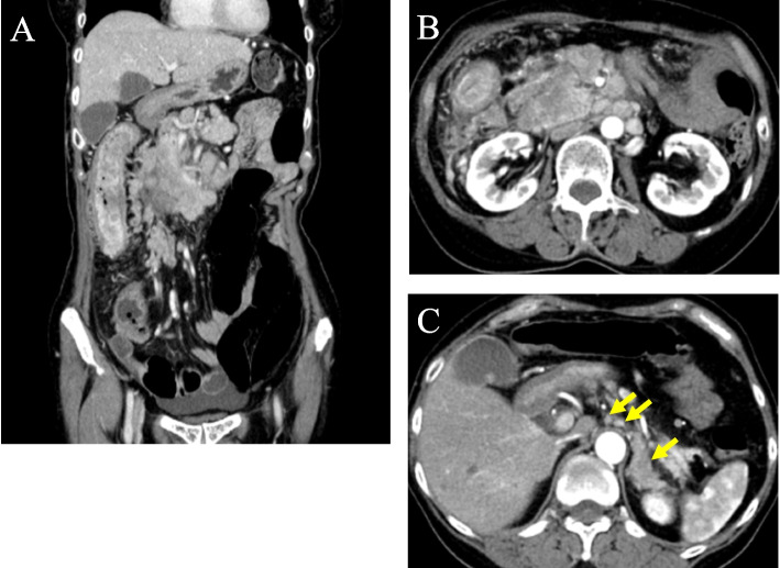
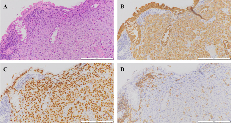
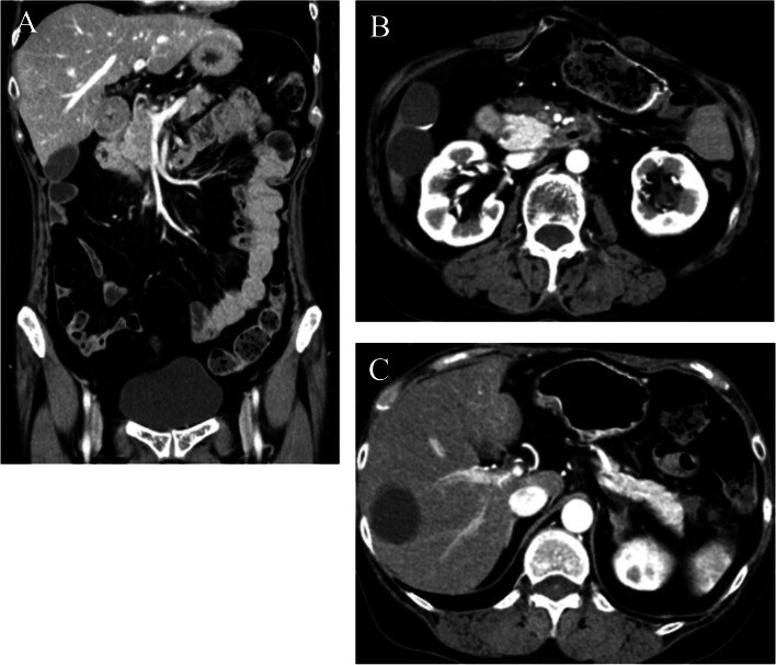
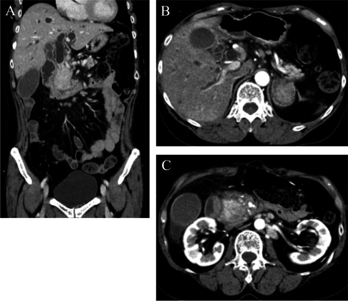
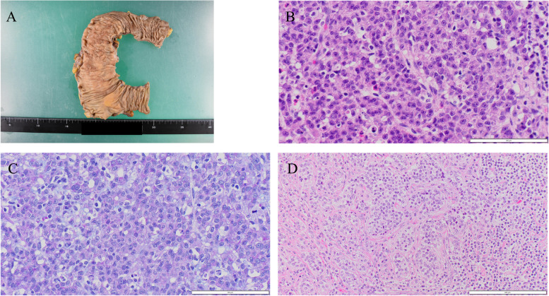
Case presentation: A 59-year-old woman visited our hospital with whole abdominal pain and body weight loss and was diagnosed with scirrhous-type poorly differentiated adenocarcinoma of the ascending colon with aggressive lymph node metastases. The intrinsic chemosensitivity of the tumors was evident upon initiation of mFOLFOX6 plus cetuximab therapy, and right hemicolectomy was performed, and the tumor obviously remained in the peripancreatic area, paraaortic region, or other retroperitoneal areas. The ascending colon tumors mainly consisted of poorly differentiated adenocarcinoma and were not associated with signet-ring cell components except for minute clusters in a few lymphatic emboli in the main tumor. Chemotherapy was continued, and metastases were eliminated at 8 months after the operation; this response was maintained for an additional 4 months. Discontinuation of chemotherapy plus cetuximab resulted in immediate tumor recurrence and rapid expansion, and the patient died of the recurrent tumor 1 year and 2 months after the operation. Autopsy specimens revealed that almost all of the recurrent tumors exhibited transformation and consisted of signet-ring cell histology.

Conclusion: This case might suggest that various oncogene mutations or epigenetic changes resulting from chemotherapy, especially regimens that include cetuximab, contribute to the transformation of non-signet-ring cell colorectal carcinoma to signet-ring cell carcinoma histology and can promote the aggressive clinical progression characteristic of signet-ring cell carcinoma.

Keywords: Ascending colon cancer; Cetuximab; Chemotherapy; Histological transformation; Poorly differentiated adenocarcinoma; Signet-ring cell carcinoma.


Conflict of interest statement

The authors declare no competing interests.

Figures

Fig. 1
Fig. 1
Contrast-enhanced CT findings at diagnosis. A CT showed wall thickness of the whole length of the ascending colon and aggressive metastasis to locoregional lymph nodes and peripancreatic lymph nodes. B Paraaortic lymph node metastases and an enlarged pancreatic head are shown. C Lymph node metastases of the peri-left adrenal region (arrows)
Fig. 2
Fig. 2
Colonoscopy findings. Colonoscopy revealed the anal side margin of the circumferential tumor on the hepatic flexure. Biopsy was taken from the tumor, and the tumor was diagnosed as PDA of the ascending colon using immunostaining to discriminate it from malignant lymphoma
Fig. 3
Fig. 3
Histopathology and immunostaining of ascending colon biopsy specimens. Hematoxylin and eosin staining (A) and immunostaining for CK AE1/AE3 (B), CDX2 (C), and CD45 (D). Tumor cells show poorly differentiated adenocarcinoma-like findings (A). These cells are positive for CK AE1/AE3 (B) and CDX2 (C) and negative for CD45 (D). AD Original magnification × 200. Each bar displayed at the bottom right indicates 200 μm
Fig. 4
Fig. 4
Contrast-enhanced CT findings after 6 courses of chemotherapy. A Although the wall thickness of the ascending colon remained consistent, the enhancement of the colon wall was decreased, as was the enhancement of lymph nodes near the tumor. B Paraaortic lymph nodes and enlargement of the pancreas head disappeared. C Lymph node metastases in the peri-left adrenal region were also diminished
Fig. 5
Fig. 5
Contrast-enhanced CT findings after adjuvant chemotherapy was continued for 8 months after colectomy. A CT showed elimination of peripancreatic lymph node metastases. B Elimination of paraaortic lymph node metastases and enlargement of the pancreas head were maintained. C Elimination of lymph node metastases of the peri-left adrenal region was also maintained
Fig. 6
Fig. 6
Contrast-enhanced CT one month after confirmation of maintenance and no evidence of recurrence by previous CT. A CT revealed choledochal obstruction due to the mass in the pancreas head and poor extension of the duodenum due to tumor invasion from the pancreas. B Enlarged metastatic lymph nodes in the peri-left adrenal region are shown. C Swelled paraaortic lymph nodes are also revealed
Fig. 7
Fig. 7
Endoscopic findings of the recurrent tumor with obstructive jaundice. Endoscopy showed poor extension of the descending part of the duodenum and exposure of the recurrent tumor. Biopsy revealed cancer cells with PDA-like histology with scattered signet-ring-like cells
Fig. 8
Fig. 8
Macroscopic and histopathological features of right hemicolectomy specimens. Gross findings of the ascending colon (A), hematoxylin and eosin (HE) staining (B) and Alcian blue-periodic acid Schiff (AB-PAS) staining (C) of the tumor, and HE staining of the metastatic site of the lymph node (D). Accumulation of polypoid gross features of the mucosal surface and wall thickening were found (A). The histological type of the tumor is mainly poorly differentiated adenocarcinoma (PDA) (B) with scant intracytoplasmic mucin (C). Metastatic PDA without a signet-ring cell component is observed in the lymph node (D). BD Original magnification × 400. Each bar displayed at the bottom right indicates 100 μm
Fig. 9
Fig. 9
Histopathology of duodenal biopsy specimens. Hematoxylin and eosin staining (A) and Alcian blue-periodic acid Schiff (AB-PAS) staining (B). Poorly differentiated adenocarcinoma (PDA) with scattered signet-ring cell-like cells is observed (A). Signet-ring cell-like cells have enriched intracytoplasmic mucin, and most PDA-like cells also have more intracytoplasmic mucin than the original PDA cells (B). A, B Original magnification × 400. Each bar displayed at the bottom right indicates 100 μm
Fig. 10
Fig. 10
Representative histopathology and immunostaining of autopsy specimens. Hematoxylin and eosin staining (A pancreas head, B lymph node near left adrenal gland), Alcian blue-periodic acid Schiff (AB-PAS) staining (C), and immunostaining for CK20 (D), CDX2 (E), SATB2 (F), and CK7 (G). Signet-ring cell-like cells are proliferating and infiltrating (A and B). These cells have enriched intracytoplasmic mucin (C) and are positive for CK20 (D), CDX2 (E) and SATB2 (F) and negative for CK7 (G). AG Original magnification × 400. Each bar displayed at the bottom right indicates 100 μm
Fig. 11
Fig. 11
Retrospective evaluation of the right hemicolectomy specimens. Hematoxylin and eosin staining. A very small number of signet-ring-like cells are observed in lymphatic emboli. A Original magnification × 400. The bar displayed at the bottom right indicates 100 μm

References

    1. Sorbarikor P, Alan PV. Targeted therapy for colorectal cancer metastases: a review of current methods of molecularly targeted therapy and the use of tumor biomarkers in the treatment of metastatic colorectal cancer. Cancer. 2019;125:4139–4147. doi: 10.1002/cncr.32163. - DOI - PubMed
    1. MisaleYaeger SR, Hobor S. Emergence of KRAS mutations and acquired resistance to anti-EGFR therapy in colorectal cancer. Nature. 2012;486:532–536. doi: 10.1038/nature11156. - DOI - PMC - PubMed
    1. Diaz LA, Jr, Williams RT, Wu J. The molecular evolution of acquired resistance to targeted EGFR blockage in colorectal cancers. Nature. 2012;486:537–540. doi: 10.1038/nature11219. - DOI - PMC - PubMed
    1. Vidal J, Muinelo L, Dalmases F, Jones F, Edelstein D, Iglesias M, et al. Plasma ctDNA RAS mutation analysis for the diagnosis and treatment monitoring of metastatic colorectal cancer patients. Ann Oncol. 2017;28:1325–1332. doi: 10.1093/annonc/mdx125. - DOI - PMC - PubMed
    1. Bray SM, Lee J, Kim ST, Hur JY, Ebert PJ, Calley JN, et al. Genomic characterization of intrinsic and acquired resistance to cetuximab in colorectal cancer patients. Sci Rep. 2019;9:15365. doi: 10.1038/s41598-019-51981-5. - DOI - PMC - PubMed

Publication types

LinkOut - more resources