

Radiographic involvement of cervical facet joints in ankylosing spondylitis: a longitudinal analysis in correlation with vertebral body lesions
- PMID: 37280716
- PMCID: PMC10245667
- DOI: 10.1186/s41927-023-00334-x
Radiographic involvement of cervical facet joints in ankylosing spondylitis: a longitudinal analysis in correlation with vertebral body lesions
Abstract
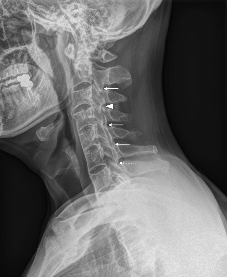
Background: The inability to assess structural changes in facet joints is a limitation of established radiographic scoring systems for ankylosing spondylitis (AS). We compared radiographic evidence of ankylosis in cervical facet joints and cervical vertebral bodies in patients with AS.
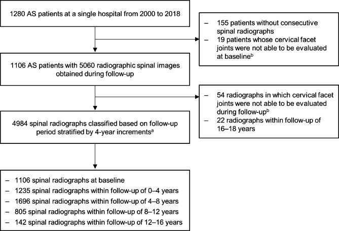
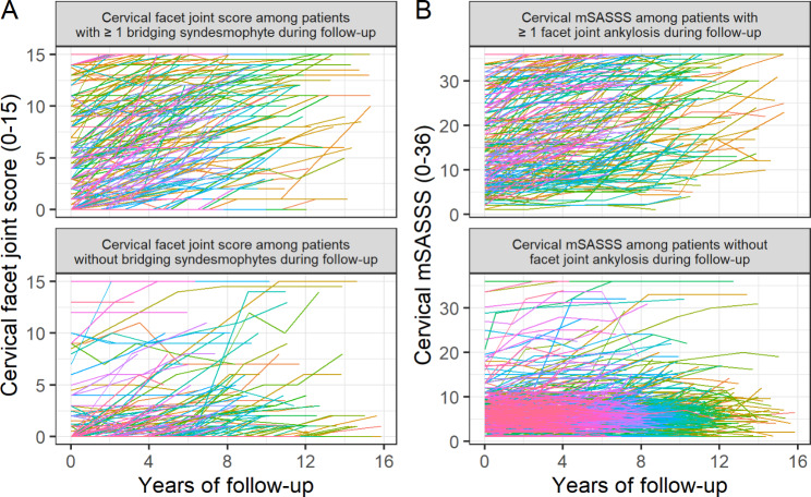
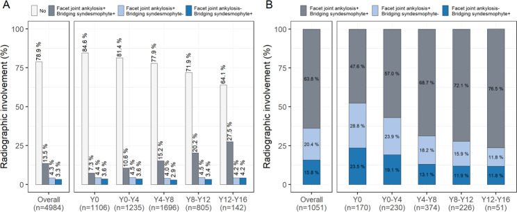
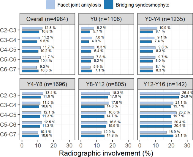
Methods: We analysed longitudinal data collected from 1106 AS patients and assessed 4984 spinal radiographs obtained up to 16 years of follow-up. Comparisons between cervical facet joints and cervical vertebral bodies focused on the presence of ankylosis, which was defined by at least one facet joint exhibiting complete ankylosis (according to the method of de Vlam) or at least one vertebral body with a bridging syndesmophyte (according to the modified Stoke Ankylosing Spondylitis Spinal Score [mSASSS]). Ankylosis was assessed over time using spinal radiographs collected during follow-up periods stratified in 4-year increments.
Results: Patients with cervical facet joint ankylosis had higher cervical mSASSS, sacroiliitis grades, and inflammatory markers, with more prevalent hip involvement and uveitis. Overall, the numbers of spinal radiographs indicating ankylosis were comparable between cervical facet joints (17.8%) and cervical vertebral bodies (16.8%), and they usually presented together (13.5%). We observed similar proportions of radiographs with ankylosis only in cervical facet joints (4.3%) and cervical vertebral bodies (3.3%). As damage progressed, configurations with both cervical facet joint ankylosis and bridging syndesmophytes became more predominant with longer follow-up times, while configurations with cervical facet joint ankylosis only or bridging syndesmophytes only were less frequently observed.
Conclusions: Evidence of cervical facet joint ankylosis appears as often as bridging syndesmophytes on routine AS spinal radiographs. Presence of cervical facet joint ankylosis should be considered because it may have a higher disease burden.
Keywords: Ankylosing spondylitis; Facet joint; Radiography; Syndesmophyte.
© 2023. The Author(s).
Conflict of interest statement
The authors declare no competing interests.
Figures
References
-
- van der Heijde D, Braun J, Deodhar A, Baraliakos X, Landewe R, Richards HB, et al. Modified stoke ankylosing spondylitis spinal score as an outcome measure to assess the impact of treatment on structural progression in ankylosing spondylitis. Rheumatology (Oxford) 2019;58(3):388–400. doi: 10.1093/rheumatology/key128. - DOI - PMC - PubMed
-
- Lee JY, Kim JI, Park JY, Choe JY, Kim CG, Chung SH, et al. Cervical spine involvement in longstanding ankylosing spondylitis. Clin Exp Rheumatol. 2005;23(3):331–8. - PubMed
LinkOut - more resources
Full Text Sources
Research Materials
