Case Reports
. 2023 May 6:15:101863.
doi: 10.1016/j.jaccas.2023.101863. eCollection 2023 Jun 7.

Inflammatory Fabry Cardiomyopathy Demonstrated Using Simultaneous [18F]-FDG PET-CMR

Affiliations
Case Reports

Inflammatory Fabry Cardiomyopathy Demonstrated Using Simultaneous [18F]-FDG PET-CMR

Christopher Orsborne et al. JACC Case Rep. .

Abstract

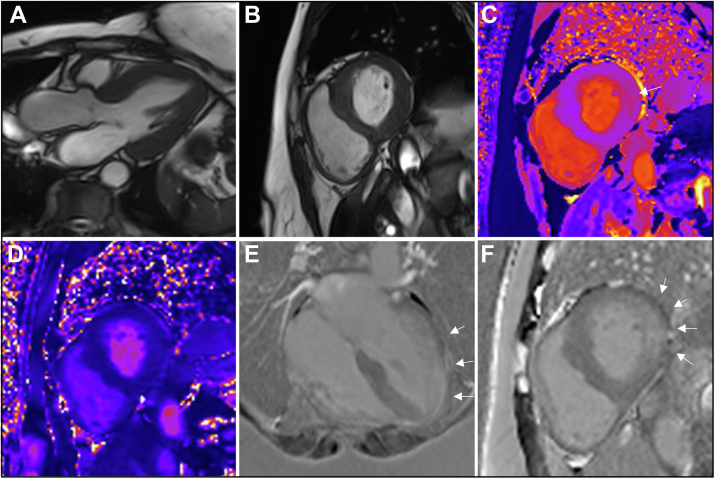
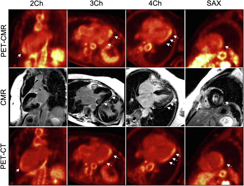
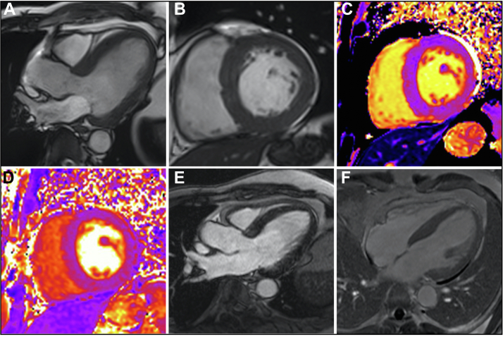
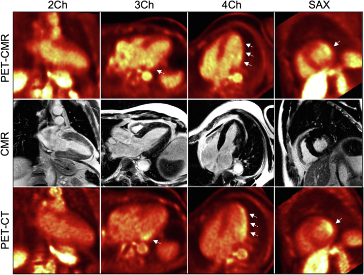
Using hybridized [18F]-fluorodeoxyglucose positron emission tomography with cardiac magnetic resonance, we identify active myocardial inflammation and demonstrate its relationship with late gadolinium enhancement, in Fabry disease. We demonstrate that late gadolinium enhancement represents, at least in part, active myocardial inflammation and identify an early inflammatory phenotype that may represent a therapeutic window before irreversible tissue injury and adaptation occur. (Level of Difficulty: Intermediate.).

Keywords: Fabry disease; [18F]-fluorodeoxyglucose positron emission tomography; cardiovascular magnetic resonance; myocardial fibrosis; myocardial inflammation.


Conflict of interest statement

This work is part of a study funded by Amicus Therapeutics. Amicus had no role in the design and conduct of the study; collection, management, analysis, and interpretation of the data; preparation or approval of the manuscript; or the decision to submit the manuscript for publication. This work was also supported in part by a British Heart Foundation Accelerator Award to the University of Manchester (AA/18/4/34221). Dr Orsborne has received research support from Amicus Therapeutics. Dr Lohezic has been formerly employed by GE Healthcare; and has served as a contractor for GE Healthcare. Dr Schmitt has received research support from Amicus Therapeutics. Dr Jovanovic has received research support from Amicus Therapeutics. Dr Miller has received funding through an Advanced Fellowship from the National Institute for Health Research (NIHR; NIHR301338) (The views expressed in this publication are those of the authors and not necessarily those of the NIHR, the National Health Service, or the UK Department of Health and Social Care); acknowledges support from the University of Manchester British Heart Foundation Accelerator Award (AA/18/4/34221) and the NIHR Manchester Biomedical Research Centre (NIHR203308); has participated on advisory boards/consulted for AstraZeneca, Boehringer Ingelheim and Lilly Alliance, Novartis and PureTech Health; has served as an advisor for HAYA Therapeutics; has received speaker fees from AstraZeneca, Boehringer Ingelheim and Novo Nordisk; has received conference attendance support from AstraZeneca and has received research support from Amicus Therapeutics, AstraZeneca, Guerbet Laboratories Limited, Roche and Univar Solutions B.V. All other authors have reported that they have no relationships relevant to the contents of this paper to disclose.

Figures

None
Graphical abstract
Figure 1
Figure 1
Case #1 Cardiac Magnetic Resonance Images (A) Three-chamber and (B) basal short-axis balanced steady-state free precession images. (C) Basal short-axis T1 map (modified look-locker inversion recovery). The arrow indicates an area of high native myocardial T1 relaxation time. (D) Basal short-axis T2 map. (E) Four-chamber phase-sensitive inversion recovery late gadolinium enhancement image acquired in the late phase following gadolinium administration. (F) Basal short-axis phase-sensitive inversion recovery late gadolinium enhancement image. The arrows indicate areas of late gadolinium enhancement. The basal short-axis T1 map, the basal short-axis T2 map, and the basal short-axis phase-sensitive inversion recovery late gadolinium enhancement images were acquired in identical imaging planes. Images are from a cardiac magnetic resonance scan conducted before the hybridized imaging.
Figure 2
Figure 2
Case #1 Hybridized Images (Top row) [18F]-fluorodeoxyglucose ([18F]-FDG) positron emission tomography (PET) with cardiac magnetic resonance (CMR). (Middle row) Cardiac magnetic resonance. (Bottom row) [18F]-FDG positron emission tomography with computed tomography (CT). On the [18F]-FDG positron emission tomography with cardiac magnetic resonance and [18F]-FDG positron emission tomography with computed tomography images, the arrows indicate areas of increased [18F]-FDG uptake. On the cardiac magnetic resonance images, the arrows indicate areas of late gadolinium enhancement. Individual modality images rather than hybridized images are presented throughout to facilitate visualization of the abnormalities identified with each modality. 2Ch = 2-chamber view; 3Ch = 3-chamber view; 4Ch = 4-chamber view; SAX = short-axis view.
Figure 3
Figure 3
Case #2 Cardiac Magnetic Resonance Images (A) Three-chamber and (B) basal short-axis balanced steady-state free precession images. (C) Basal short-axis T1 map (modified look-locker inversion recovery). (D) Basal short-axis T2 map. (E) Three-chamber phase-sensitive inversion recovery late gadolinium enhancement image acquired in the late phase following gadolinium administration. (F) Basal short-axis phase-sensitive inversion recovery late gadolinium enhancement image. Images are from a cardiac magnetic resonance scan conducted before the hybridized imaging.
Figure 4
Figure 4
Case #2 Hybridized Images (Top row) [18F]-fluorodeoxyglucose ([18F]-FDG) positron emission tomography (PET) with cardiac magnetic resonance (CMR). (Middle row) Cardiac magnetic resonance. (Bottom row) [18F]-FDG positron emission tomography with computed tomography (CT). On the [18F]-FDG positron emission tomography with cardiac magnetic resonance and [18F]-FDG positron emission tomography with computed tomography images, the arrows indicate areas of increased [18F]-FDG uptake. Individual modality images rather than hybridized images are presented throughout to facilitate visualization of the abnormalities identified with each modality. Abbreviations as in Figure 2.
Figure 5
Figure 5
Native T1, T2, and Extracellular Volume Maps (Top row) Case #1. (Bottom row) Case #2. (A and D) are native T1 maps, (B and E) are T2 maps, and (C and F) are extracellular volume maps acquired from the basal short-axis slice. Regions of interest are annotated on the relevant maps. Native myocardial T1 relaxation time, myocardial T2 relaxation time, and extracellular volume fraction within the [18F]-fluorodeoxyglucose–enhancing segments were as follows: case #1: 1,350.4 ms, 45.9 ms, and 42.1%, respectively, within the inferolateral segment; case #2, 1,224.9 ms, 40.1 ms, and 23.2%, respectively, within the anterolateral segment and 1,255.0 ms, 41.5 ms, and 24.9%, respectively, within the inferolateral segment.
Figure 6
Figure 6
Schematic of the Simultaneous Hybridized [18F]-FDG-PET-CMR Workflow CH2 = 2-chamber; CH3 = 3-chamber; CH4 = 4-chamber; Gd = gadolinium; LGE = late gadolinium enhancement; MR = magnetic resonance; pre-con = precontrast; SA = short-axis; other abbreviations as in Figure 2.

References

    1. Orsborne C., Bradley J., Bonnett L.J. Validated model for prediction of adverse cardiac outcome in patients with Fabry disease. J Am Coll Cardiol. 2022;80(10):982–994. - PubMed
    1. Rozenfeld P., Feriozzi S. Contribution of inflammatory pathways to Fabry disease pathogenesis. Mol Genet Metab. 2017;122(3):19–27. - PubMed
    1. Moon J.C., Sheppard M., Reed E., Lee P., Elliott P.M., Pennell D.J. The histological basis of late gadolinium enhancement cardiovascular magnetic resonance in a patient with Anderson-Fabry disease. J Cardiovasc Magn Reson. 2006;8(3):479–482. - PubMed
    1. Nappi C., Ponsiglione A., Pisani A. Role of serial cardiac (18)F-FDG PET-MRI in Anderson-Fabry disease. Insights Imaging. 2021;12(1):124. - PMC - PubMed
    1. Spinelli L., Imbriaco M., Nappi C. Early cardiac involvement affects left ventricular longitudinal function in females carrying alpha-galactosidase A mutation. Circ Cardiovasc Imaging. 2018;11(4) - PubMed

Publication types

LinkOut - more resources