. 2023 Mar 20:4:4.
doi: 10.17879/freeneuropathology-2023-4692. eCollection 2023 Jan.

Neurooncology: 2023 update

Affiliations

Neurooncology: 2023 update

Michel Mittelbronn. Free Neuropathol. .

Abstract

This article presents some of the author's neuropathological highlights in the field on neuro-oncology research encountered in 2022. Major advances were made in the development of more precise, faster, easier, less invasive and unbiased diagnostic tools ranging from immunohistochemical prediction of 1p/19q loss in diffuse glioma, methylation analyses in CSF samples, molecular profiling for CNS lymphoma, proteomic analyses of recurrent glioblastoma, integrated molecular diagnostics for better stratification in meningioma, intraoperative profiling making use of Raman effect or methylation analysis, to finally, the assessment of histological slides by means of machine learning for the prediction of molecular tumor features. In addition, as the discovery of a new tumor entity may also be a highlight for the neuropathology community, the newly described high-grade glioma with pleomorphic and pseudopapillary features (HPAP) has been selected for this article. Regarding new innovative treatment approaches, a drug screening platform for brain metastasis is presented. Although diagnostic speed and precision is steadily increasing, clinical prognosis for patients with malignant tumors affecting the nervous system remains largely unchanged over the last decade, therefore future neuro-oncological research focus should be put on how the amazing developments presented in this article can be more sustainably applied to positively impact patient prognosis.

Keywords: Brain metastasis; Brain tumors; Glioblastoma; Meningioma; Neuro-oncology; Neuropathology.


Conflict of interest statement

The author declares to not have any competing interests.

Figures

Figure 1
Figure 1
Algorithm for a 2-step immunohistochemical prediction of the 1p/19q status in IDH-mutant gliomas (positivity of tumor cells is evaluated). For each staining, four different expression levels were defined. HIP1R staining was considered positive if expression was observed in the cytoplasm or at the cell membrane. Scores H0 to H3 were defined as follows: H0: less than 10% of tumor cells are weakly positive; H1: 10%-75% of tumor cells are positive, however, less than 30% are strongly positive; H2: more than 50% of the tumor cells are positive, and more than 30% are strongly positive, however, still showing unstained spaces between the tumor cells. The score H2 is also given if the tumor was only moderately positive, however, approximately 100% of the cells showed moderate immunoreactivity in a densely packed manner; H3: approximately 100% of the tumor cells are strongly positive in a diffuse and densely packed manner, with only blood vessels being spared. For the assessment of vimentin (scores V0 to V3 were given), staining of blood vessels was excluded, however, no distinction could be made between reactive and neoplastic glial cells. V0: less than 10% of tumor cells are positive (however, blood vessels and reactive astrocytes could be positive); V1: 10%-20% of tumor cells or tumor cell processes are positive; V2: 20%-70% of the tumor cells with many tumor cell processes are positively labeled, however still 30% or more of the tumor cells are negative; V3: more than 70% of the tumor cells are strongly positive, thereby the tumors exhibit a strong diffuse staining pattern. After this individual assessment, the comparison between HIP1R and vimentin staining was done as shown in Figure 1.
Figure 2
Figure 2
Algorithm for integrated grading for recurrence risk prediction in meningioma (mitotic index defined as mitoses per 10 high-power fields).
Figure 3
Figure 3
Potential intraoperative applications of Raman effect-based techniques combined with machine learning tools. Raman spectroscopy is a label-free, non-disruptive method based on an inelastic scattering effect of photons allowing for the quantitative molecular assessment of biological samples resulting in different spectra. Stimulated Raman histology (SRH) is a variant of Raman spectroscopy allowing for the creation of hematoxylin and eosin (HE) slide mimics based on quantification of distinct molecules (mainly lipids, proteins and nucleic acids).
Figure 4
Figure 4
Scheme for fast, precise and unbiased molecular diagnostics using Nanopore sequencing, suitable for mutational, transcriptome and methylome analyses.
Figure 5
Figure 5
Potential implementation of personalized drug-screening approaches in the clinical setting.

References

    1. Falagas ME, Pitsouni EI, Malietzis GA, Pappas G. Comparison of PubMed, Scopus, Web of Science, and Google Scholar: strengths and weaknesses. FASEB J. 2008 Feb;22(2):338-42. 10.1096/fj.07-9492LSF. Epub 2007 Sep 20. PMID: 17884971. - DOI - PubMed
    1. Shariff SZ, Bejaimal SA, Sontrop JM, Iansavichus AV, Haynes RB, Weir MA, Garg AX. Retrieving clinical evidence: a comparison of PubMed and Google Scholar for quick clinical searches. J Med Internet Res. 2013 Aug 15;15(8):e164. 10.2196/jmir.2624. PMID: 23948488. - DOI - PMC - PubMed
    1. Pratt D, Abdullaev Z, Papanicolau-Sengos A, Ketchum C, Panneer Selvam P, Chung HJ, Lee I, Raffeld M, Gilbert MR, Armstrong TS, Pytel P, Borys E, Klonoski JM, McCord M, Horbinski C, Brat D, Perry A, Solomon D, Eberhart C, Giannini C, Quezado M, Aldape K. High-grade glioma with pleomorphic and pseudopapillary features (HPAP): a proposed type of circumscribed glioma in adults harboring frequent TP53 mutations and recurrent monosomy 13. Acta Neuropathol. 2022 Mar;143(3):403-414. 10.1007/s00401-022-02404-9. Epub 2022 Feb 1. PMID: 35103816. - DOI - PMC - PubMed
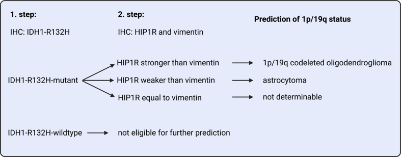
    1. Felix M, Friedel D, Jayavelu AK, Filipski K, Reinhardt A, Warnken U, Stichel D, Schrimpf D, Korshunov A, Wang Y, Kessler T, Etminan N, Unterberg A, Herold-Mende C, Heikaus L, Sahm F, Wick W, Harter PN, von Deimling A, Reuss DE. HIP1R and vimentin immunohistochemistry predict 1p/19q status in IDH-mutant glioma. Neuro Oncol. 2022 Dec 1;24(12):2121-2132. 10.1093/neuonc/noac111. PMID: 35511748. - DOI - PMC - PubMed
    1. Liechty B, Xu Z, Zhang Z, Slocum C, Bahadir CD, Sabuncu MR, Pisapia DJ. Machine learning can aid in prediction of IDH mutation from H&E-stained histology slides in infiltrating gliomas. Sci Rep. 2022 Dec 31;12(1):22623. 10.1038/s41598-022-26170-6. PMID: 36587030. - DOI - PMC - PubMed

LinkOut - more resources