. 2022 Jul 8:3:16.
doi: 10.17879/freeneuropathology-2022-4076. eCollection 2022 Jan.

Pigmented ependymoma, a tumor with predilection for the middle-aged adult: case report with methylation classification and review of 16 literature cases

Affiliations

Pigmented ependymoma, a tumor with predilection for the middle-aged adult: case report with methylation classification and review of 16 literature cases

Alexander S Himstead et al. Free Neuropathol. .

Abstract

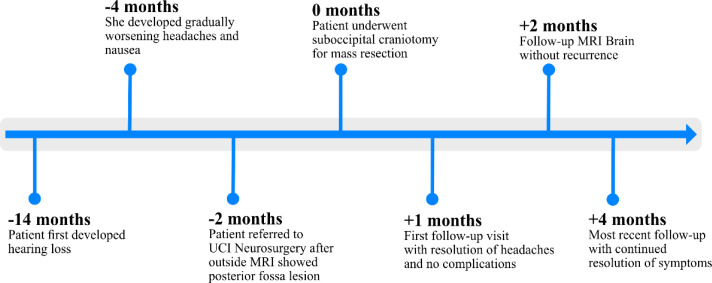
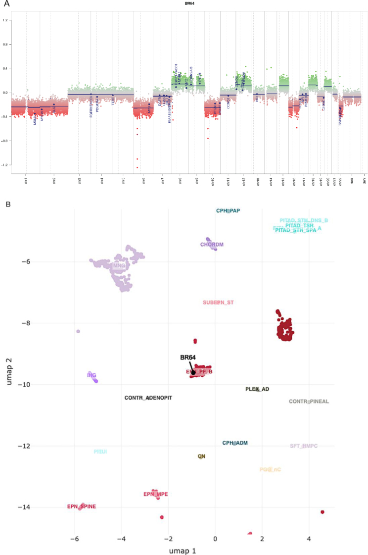
Ependymomas have rarely been described to contain pigment other than melanin, neuromelanin, lipofuscin or a combination. In this case report, we present a pigmented ependymoma in the fourth ventricle of an adult patient and review 16 additional cases of pigmented ependymoma from the literature. A 46-year-old female showed up with hearing loss, headaches, and nausea. Magnetic resonance imaging revealed a 2.5 cm contrast-enhancing cystic mass in the fourth ventricle, which was resected. Intraoperatively, the tumor appeared grey-brown, cystic, and was adherent to the brainstem. Routine histology revealed a tumor with true rosettes, perivascular pseudorosettes and ependymal canals consistent with ependymoma, but also showed chronic inflammation and abundant distended pigmented tumor cells that mimicked macrophages in frozen and permanent sections. The pigmented cells were positive for GFAP and negative for CD163 consonant with glial tumor cells. The pigment was negative for Fontana-Masson, positive for Periodic-acid Schiff and autofluorescent, which coincide with characteristics of lipofuscin. Proliferation indices were low and H3K27me3 showed partial loss. H3K27me 3 is an epigenetic modification to the DNA packaging protein Histone H3 that indicates the tri-methylation of lysine 27 on histone H3 protein. This methylation classification was compatible with a posterior fossa group B ependymoma (EPN_PFB). The patient was clinically well without recurrence at three-month post-operative follow-up appointment. Our analysis of all 17 cases, including the one presented, shows that pigmented ependymomas are most common in the middle-aged with a median age of 42 years and most have a favorable outcome. However, one patient that also developed secondary leptomeningeal melanin accumulations died. Most (58.8%) arise in the 4th ventricle, while spinal cord (17.6%) and supratentorial locations (17.6%) were less common. The age of presentation and generally good prognosis raise the question of whether most other posterior fossa pigmented ependymomas may also fall into the EPN_PFB group, but additional study is required to address that question.

Keywords: Case report; Ependymoma; Fourth ventricle; Lipofuscin; Methylation; Pigmented; Posterior fossa.


Figures

Figure 1
Figure 1
A. Sagittal pre-contrast T1 demonstrates an intraventricular mass involving the floor of the fourth ventricle and heterogenous T1 signal with areas of prominent intrinsic T1 hyperintensity (blue arrow), areas isointense to brain (red arrows), and scattered areas of low signal. B. Fat saturated sagittal post-contrast T1 demonstrates enhancement of the previously isointense portions (red arrows) with persistent areas of low signal. C. Axial fat saturated T2 at the level of the lesion demonstrates cystic areas of high T2 signal (red arrow) which correspond to low intensity of pre-contrast T1. D. A prominent low signal area on T2 is intrinsically bright on T1 (blue arrow), suggesting high protein and/or lipid contents. E. Axial FLAIR demonstrates predominantly iso- to hyperintense signal.
Figure 2
Figure 2
Intraoperative photographs of the tumor demonstrating: A. dissection of arachnoid bands tethering the tumor to the floor of the fourth ventricle and B. the macroscopic appearance of the lesion with notable grey-brown color (blue arrows).
Figure 3
Figure 3
Patient care timeline.
Figure 4
Figure 4
Staining is with hematoxylin and eosin (H&E) unless otherwise specified. A. (400X) Smear- swollen pigmented tumor cells. B. (400X) Smear- unpigmented and pigmented tumor cells. C. (100X) Frozen section- “epithelial” surfaces resembling glands (red arrowhead). D. (400X) Frozen section- Fine, grey-brown pigment. Figures E-I are from permanent sections. E. (40X) Cellular tumor (right fragment) and areas reminiscent of subependymoma gliopil (left fragment). F. (100X) Ependymal canal (blue arrowhead) and pseudopapillary configuration (red arrowhead). G. (200X) Perivascular pseudorosette (red arrowhead). H. (100X), I. (400X) Swollen pigmented cells. J. (200X) Fontana-Masson stain- tumor cells negative; few perivascular macrophages positive (blue arrowhead). K. (600X) PAS stain. L. (600X) Fluorescence microscopy shows autofluorescence. M. (200X), N. (400X) GFAP- pigmented cells immunopositive (red arrowheads). O. (200X) CD163- tumor cells negative; scattered microglia immunopositive. P. (200X) H3K27me3- loss in subset of tumor cells. Clicking into the figure will lead you to the full virtual slide.
Figure 5
Figure 5
Methylation analysis results. A. Genome-wide copy number profile generated from DNA methylation array signal intensities. The copy number profile shows numeric whole chromosome changes, which are typical for EPN_PFB group. B. The UMAP embedding of DNA methylation array data for select tumor groups. Sample tumor embeds with EPN_PFB tumor cluster.
Figure 6
Figure 6
Histogram demonstrating age distribution of patients with pigmented ependymomas.

References

    1. Pigmented lesions of the central nervous system: radiologic-pathologic correlation. Smith AB, Rushing EJ, Smirniotopoulos JG, et al. Radiographics. 2009;29(5) doi: 10.1148/rg.295095109. - DOI - PubMed
    1. Pigmented ependymoma of the fourth ventricle-a curious entity: report of a rare case with review of literature. Malhotra A, Rao S, Santhoshkumar R, Muralidharan N, Mitra S, Shetty S. International Journal of Surgical Pathology. 2021;29(1) doi: 10.1177/1066896920926700. - DOI - PubMed
    1. Melanotic astrocytoma. Vajtai I, Yonekawa Y, Schäuble B, Paulus W. Acta Neuropathol. 1996;91(5) doi: 10.1007/s004010050465. - DOI - PubMed
    1. Pigmented central neurocytoma: case report and literature review. Ng TH, Wong AY, Boadle R, Compton JS. The American Journal of Surgical Pathology. 1999;23(9) doi: 10.1097/00000478-199909000-00019. - DOI - PubMed
    1. Citation analysis of the most influential ependymoma research articles illustrates improved knowledge of the molecular biology of ependymoma. Brown NJ, Wilson B, Lien BV, et al. Neurosurgical Review. 2021;45 doi: 10.1007/s10143-021-01579-1. - DOI - PMC - PubMed