. 2023 Jun:30:101597.
doi: 10.1016/j.mtchem.2023.101597. Epub 2023 Jun 2.

COVID-19 impedimetric biosensor based on polypyrrole nanotubes, nickel hydroxide and VHH antibody fragment: specific, sensitive, and rapid viral detection in saliva samples

Affiliations

COVID-19 impedimetric biosensor based on polypyrrole nanotubes, nickel hydroxide and VHH antibody fragment: specific, sensitive, and rapid viral detection in saliva samples

A Santos et al. Mater Today Chem. 2023 Jun.

Abstract

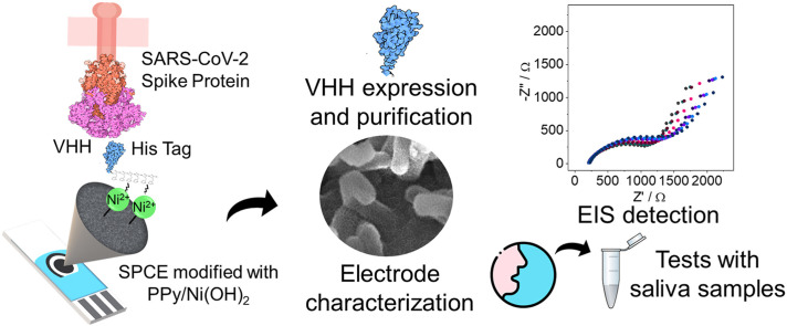
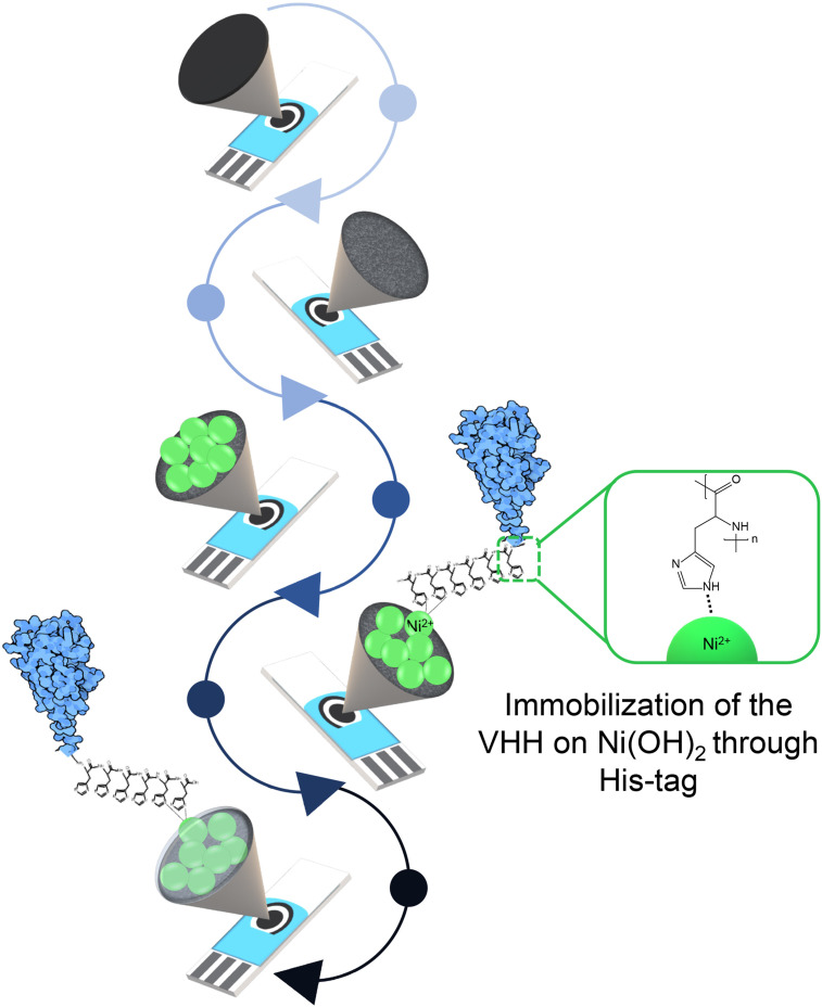
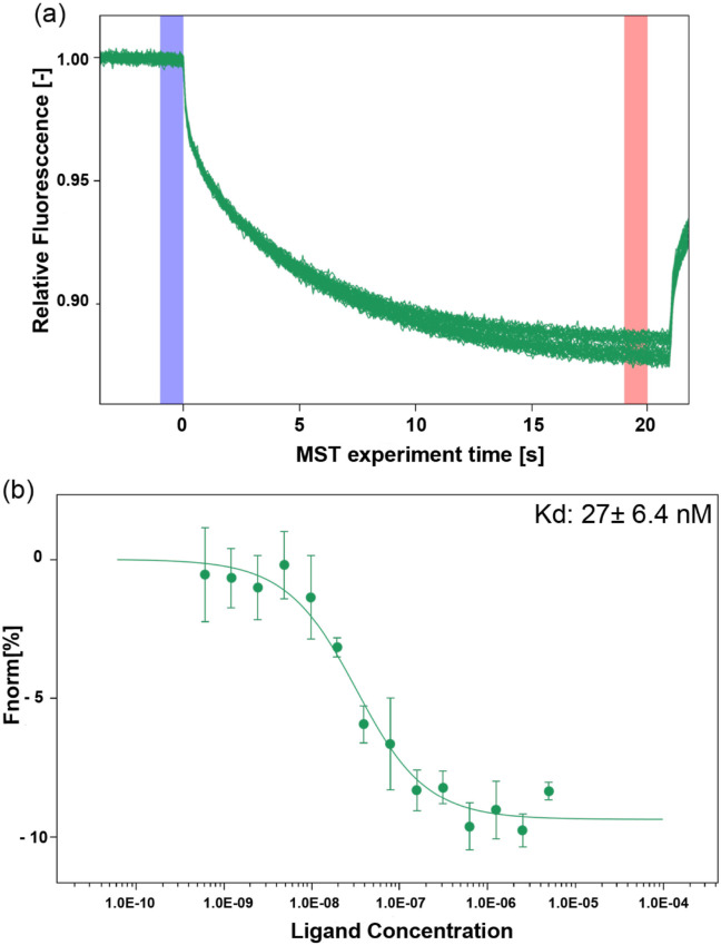
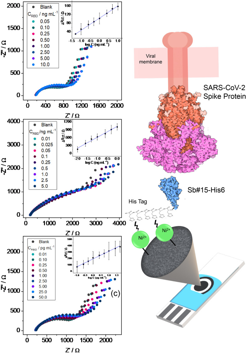
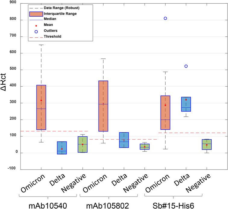
SARS-CoV-2 rapid spread required urgent, accurate, and prompt diagnosis to control the virus dissemination and pandemic management. Several sensors were developed using different biorecognition elements to obtain high specificity and sensitivity. However, the task to achieve these parameters in combination with fast detection, simplicity, and portability to identify the biorecognition element even in low concentration remains a challenge. Therefore, we developed an electrochemical biosensor based on polypyrrole nanotubes coupled via Ni(OH)2 ligation to an engineered antigen-binding fragment of heavy chain-only antibodies (VHH) termed Sb#15. Herein we report Sb#15-His6 expression, purification, and characterization of its interaction with the receptor-binding domain (RBD) of SARS-CoV-2 in addition to the construction and validation of a biosensor. The recombinant Sb#15 is correctly folded and interacts with the RBD with a dissociation constant (KD) of 27.1 ± 6.4 nmol/L. The biosensing platform was developed using polypyrrole nanotubes and Ni(OH)2, which can properly orientate the immobilization of Sb#15-His6 at the electrode surface through His-tag interaction for the sensitive SARS-CoV-2 antigen detection. The quantification limit was determined as 0.01 pg/mL using recombinant RBD, which was expressively lower than commercial monoclonal antibodies. In pre-characterized saliva, both Omicron and Delta SARS-CoV-2 were accurately detected only in positive samples, meeting all the requirements recommended by the World Health Organization for in vitro diagnostics. A low sample volume of saliva is needed to perform the detection, providing results within 15 min without further sample preparations. In summary, a new perspective allying recombinant VHHs with biosensor development and real sample detection was explored, addressing the need for accurate, rapid, and sensitive biosensors.

Keywords: COVID-19; Chemometrics; Impedimetric biosensor; Nanostructured electrode; Saliva sample.


Conflict of interest statement

The authors declare that they have no known competing financial interests or personal relationships that could have appeared to influence the work reported in this paper.

Figures

Image 1
Graphical abstract
Fig. 1
Fig. 1
Schematic representation of the biosensor development.
Fig. 2
Fig. 2
Purification of His6-Sb#15, Sb#15-His6, and SARS-CoV-2 RBD. (a), (b), (g) SDS-PAGE analysis of the indicated proteins purified by affinity chromatography. (c), (e), (h) Size exclusion chromatography profiles of the indicated proteins. Sb#15 shows a major peak eluting at 78 mL, Sb#15-His6 elutes in two peaks eluting, respectively, at 64 and 81 mL, and SARS-CoV-2 RBD elutes in a major peak with 59 mL and a shoulder eluting earlier with 56.5 mL. (d), (f), (i) SDS-PAGE analysis of the indicated proteins after fractionation by size exclusion chromatography. L, molecular mass marker. In, input. PC, precolumn. FT, flow through. Bc, before concentrate. RBD, receptor-binding domain.
Fig. 3
Fig. 3
Biophysical analysis of Sb#15, Sb#15-His6, and SARS-CoV-2 RBD. Top panels show DLS intensity distribution curves. Sb#15 shows a cumulant hydrodynamic radius (CR) of 1.43 ± 0.12 nm and polydispersity index (PDI) of 0.11 ± 0.02%, His6-Sb#15 shows CR of 2.37 ± 0.04 nm and PDI of 0.11 ± 0.02%, and SARS-CoV-2 RBD shows CR of 6.43 ± 0.05 nm and PDI of 0.34 ± 0.01%. Center panels show the 350nm/330nm fluorescence intensity ratio along the thermal denaturation curves of His6-Sb#15, Sb#15-His6, and SARS-CoV-2 RBD. Lower panels show the first derivative of the 350nm/330nm fluorescence intensity ratio to determine the transition midpoints (Tm) along the thermal denaturation curves for Sb#15 (Tm = 50.9 °C), Sb#15-His6 (Tm = 49.8 °C), and SARS-CoV-2 RBD (Tm = 47.6 °C). DLS, dynamic light scattering.
Fig. 4
Fig. 4
Interaction analysis between Sb#15 and SARS-CoV-2 RBD. (a) Representative MST time-trace of labeled RBD tested with serial dilutions of Sb#15. Cold (blue) and hot (red) regions as defined in central data analysis to calculate ΔFnorm. (b) Representative curve for the interaction of SARS-CoV-2 RBD at 10 nmol/L with Sb#15 using serial dilutions of Sb#15 starting at 10 μmol/L. Each curve was measured in quadruplicates. The binding constant (KD) was 27 ± 6.4 nmol/L. RBD, receptor-binding domain. (For interpretation of the references to color in this figure legend, the reader is referred to the Web version of this article.)
Fig. 5
Fig. 5
SEM images of (a) PPy-NTs and (b) PPy-NTs/Ni(OH)2 modified SPCEs (accelerating voltage = 20 kV; Detector: In-Bean SE). Electrochemical characterization of the modified electrodes using (c) CV at a scan rate of 20 mV/s and (d) EIS in ocp. Electrolyte = PBS pH 7.4. (e) Single-channel TL with distributed elements of impedance representing the solid network (χ) and the charge accumulation in the polymeric matrix (ζ). (f) TL and (g) equivalent circuit models used to adjust the EIS data. CV, cyclic voltammetry; EIS, electrochemical impedance spectroscopy; PPy-NT, polypyrrole nanotube.
Fig. 6
Fig. 6
Step-by-step characterization of the biosensor construction by (a) FTIR and (b) EIS measurements. I: PPy-NTs, II: PPy-NTs/Ni(OH)2, III: PPy-NTs/Ni(OH)2/Sb#15-His6, IV: PPy-NTs/Ni(OH)2/Sb#15-His6/BSA. EIS, electrochemical impedance spectroscopy; FTIR, Fourier transform infrared.
Fig. 7
Fig. 7
EIS response to different spike protein RBD concentrations in PBS for (a) mAb10540, (b) mAb105802, and (c) Sb#15-His6 biosensors. The corresponding analytical curves are shown inset in the figures. (d) Representation of the fabricated biosensor using Sb#15-His6 (not-to-scale). PBS, phosphate buffer saline; RBD, receptor-binding domain.
Fig. 8
Fig. 8
The statistical analyses and threshold determination obtained for biosensors in the detection of SARS-CoV-2 variants (Delta and Omicron) and negative saliva samples.

References

    1. McFee R.B. SARS 2 human coronavirus (COVID -19, SARS CoV2) Disease-a-Month. 2020;66 doi: 10.1016/j.disamonth.2020.101063. - DOI - PMC - PubMed
    1. Gruell H., Vanshylla K., Korenkov M., Kurth F., Kreer C., Klein F. SARS-CoV-2 Omicron sublineages exhibit distinct antibody escape patterns short article SARS-CoV-2 Omicron sublineages exhibit distinct antibody escape patterns. Cell Host Microbe. 2022;30:1–11. doi: 10.1016/j.chom.2022.07.002. - DOI - PMC - PubMed
    1. Wilhelm A., Widera M., Grikscheit K., Toptan T., Schenk B., Pallas C., Metzler M., Kohmer N., Hoehl S., Marschalek R., Herrmann E., Helfritz F.A., Wolf T., Goetsch U., Ciesek S. Limited neutralisation of the SARS-CoV-2 Omicron subvariants BA.1 and BA.2 by convalescent and vaccine serum and monoclonal antibodies. EBioMedicine. 2022;82 doi: 10.1016/j.ebiom.2022.104158. - DOI - PMC - PubMed
    1. Yao L., Zhu K.-L., Jiang X.-L., Wang X.-J., Zhan B.-D., Gao H.-X., Geng X.-Y., Duan L.-J., Dai E.-H., Ma M.-J. Omicron subvariants escape antibodies elicited by vaccination and BA.2.2 infection. Lancet Infect. Dis. 2022;22:1116–1117. doi: 10.1016/s1473-3099(22)00410-8. - DOI - PMC - PubMed
    1. Hu F., Shang X., Chen M., Zhang C. Joint detection of serum IgM/IgG antibody is an important key to clinical diagnosis of SARS-CoV-2 infection. Can. J. Infect Dis. Med. Microbiol. 2020:2020. doi: 10.1155/2020/1020843. - DOI - PMC - PubMed

LinkOut - more resources