Case Reports
. 2023 May 6;15(5):e38645.
doi: 10.7759/cureus.38645. eCollection 2023 May.

Local Control Failure After Five-Fraction Stereotactic Radiosurgery Alone for Symptomatic Brain Metastasis From Squamous Cell Lung Carcinoma Despite 43 Gy to Gross Tumor Margin With Internal Steep Dose Increase and Tumor Shrinkage During Irradiation

Affiliations
Case Reports

Local Control Failure After Five-Fraction Stereotactic Radiosurgery Alone for Symptomatic Brain Metastasis From Squamous Cell Lung Carcinoma Despite 43 Gy to Gross Tumor Margin With Internal Steep Dose Increase and Tumor Shrinkage During Irradiation

Kazuhiro Ohtakara et al. Cureus. .

Abstract

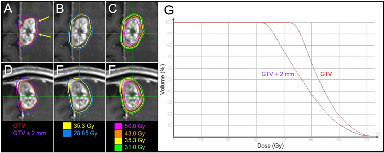
Five-fraction (fr) stereotactic radiosurgery (SRS) is increasingly being applied to large brain metastases (BMs) >2-3 cm in diameter, for which 30-35 Gy is the commonly prescribed dose. Since 2018, to further enhance both safety and efficacy, we have limited the five-fr SRS to approximately ≤3 cm BMs and adopted our own modified dose prescription and distribution: 43 and 31 Gy cover the boundaries of the gross tumor volume (GTV) and 2 mm outside the GTV, respectively, along with a steep dose increase inside the GTV boundary, that is, an intentionally very inhomogeneous GTV dose. Herein, we describe a case of symptomatic BM treated with five-fr SRS using the above policy, which resulted in a maximum tumor response with nearly complete remission (nCR) followed by gradual tumor regrowth despite obvious tumor shrinkage during irradiation. A 71-year-old man who had previously undergone surgery for squamous cell carcinoma (SCC) of the lungs presented with right-sided hemiparesis attributed to the para-falcine BM (27 mm in maximum diameter, 5.38 cm3). The BM was treated with five-fr SRS, with 99.2% of the GTV covered with 43 Gy and 59% isodose. Neurological symptoms improved during SRS, and obvious tumor shrinkage and mitigation of perilesional edema were observed upon completion of SRS. No subsequent anti-cancer pharmacotherapy was administered due to idiopathic pulmonary fibrosis (IPF). Despite a maximum response with nCR at four months, the tiny residual enhancing lesion gradually enlarged from 7.7 months to 22.7 months without neurological worsening. Although a consistent T1/T2 mismatch suggested the dominance of brain radionecrosis, 11C-methionine positron emission tomography showed increased uptake in the enhancing lesion. Pathological examination after total lesionectomy at 24.6 months revealed viable tumor tissue. Post-SRS administration of nintedanib for IPF may have provided some anti-tumor efficacy for lung SCC and may mitigate the adverse effects of SRS. The present case suggests that even ≥43 Gy with ≤60% isodose to the GTV boundary and ≥31-35 Gy to the 2 mm outside the GTV are insufficient to achieve long-term local tumor control by five-fr SRS alone in some large BM from lung SCC.

Keywords: biological effective dose; brain metastasis; five fraction; fractionation; linear-quadratic model; lung cancer; neuroendoscopy; nintedanib; squamous cell carcinoma; stereotactic radiosurgery.


Conflict of interest statement

The authors have declared that no competing interests exist.

Figures

Figure 1
Figure 1. Magnetic resonance images before five-fraction stereotactic radiosurgery
The images show contrast-enhanced (CE) axial T1-weighted images (WI) (A-C); axial T2-WI (D-F); 1.5-Tesla images for conventional diagnosis (A, B, D, and E); 3-Tesla images for treatment planning (C, F); 5.1 months (mo) (A, D), one week (w) (B, E), and three days (d) (C, F) before stereotactic radiosurgery (SRS). (A–F): All images are shown at the same magnification and coordinates under co-registration and fusion. (A, D): The normal morphology of the left superior and median frontal gyri can be seen before the development of brain metastasis (BM). (B, C, E, F): A heterogeneously enhanced mass lesion (arrows in B and C) is observed in the left frontal lobe, with broad attachment to the falcine dura, concomitant with extensive perilesional edema involving the primary and supplementary motor areas. (E, F): An irregularly-shaped mass lesion (arrows in E and F) with slightly high heterogeneous intensity is surrounded by a single-layered structure (dashed arrows in E and F) with slightly high intensity. A high-intensity component intervenes between them. The faint contrast exudation (dashed arrows in B and C) spreading outside the visible mass on T2-WI does not extend to the layered structure on T2-WI.
Figure 2
Figure 2. Difference in treatment planning for 5-fraction stereotactic radiosurgery
The schemas show (A-C) a gross tumor volume (GTV) covered by the isodose surface (IDS) for dose prescription; our previous method until 2017 (A); the method reported by Kogo et al. in 2013 (B); and our current method since 2018 (C). (A-C) DX% means a minimum dose covering ≥X% of the target volume. The percentage of the IDS is normalized to 100% at the maximum dose or isocenter dose. (A) In our previous protocol, the planning target volume (PTV) was generated by adding a uniform 1 mm margin to the GTV. The D≥95% of the PTV was covered by 35 Gy with 80% IDS. (B) According to Kogo et al., the PTV was defined as the GTV + 1 mm, while 2 mm margin was added to the GTV for >15 cm3 and/or cystic tumors. The D95% of the PTV was covered by 43 Gy with 90% IDS. (C) The GTV boundary is the base for dose prescription and planning. Compared to the method by Kogo et al., the normal tissue dose outside the GTV boundary is generally low, while the internal dose of the GTV is high.
Figure 3
Figure 3. Target definition, dose distribution, and dose-volume histograms for five-fraction stereotactic radiosurgery
The images show (A-F) CE-T1-WIs with target volumes (A, D) and dose distributions (B, C, E, F); axial views (A-C); coronal views (D-F); and dose-volume histograms (G). (A, D) Gross tumor volume (GTV) and 2-mm margin-added object volume (GTV + 2 mm). (A) Faint exudation (arrows in A) beyond the solid enhancing mass was excluded from the GTV, but included in the GTV + 2 mm volume. (D) In the coronal view, the contour line of the GTV is not smooth and jagged. CE: contrast-enhanced; WIs: weighted images
Figure 4
Figure 4. T2-weighted images before and at the completion of five-fraction stereotactic radiosurgery
The images show axial images (A, D); coronal images (B, E); sagittal images (C, F); three days before initiation of SRS (A-C); and at the completion of five-fr SRS (seven days after initiation of SRS) (D-F). (A-C) The para-falcine mass lesion (arrows in A-C) is seen. (D-F) At the completion of SRS, shrinkage of the mass lesion along with mitigation of the perilesional edema and mass effect was observed. SRS: stereotactic radiosurgery; fr: fraction
Figure 5
Figure 5. Magnetic resonance images before and after five-fraction stereotactic radiosurgery
The images show axial CE-T1-WIs (A-F); axial T2-WIs (G-L); coronal CE-T1-WIs (M-R); 3 days before (pre) SRS (A, G, M); at four months (mo) after SRS (B, H, N); at 7.7 months (C, I, O); at 11.8 months (D, J, P); at 15.2 months (E, K, Q); and at 22.7 months (F, L, R). (A-R) These images are shown at the same magnification and coordinates under co-registration and fusion. (B, H, N) The BM (arrows in A, G, and M) showed remarkable shrinkage at four months, with the tumor remnant being seen as a tiny lesion (arrows in B, H, and N). (C-F, I-L, O-R) The tiny enhancing lesion gradually increased from 7.7 months to 22.7 months. Although no corresponding mass lesion was observed on T2-WI until 15.2 months, i.e., T1/T2 mismatch, the irregularly-shaped mass lesion (arrows in L) appeared at 22.7 months, which was still much smaller than the enhancing lesion (arrows in F, R) (T1/T2 mismatch). CE: contrast-enhanced; WI: weighted image; SRS: stereotactic radiosurgery; BM: brain metastasis
Figure 6
Figure 6. Axial images before and after the lesionectomy
The images show CE-T1-WI at 22.7 months (mo) after SRS (A); a fusion image of CE-T1-WI at 22.7 months and an 11C-methionine (Met) positron emission tomography (PET) image at 23.1 months (B); 11C-Met-PET image at 23.1 months (C); CE-T1-WI at 24.7 months after SRS (three days after the lesionectomy) (D); and a fluid-attenuated inversion recovery image at 34.7 months after SRS (6.6 months after the lesionectomy) (E). (A-E) These images are shown at the same magnification and coordinates under co-registration and fusion. (B, C) 11C-Met-PET showed increased uptake in the enhancing lesion. CE: contrast-enhanced; WI: weighted image; SRS: stereotactic radiosurgery

References

    1. Tumor control probability of radiosurgery and fractionated stereotactic radiosurgery for brain metastases. Redmond KJ, Gui C, Benedict S, et al. Int J Radiat Oncol Biol Phys. 2021;110:53–67. - PubMed
    1. Five-year sustained complete remission with minimal adverse effects following radiosurgery for 2-cm brain metastasis with deep eloquent location from lung adenocarcinoma despite low marginal dose and high 12 Gy volume. Ohtakara K, Nakao M, Muramatsu H, Suzuki K. Cureus. 2023;15:0. - PMC - PubMed
    1. Gradual and remarkable tumor shrinkage following seven-fraction stereotactic radiosurgery alone with a marginal dose of 48.3 Gy for large lobar possibly intra-sulcal brain metastasis from renal cell carcinoma. Ohtakara K, Aoki S, Tajima M, Ohno T, Suzuki K. Cureus. 2023;15:0. - PMC - PubMed
    1. Correlation of brain metastasis shrinking and deviation during 10-fraction stereotactic radiosurgery with late sequela: Suggesting dose ramification between tumor eradication and symptomatic radionecrosis. Ohtakara K, Tanahashi K, Kamomae T, Miyata K, Suzuki K. Cureus. 2023;15:0. - PMC - PubMed
    1. Modified dynamic conformal arcs with forward planning for radiosurgery of small brain metastasis: Each double arc and different to-and-fro leaf margins to optimize dose gradient inside and outside the gross tumor boundary. Ohtakara K, Suzuki K. Cureus. 2023;15:0. - PMC - PubMed

Publication types

LinkOut - more resources