. 2023 Jun 9;9(23):eadg0330.
doi: 10.1126/sciadv.adg0330. Epub 2023 Jun 7.

Structural and computational design of a SARS-CoV-2 spike antigen with improved expression and immunogenicity

Affiliations

Structural and computational design of a SARS-CoV-2 spike antigen with improved expression and immunogenicity

James A Williams et al. Sci Adv. .

Abstract

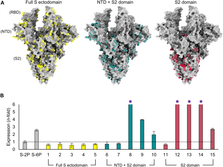
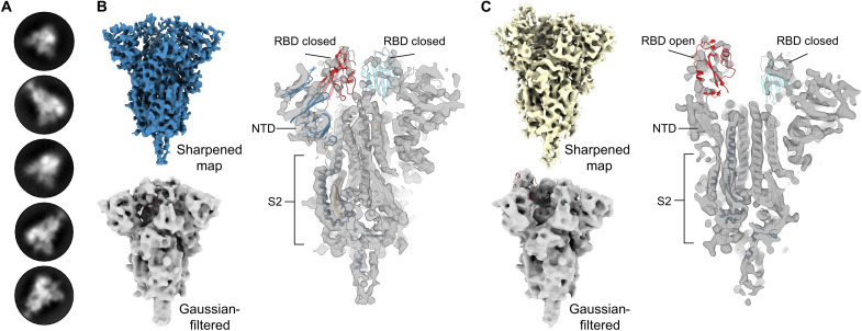
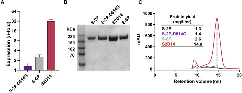
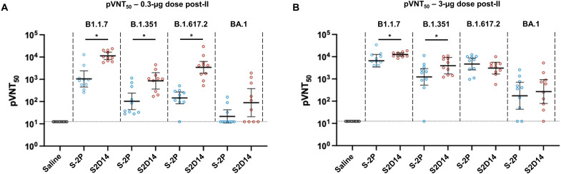
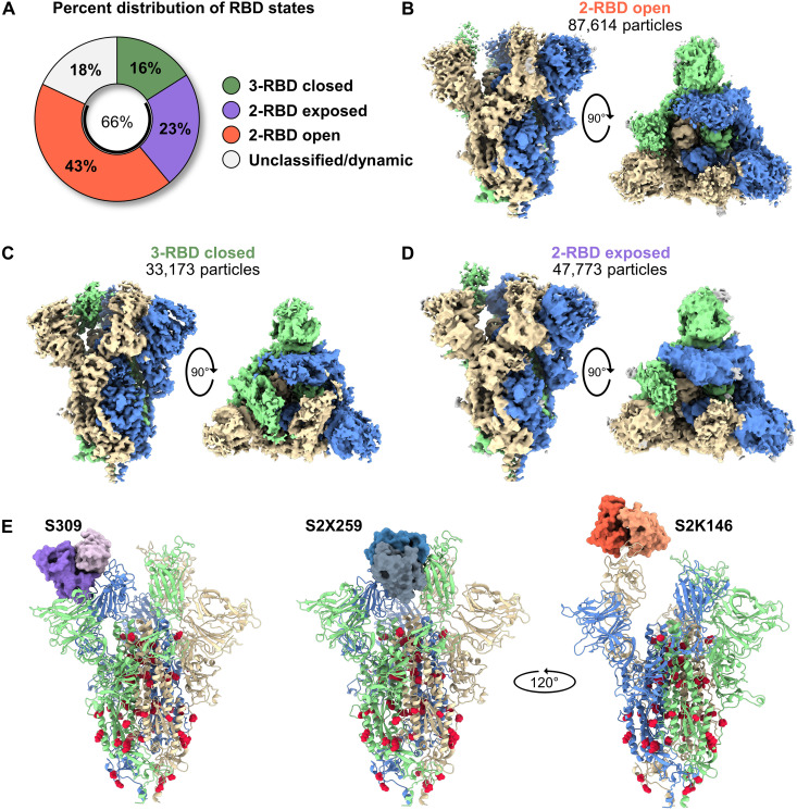
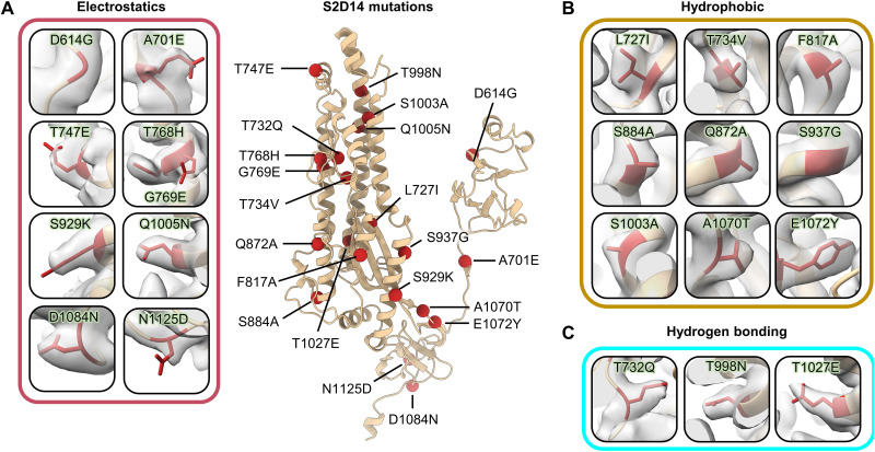
Severe acute respiratory syndrome coronavirus 2 (SARS-CoV-2) variants of concern challenge the efficacy of approved vaccines, emphasizing the need for updated spike antigens. Here, we use an evolutionary-based design aimed at boosting protein expression levels of S-2P and improving immunogenic outcomes in mice. Thirty-six prototype antigens were generated in silico and 15 were produced for biochemical analysis. S2D14, which contains 20 computationally designed mutations within the S2 domain and a rationally engineered D614G mutation in the SD2 domain, has an ~11-fold increase in protein yield and retains RBD antigenicity. Cryo-electron microscopy structures reveal a mixture of populations in various RBD conformational states. Vaccination of mice with adjuvanted S2D14 elicited higher cross-neutralizing antibody titers than adjuvanted S-2P against the SARS-CoV-2 Wuhan strain and four variants of concern. S2D14 may be a useful scaffold or tool for the design of future coronavirus vaccines, and the approaches used for the design of S2D14 may be broadly applicable to streamline vaccine discovery.


Figures

Fig. 1.
Fig. 1.. Evolutionary-based design strategy and expression levels of spike mutants.
(A) Model of the SARS-CoV-2 spike (S) protein structure with all three RBDs in the open conformation. The first design strategy (left) allowed for mutations (yellow) across the full S ectodomain. The second design strategy (center) limited the design landscape to the NTD + S2 domain (mutations in teal). The third design strategy (right) only modified the S2 domain and mutations are colored red. (B) Protein expression level (determined by biolayer interferometry using anti-HIS biosensors) of spike mutants normalized to S-2P (shown by dotted line) and grouped according to domain-specific design strategies in (A). S-6P (HexaPro) was used as an additional comparator. Error bars were determined from duplicate measurements, and asterisks indicate designs where expression exceeded the assay’s limit of quantitation.
Fig. 2.
Fig. 2.. S2D14 (design 14) adopts the trimeric prefusion S conformation and exhibits RBD conformational heterogeneity.
(A) Representative two-dimensional classes of S2 domain design S2D14 from cryo-EM micrographs, confirming the expected S protein trimers. (B) The cryo-EM map of S2D14 in a three-RBD closed state at 7.1-Å resolution is shown above and colored blue. A Gaussian-filtered volume with a rigid body fit of the S protein in the closed conformation [Protein Data Bank (PDB) accession number: 6VXX] is shown below the cryo-EM map and more clearly displays the three-RBD closed state. Shown on the right is a cross section of the sharpened map with a rigid body fit of the S protein in the closed conformation (PDB accession number: 6VXX). (C) The cryo-EM map of S2D14 with one RBD in the open conformation at 8.5-Å resolution and obtained from the same dataset as (B) is shown above and colored yellow. A Gaussian-filtered volume with a rigid body fit of the S protein in the one-RBD open state is shown below the cryo-EM map and, more clearly, displays the one-RBD open state. Shown on the right is a cross section of the EM map with a rigid body fit of the spike protein in the one-RBD open state (PDB accession number: 6VSB).
Fig. 3.
Fig. 3.. S2 domain mutations in S2D14 enhance expression of functional prefusion trimer.
(A) Total protein expression in cell culture supernatant for S proteins showing an n-fold increase relative to S-2P. A total of six replicate measurements were taken, and error bars represent SD from the means. (B) SDS–polyacrylamide gel electrophoresis analysis of size exclusion chromatography (SEC)–purified S proteins. Relevant bands for the molecular weight ladder (lane 1) are labeled in kilodaltons. (C) Overlay of the SEC chromatograms for S protein antigens. The dashed vertical line indicates the peak retention volume for the S2D14 trimer. The final yield of purified S protein trimers is shown in the inset.
Fig. 4.
Fig. 4.. IgG and neutralizing antibody titers from mice immunized with adjuvanted S2D14 or S-2P.
(A) Mouse immunization schedule. Mice were immunized with either a 0.3- or 3.0-μg doses containing AS03 adjuvanted S2D14 or S-2P. Serum was collected 3 weeks after the first immunization (post-I) and 2 weeks after the second immunization (post-II). (B and C) Enzyme-linked immunosorbent assay immunoglobulin G (IgG) titers of S2D14 compared to S-2P at both 0.3-μg (B) and 3.0-μg (C) doses for both post-I and post-II serum collections. Individual data with geometric mean titers (GMT) and 95% confidence intervals (CIs) are presented. (D and E) 50% pseudovirus neutralization titers (pVNT50) against the Wuhan strain were analyzed 2 weeks post-II at two antigen dosages, 0.3 (D) and 3.0 μg (E). Neutralizing antibody titers at either dose post-II were noticeably greater than for human convalescent sera (HCS). Individual data with GMT and 95% CIs are presented. For HCS samples, GMT and 95% CIs were calculated separately from vaccination groups in GraphPad Prism. Asterisks, *, indicate a statistically significant increase in neutralization response based on geometric mean ratio (GMR) comparisons with a two-sided 90% CI. Ratios for which the CI does not include 1 are considered statistically significant. See Materials and Methods for a description of the sera panel.
Fig. 5.
Fig. 5.. S2D14 elicits neutralizing antibody titers in mice capable of neutralizing variants of concern.
(A) Neutralization of VoCs compared between S2D14 and S-2P at low doses for post-II serum collection. S2D14 induced statistically significant higher neutralization responses against the Alpha (B1.1.7), Beta (B.1.351), and Delta (B.1.617.2) variants compared to S-2P. (B) Neutralization of VoCs compared between S2D14 and S-2P at high doses for post-II serum collection. S2D14 induced statistically significant higher neutralization responses against the Alpha (B1.1.7) and Beta (B.1.351) variants compared to S-2P. Individual data with GMT and 95% CIs are presented. Asterisks, *, indicate a statistically significant increase in neutralization response based on GMR comparisons with a two-sided 90% CIs. Ratios for which the CI does not include 1 are considered statistically significant.
Fig. 6.
Fig. 6.. S2D14 displays RBD open conformational states and is accessible for binding RBD nAbs.
(A) Pie chart illustrating the percent distribution of RBD conformational states. (B) The cryo-EM structure of S2D14 in the two-RBD open conformations (3.1-Å resolution) was the dominant population and represents 43% of total particles. (C) The cryo-EM structure of S2D14 in the three-RBD closed conformation (2.8-Å resolution) was the minority population with 16% of total particles. (D) The cryo-EM structure (3.3-Å resolution) of S2D14 in the two-RBD exposed state was representative of 23% of the total population. Eighteen percent of the particles could not be classified into a distinct conformation. Side and top views are shown for each structure, and monomers are individually colored for clarity. (E) Docking of Fabs of S309 (left), S2X259 (center), and S2K146 (right) onto S2D14 in the two-RBD open conformation confirms the accessibility of these broadly neutralizing RBD epitopes. S2D14 protomers are colored blue, green, or tan, and mutations are shown as red spheres.
Fig. 7.
Fig. 7.. PROSS mutations incorporated in S2D14 are well resolved by cryo-EM.
A single protomer of S2D14 residues 600 to 1147 is shown in the center in ribbon representation with mutations indicated as red spheres. Mutations are grouped on the basis of changes in either (A) electrostatic complementarity, (B) alterations in hydrophobicity, or (C) the introduction of hydrogen bonds. Residue side chains are depicted as sticks and cryo-EM density is shown as a transparent surface.
Fig. 8.
Fig. 8.. Mutations within S2D14 that may influence S2 domain stability.
(A) S2D14 is shown with two protomers as transparent surfaces, and a single protomer is depicted as a cartoon with domains colored as labeled in the figure. Mutations that may stabilize the S2 domain are colored red and shown as spheres. (B) A T998N mutation in the CH domain introduces a hydrogen bond with Y756 within the same protomer. (C) A T1027E mutation in the S2 core forms an interprotomer hydrogen bond network between R1039 residues. (D) An A701E mutation creates a negative patch at the S1/S2 linker region in proximity to a positively charged lysine residue K786 on an adjacent protomer. (E) The E1072Y mutation creates a hydrophobic surface that displaces an adjacent loop away from the trimer interior by ~7.2 Å. The positioning of this loop for S-2P is shown for comparison (light blue).

Similar articles

Cited by

References

    1. E. Volz, S. Mishra, M. Chand, J. C. Barrett, R. Johnson, L. Geidelberg, W. R. Hinsley, D. J. Laydon, G. Dabrera, Á. O’Toole, R. Amato, M. Ragonnet-Cronin, I. Harrison, B. Jackson, C. V. Ariani, O. Boyd, N. J. Loman, J. T. McCrone, S. Gonçalves, D. Jorgensen, R. Myers, V. Hill, D. K. Jackson, K. Gaythorpe, N. Groves, J. Sillitoe, D. P. Kwiatkowski; COVID-19 Genomics UK (COG-UK) consortium, S. Flaxman, O. Ratmann, S. Bhatt, S. Hopkins, A. Gandy, A. Rambaut, N. M. Ferguson, Assessing transmissibility of SARS-CoV-2 lineage B.1.1.7 in England. Nature 593, 266–269 (2021). - PubMed
    1. H. Tegally, E. Wilkinson, M. Giovanetti, A. Iranzadeh, V. Fonseca, J. Giandhari, D. Doolabh, S. Pillay, E. J. San, N. Msomi, K. Mlisana, A. von Gottberg, S. Walaza, M. Allam, A. Ismail, T. Mohale, A. J. Glass, S. Engelbrecht, G. van Zyl, W. Preiser, F. Petruccione, A. Sigal, D. Hardie, G. Marais, N. Y. Hsiao, S. Korsman, M. A. Davies, L. Tyers, I. Mudau, D. York, C. Maslo, D. Goedhals, S. Abrahams, O. Laguda-Akingba, A. Alisoltani-Dehkordi, A. Godzik, C. K. Wibmer, B. T. Sewell, J. Lourenço, L. C. J. Alcantara, S. L. Kosakovsky Pond, S. Weaver, D. Martin, R. J. Lessells, J. N. Bhiman, C. Williamson, T. de Oliveira, Detection of a SARS-CoV-2 variant of concern in South Africa. Nature 592, 438–443 (2021). - PubMed
    1. H. Jhun, H. Y. Park, Y. Hisham, C. S. Song, S. Kim, SARS-CoV-2 Delta (B.1.617.2) variant: A unique T478K mutation in receptor binding motif (RBM) of spike gene. Immune Netw. 21, e32 (2021). - PMC - PubMed
    1. H. Zhou, B. M. Dcosta, N. R. Landau, T. Tada, Resistance of SARS-CoV-2 omicron BA.1 and BA.2 variants to vaccine-elicited sera and therapeutic monoclonal antibodies. Viruses 14, 1334 (2022). - PMC - PubMed
    1. S. Jangra, C. Ye, R. Rathnasinghe, D. Stadlbauer, F. Krammer, V. Simon, L. Martinez-Sobrido, A. García-Sastre, M. Schotsaert, H. Alshammary, A. A. Amoako, M. H. Awawda, K. F. Beach, M. C. Bermúdez-González, R. L. Chernet, L. Q. Eaker, E. D. Ferreri, D. L. Floda, C. R. Gleason, G. Kleiner, D. Jurczyszak, J. C. Matthews, W. A. Mendez, L. C. F. Mulder, K. T. Russo, A. B. T. Salimbangon, M. Saksena, A. S. Shin, L. A. Sominsky, K. Srivastava, SARS-CoV-2 spike E484K mutation reduces antibody neutralisation. Lancet Microbe 2, e283–e284 (2021). - PMC - PubMed

Substances