. 2023 Jun 9;9(23):eadh8502.
doi: 10.1126/sciadv.adh8502. Epub 2023 Jun 7.

Human SAMD9 is a poxvirus-activatable anticodon nuclease inhibiting codon-specific protein synthesis

Affiliations

Human SAMD9 is a poxvirus-activatable anticodon nuclease inhibiting codon-specific protein synthesis

Fushun Zhang et al. Sci Adv. .

Abstract

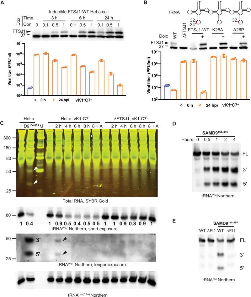
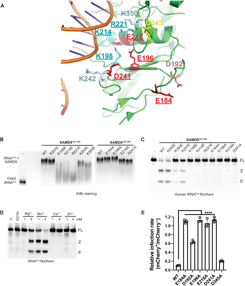
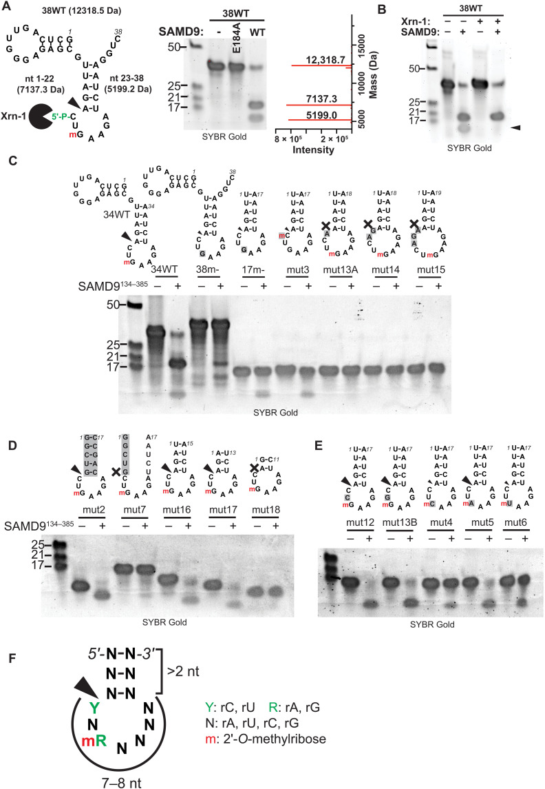
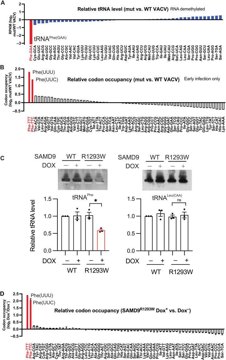
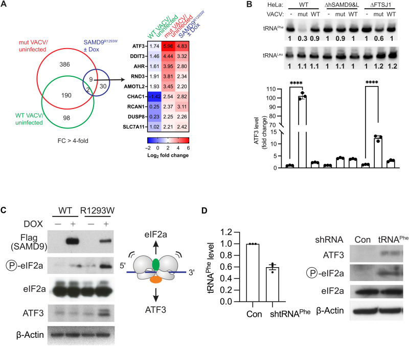
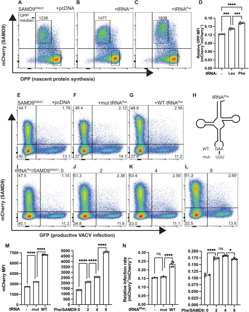
As a defense strategy against viruses or competitors, some microbes use anticodon nucleases (ACNases) to deplete essential tRNAs, effectively halting global protein synthesis. However, this mechanism has not been observed in multicellular eukaryotes. Here, we report that human SAMD9 is an ACNase that specifically cleaves phenylalanine tRNA (tRNAPhe), resulting in codon-specific ribosomal pausing and stress signaling. While SAMD9 ACNase activity is normally latent in cells, it can be activated by poxvirus infection or rendered constitutively active by SAMD9 mutations associated with various human disorders, revealing tRNAPhe depletion as an antiviral mechanism and a pathogenic condition in SAMD9 disorders. We identified the N-terminal effector domain of SAMD9 as the ACNase, with substrate specificity primarily determined by a eukaryotic tRNAPhe-specific 2'-O-methylation at the wobble position, making virtually all eukaryotic tRNAPhe susceptible to SAMD9 cleavage. Notably, the structure and substrate specificity of SAMD9 ACNase differ from known microbial ACNases, suggesting convergent evolution of a common immune defense strategy targeting tRNAs.


Figures

Fig. 1.
Fig. 1.. SAMD9 is a VACV-activatable endoribonuclease specifically targeting tRNAPhe.
(A) HeLa cells with a Dox-inducible FTSJ1–wild type (WT) allele were induced to express different amount of FTSJ1 with varied Dox concentrations (0.1 to 1 μg/ml) for various time (3 to 24 hours). The FTSJ1 protein (arrowhead) and a nonspecific protein were detected by the anti-FTSJ1 antibody. The position of the 37-kDa molecular weight (MW) marker is shown. The abilities of the cells to restrict a SAMD9-sensitive mutant VACV (vK1C7) were determined by measuring the viral titers [plaque-forming units (PFU) per milliliter] at 0 and 24 hours postinfection (hpi) with plaque assays on Vero cells. (B) HeLa cells were engineered to express no FSTJ1 (ΔFTSJ1) or different FTSJ1 mutants under a Dox-inducible promoter. FTSJ1-K28A mutant is catalytically inactive, while FTSJ1-A26P mutant is defective of methylating at position 34 of certain tRNAs. 2′-O-methylations are indicated by the red lines in the tRNA schematics. (C) WT and ΔFTSJ1 HeLa cells were infected with vK1C7 for the indicated time (2 to 8 hours). 8 + A, infection for 8 hours in the presence of cytosine arabinoside (AraC). Separately, total RNAs from uninfected HeLa cells were incubated with recombinant SAMD9134–385 protein. RNAs resolved on a denaturing gel were stained with SYBR Gold. Lane M contains an RNA ladder, the sizes of which (in bases) are indicated on the left. Northern blots were performed with two probes complementary to 5′ or 3′ end of tRNAPhe or one probe complementary to 3′ end of tRNALeu(CAA). Numbers below the gel are relative band intensities with respect to that in uninfected cells. Black or white arrowheads point to the tRNAPhe cleavage products. (D) Total RNAs from HeLa cells were incubated with SAMD9134–385 protein for the indicated time (0.5 to 4 hours). FL, full-length tRNA. (E) Total RNAs from HeLa (WT) or ΔFTSJ1 (ΔFt1) cells were incubated with SAMD9134–385 protein for 1 hour.
Fig. 2.
Fig. 2.. tRNA binding and active site of SAMD9134–385.
(A) Part of the crystal structure of SAMD9156–385/DNA complex (Protein Data Bank, 7ksp) is shown with residues that have been targeted for mutagenesis depicted. Residues that are essential for SAMD9134–385 cleavage of tRNAPhe are underlined with putative binding and active site residues colored in cyan and red, respectively. (B) Effects of the mutations on SAMD9134–385 binding of yeast tRNAPhe in a gel mobility shift assay. Purified SAMD9134–385 proteins were incubated with yeast tRNAPhe at 2:1 molar ratio and ran on an agarose gel. Ethidium bromide (EtBr)–stained gel is shown. (C) Effects of the mutations on SAMD9134–385 cleavage of human tRNAPhe. Total RNAs from HeLa cells were incubated with SAMD9134–385 protein for 1 hour. tRNAPhe fragments were detected by Northern blot. (D) Divalent cation requirements for SAMD9134–385 activities. Total RNAs from HeLa cells were incubated with SAMD9134–385 in the presence of the indicated concentrations (1 or 4 mM) of cations for 1 hour. tRNAPhe fragments were detected by Northern blot. FL, full-length tRNA. (E) The effect of the mutations on SAMD9 antiviral activities. Human embryonic kidney (HEK) 293T cells were transfected with mCherry-SAMD9 fusions for 36 hours and infected with vK1C7/GFP+ for 15 hours. Infection rates (GFP+%) among SAMD9-expressing and nontransfected control cells from the same culture well were simultaneously determined with flow cytometry. Relative infection rates between SAMD9-expressing and nontransfected cells are derived from the flow cytometry data shown in fig. S2B. Statistics: one-way ANOVA compared to the WT (****P <  0.0001).
Fig. 3.
Fig. 3.. SAMD9134–385 is an ACNase with a specificity for the eukaryotic tRNAPhe.
(A) A 38-nt synthetic RNA with the depicted secondary structure was incubated with SAMD9134–385 (WT or E184A) for 1 hour. The black arrowhead points to the cleavage site. MWs of different fragments are listed. The Xrn-1 symbol indicates that the 3′ fragment can be degraded by Xrn-1. m, 2′-O-methylation. The cleavage products were resolved on a denaturing gel and visualized after SYBR Gold staining. The sizes of the RNA ladder (in bases) are shown on the left of the gel. The cleavage products were analyzed with electrospray ionization liquid chromatography mass spectroscopy. The relative intensity and the MWs of the RNA species are shown. (B) The cleavage reaction was performed in the absence or presence of Xrn-1. The black arrowhead points to the 3′ product that disappeared in the presence of Xrn-1. Note that the RNA was synthesized with a 5'-OH group. (C to E) Synthetic RNAs with the depicted sequences were incubated with SAMD9134–385 for 1 hour. Sequences that differ from that of tRNAPhe were shown in gray background. The size of the arrows reflects the relative cleavage efficiency. “X” indicates no cleavage. (F) The consensus RNA structure cleaved by SAMD9134–385.
Fig. 4.
Fig. 4.. SAMD9 activated by VACV or through a GoF mutation specifically depletes cellular tRNAPhe and causes ribosomal pausing at Phe codons.
(A) HeLa cells were uninfected or infected with either WT or vK1C7 VACV for 8 hours. tRNAs were isolated from total RNAs resolved on a denaturing gel, treated with a tRNA demethylase and subjected to tRNA-seq. Levels of different tRNA species in vK1C7-infected cells with respect to that in WT VACV–infected cells are ranked from the lowest to the highest. tRNAPhe is highlighted in red. (B) HeLa cells were infected as in (A) but in the presence of AraC to limit the infection to the early phase. Ribosome sequencing (Ribo-seq) was conducted, and global codon occupancy of all cellular transcripts was analyzed. Codon occupancies in vK1C7-infected cells with respect to that in WT VACV–infected cells are ranked from the highest to the lowest. Phe codons are shown in red. (C) BT20 cell lines with a Dox-inducible SAMD9 allele (WT or SAMD9R1293W) were either uninduced or induced with Dox for 24 hours. tRNAPhe and tRNALeu(CAA) levels in the cells were determined by Northern blot. The graph summarizes the results from three biological replicates. ns, not significant. (D) BT20 cells with a Dox-inducible SAMD9R1293W allele were either uninduced or induced with Dox for 24 hours. Ribo-seq was conducted, and global codon occupancy of all cellular transcripts was analyzed. Codon occupancies in Dox-induced cells with respect to that in uninduced cells are ranked from the highest to the lowest. Phe codons are shown in red.
Fig. 5.
Fig. 5.. Activated SAMD9 causes stress response associated with ATF3 induction.
(A) RNA-seq was performed on HeLa cells that were uninfected or infected with either WT or vK1C7 (mut) VACV for 8 hours. Numbers of genes in mut VACV and WT VACV–infected cells that showed RPKM (reads per kilobase of transcript, per million mapped reads) fold change (FC) greater than 4 with respect to uninfected cells are shown. A common set of nine genes were induced by SAMD9R1293W in BT20 cells and by infection with the mut VACV (but not the WT VACV) in HeLa cells. The fold changes are shown in the heatmap. (B) Depletion of cellular tRNAPhe and induction of ATF3 in VACV-infected cells in a SAMD9- and FTSJ1-dependent manner. HeLa cells with SAMD9 and SAMD9L KO (ΔhSAMD9&L) or FTSJ1 KO (ΔFTSJ1) were infected with vK1C7 or WT VACV for 8 hours. tRNAPhe and ATF3 levels in the cells were determined by Northern blot and reverse transcription quantitative polymerase chain reaction (RT-qPCR), respectively. ATF3 level is normalized with a mitochondrial mRNA level in the cells. (C) SAMD9R1293W induces ATF3 expression and eIF2a phosphorylation. BT20 cell lines with a Dox-inducible SAMD9 allele (WT or SAMD9R1293W) were either uninduced or induced with Dox for 24 hours. Proteins were detected with the indicated antibodies in immunoblots. (D) Knockdown of cellular tRNAPhe induces ATF3 expression. Short hairpin RNA (shRNA) against tRNAPhe was transduced into BT20 cells. tRNAPhe level was determined by RT-qPCR, and ATF3 and phosphorylated eIF2a levels were determined by immunoblots.
Fig. 6.
Fig. 6.. Overexpression of tRNAPhe reduces SAMD9 activities.
(A to C) SAMD9 inhibition of global protein synthesis was reduced by tRNAPhe overexpression. HEK 293T cells were transfected with mCherry-SAMD9R982C and a plasmid expressing tRNAPhe or tRNALeu and labeled with O-propargyl-puromycin (OPP) for 30 min. Representative flow cytometry plots of OPP level relative to cellular mCherry level are shown. The OPP median fluorescence intensities in SAMD9-expressing cells are listed. (D) Nascent protein synthesis levels in SAMD9R982C-expressing cells relative to nontransfected cells from the same culture wells are derived from the flow cytometry. (E to L) SAMD9 inhibitions of protein synthesis and viral replication were reduced by tRNAPhe in a dose-dependent manner. HEK 293T cells were cotransfected with a plasmid expressing mCherry-SAMD9R982C and a plasmid expressing tRNAPhe or a mutant tRNAPhe and infected with vK1C7/GFP+. Infections among SAMD9-expressing and nontransfected control cells from the same culture well were simultaneously determined with flow cytometry. Representative flow cytometry plots are shown. (M) The mean fluorescence intensity (MFI) of mCherry-SAMD9 in transfected cells and (N) the relative infection rates between SAMD9-expressing and nontransfected cells are derived from the flow cytometry data. Statistics: one-way ANOVA (ns, not significant; *P <  0.1; ***P <  0.001; ****P <  0.0001).

References

    1. H. Masaki, T. Ogawa, The modes of action of colicins E5 and D, and related cytotoxic tRNases. Biochimie 84, 433–438 (2002). - PubMed
    1. G. Kaufmann, Anticodon nucleases. Trends Biochem. Sci. 25, 70–74 (2000). - PubMed
    1. J. Lu, B. Huang, A. Esberg, M. J. O. Johansson, A. S. Bystrom, The Kluyveromyces lactis γ-toxin targets tRNA anticodons. RNA 11, 1648–1654 (2005). - PMC - PubMed
    1. D. M. Thompson, R. Parker, Stressing out over tRNA cleavage. Cell 138, 215–219 (2009). - PubMed
    1. J. Liu, S. Wennier, L. Zhang, G. McFadden, M062 is a host range factor essential for myxoma virus pathogenesis and functions as an antagonist of host SAMD9 in human cells. J. Virol. 85, 3270–3282 (2011). - PMC - PubMed

MeSH terms