

DECIPHER: Improving Genetic Diagnosis Through Dynamic Integration of Genomic and Clinical Data
- PMID: 37285546
- PMCID: PMC7615097
- DOI: 10.1146/annurev-genom-102822-100509
DECIPHER: Improving Genetic Diagnosis Through Dynamic Integration of Genomic and Clinical Data
Abstract
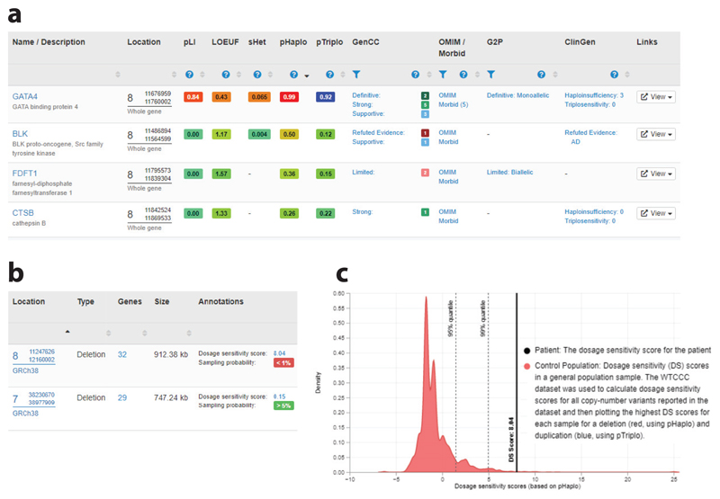
DECIPHER (
Keywords: genetic diagnosis; genetic disorders; genotype–phenotype correlation; rare diseases; variant interpretation.
Figures


References
-
- Adams D, Altucci L, Antonarakis SE, Ballesteros J, Beck S, et al. BLUEPRINT to decode the epigenetic signature written in blood. Nat Biotechnol. 2012;30:224–26. - PubMed
-
- Ars E, Serra E, Garcia J, Kruyer H, Gaona A, et al. Mutations affecting mRNA splicing are the most common molecular defects in patients with neurofibromatosis type 1. Hum Mol Genet. 2000;9:237–47. - PubMed
Publication types
MeSH terms
Grants and funding
LinkOut - more resources
Full Text Sources
