Review
. 2023 Aug:81:102619.
doi: 10.1016/j.sbi.2023.102619. Epub 2023 May 15.

Mechanism and evolution of human ACE2 binding by SARS-CoV-2 spike

Affiliations
Review

Mechanism and evolution of human ACE2 binding by SARS-CoV-2 spike

Antoni G Wrobel. Curr Opin Struct Biol. 2023 Aug.

Abstract

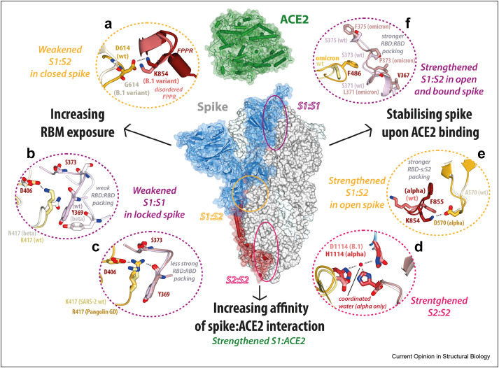
Spike glycoprotein of SARS-CoV-2 mediates viral entry into host cells by facilitating virus attachment and membrane fusion. ACE2 is the main receptor of SARS-CoV-2 and its interaction with spike has shaped the virus' emergence from an animal reservoir and subsequent evolution in the human host. Many structural studies on the spike:ACE2 interaction have provided insights into mechanisms driving viral evolution during the on-going pandemic. This review describes the molecular basis of spike binding to ACE2, outlines mechanisms that have optimised this interaction during viral evolution, and suggests directions for future research.


Conflict of interest statement

Declaration of competing interest The authors declare that they have no known competing financial interests or personal relationships that could have appeared to influence the work reported in this article.

Figures

Figure 1
Figure 1
General architecture of SARS-CoV-2 spike(a) Main conformations of SARS-CoV-2 spike trimer: pre-fusion (left, S1 and S2 subunits, PDB ID: 6zge) and postfusion (just S2 subunits, PDB ID: 6xra), with each protomer shown in different colour (b) Domains of S1 subunit with matching residue boundaries on the top: N-terminal domain (NTD aka domain A) in yellow, receptor-binding domain (RBD, aka domain B) in rosy brown, NTD-associated subdomain (NTD-s aka SD2, CTD2, domain D) in blue, RBD-associated subdomain (RBD-s aka SD1, CTD1, domain C) in plum (c) Conformational changes in S1 (blue) and S2 (red) subunits of one spike protomer associated with a transition from closed (PDB ID: 6zge, left) to open (PDB ID: 6zgg, middle) to ACE2-bound (PDB ID: 7a94, right; ACE2 in green) conformations.
Figure 2
Figure 2
Conformational rearrangements in pre-fusion spike trimer associated with binding to ACE2(a) Representative conformations of sarbecovirus spikes, from left to right: ‘helix-turn-helix-locked’ (aka locked-2) (RaTG13, PDB ID: 6zgf), SARS-CoV-2 locked (aka locked-1) (prototypic SARS-CoV-2, PDB ID: 6zge), closed (D614G SARS-CoV-2, PDB ID: 7bnm), open with one RBD erect (prototypic SARS-CoV-2, PDB ID: 6zgg), open with two RBDs erect (D614G SARS-CoV-2, PDB ID: 7bno), with one ACE2 bound (prototypic SARS-CoV-2, PDB ID: 7a94), with one ACE2 bound and one RBD erect but unsatisfied (prototypic SARS-CoV-2, PDB ID: 7a96), with two ACE2s bound (prototypic SARS-CoV-2, PDB ID: 7a97), with three ACE2s bound (prototypic SARS-CoV-2, PDB ID: 7a98); all coloured as in Fig. 1a (b) Conformational rearrangements in the interface between S1 (blue) and S2′ of the neighbouring protomer (rosy brown with S1-buried surface in red): top panels show movements of two subdomains associated with locked-to-locked (left), locked-to-closed (middle), and closed-to-bound transitions; bottom panels show interactions between NTD-s (lavender for 6zgf, goldenrod for 6zge, green for 7bnm, and blue for 7a94) and neighbouring S2’ (red, with FPPR in pink) stabilised mainly by 615–640 loop and FPPR (Open unbound conformation was omitted as it can be considered an intermediate between bound and closed for the purpose of the analysis shown here.) (c) Conformational rearrangements in S1:ACE2 complex upon its dissociation from spike trimer: the dissociated complex (bottom) is sterically incompatible with the trimer (top); S1 domains of a single protomer coloured as in Fig. 1b, ACE2 in green, remaining spike subunits in gray.
Figure 3
Figure 3
Mechanisms optimising spike:ACE2 binding. A model outlining proposed mechanisms that influenced the evolution of the SARS-CoV-2 and its interaction with human ACE2: increasing RBM exposure (left, panels A–C), stabilising spike upon ACE2 binding (right, panels D–F), and increasing affinity of spike:ACE2 interaction (see Fig. 4 for details) (a) Interface between NTD-s in yellow (wt, PDB ID: 6zge)/grey (D614G, PDB ID: 7bnm) and FPPR of an adjacent protomer in red (wt, PDB ID: 6zge)/pink (D614G, PDB ID: 7bnm); (b) RBD:RBD interface with protomers in light yellow (wt, PDB ID: 6zge)/grey (beta, PDB ID: 7r16) and lavender (wt, PDB ID: 6zge)/grey (beta, PDB ID: 7r16); (c) RBD:RBD interface with protomers in light yellow (wt, PDB ID: 6zge)/yellow (GD pangolin, PDB ID: 7bbh) and lavender (wt, PDB ID: 6zge)/pink (GD pangolin, PDB ID: 7bbh); (d) Histidine triad coordinating a water molecule and stabilising S2 core in red (alpha variant, PDB ID: 7r13) vs repulsion of the corresponding D1114 residues in pink (wt, PDB ID: 6zge); (e) Interface between RBD-s in yellow (alpha, PDB ID: 7r13)/light grey (wt, PDB ID: 6zge) and FPPR of an adjacent protomer in red (alpha, PDB ID: 7r13)/pink (wt, PDB ID: 6zge); (f) RBD:RBD interface with protomers in light yellow (wt, PDB ID: 6zge)/yellow (omicron, PDB ID: 7tgw) and lavender (wt, PDB ID: 6zge)/pink (omicron, PDB ID: 7tgw).
Figure 4
Figure 4
Substitutions affecting ACE2:spike interface(a) Interactions between human ACE2 (green) and prototypic (wt) SARS-CoV-2 RBM (magenta). All residues mentioned in the text are shown as sticks; the three main hotspots on ACE2 are boxed (PDB ID: 7a91) (b) Interface around residue 501 between ACE2 in green (wt, PDB ID: 7a91)/olive (alpha, PDB ID: 7r0z) and RBM in magenta (wt, PDB ID: 7a91)/brown (alpha, PDB ID: 7r0z) (c) Interface around two hotspots between between ACE2 in green (wt, PDB ID: 6m0j)/olive (omicron, PDB ID: 7zf7) and RBM in magenta (wt, PDB ID: 6m0j)/brown (alpha, PDB ID: 7zf7) (d) Comparison of ACE2 binding by omicron spike RBM residues (PDB ID: 7zf7, brown) and RaTG13 (PDB ID: 7drv, lilac).

References

    1. Lu G., Wang Q., Gao G.F. Bat-to-human: spike features determining ‘host jump’ of coronaviruses SARS-CoV, MERS-CoV, and beyond. Trends Microbiol. 2015;23:468–478. - PMC - PubMed
    1. Lau S.K.P., et al. Genetic characterization of betacoronavirus lineage C viruses in bats reveals marked sequence divergence in the spike protein of Pipistrellus bat coronavirus HKU5 in Japanese pipistrelle: implications for the origin of the novel Middle East Respiratory Syndrome Coronavirus. J Virol. 2013;87:8638–8650. - PMC - PubMed
    1. Walls A.C., et al. Structure, function, and antigenicity of the SARS-CoV-2 spike glycoprotein. Cell. 2020;181 - PMC - PubMed
    2. This study presents one of the first cryoEM structures of SARS-CoV-2 in open and closed conformations and measures its binding to ACE2.

    1. Hoffmann M., et al. SARS-CoV-2 cell entry depends on ACE2 and TMPRSS2 and is blocked by a clinically proven protease inhibitor. Cell. 2020;181 - PMC - PubMed
    1. Harvey W.T., et al. SARS-CoV-2 variants, spike mutations and immune escape. Nat Rev Microbiol. 2021;19:409–424. - PMC - PubMed

Publication types

MeSH terms

Substances