. 2023 Jun 7;14(1):3337.
doi: 10.1038/s41467-023-38941-4.

Coherent Stokes Raman scattering microscopy (CSRS)

Affiliations

Coherent Stokes Raman scattering microscopy (CSRS)

Sandro Heuke et al. Nat Commun. .

Abstract

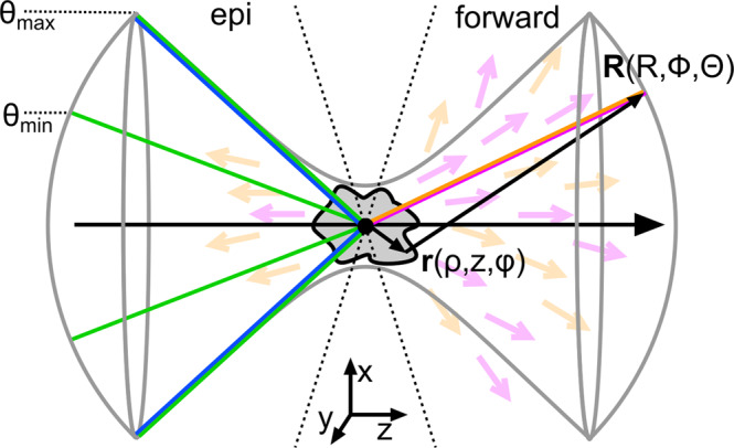
We report the first implementation of laser scanning coherent Stokes Raman scattering (CSRS) microscopy. To overcome the major challenge in CSRS imaging, we show how to suppress the fluorescence background by narrow bandpass filter and a lock-in based demodulation. Near background free CSRS imaging of polymer beads, human skin, onion cells, avocado flesh and the wing disc of a drosphila larva are presented. Finally, we explain and demonstrate numerically that CSRS solves a major obstacle of other coherent Raman techniques by sending a significant part (up to 100%) of the CSRS photons into the backward direction under tight focusing conditions. We believe that this discovery will pave the way for numerous technological advances, e.g., in epi-detected coherent Raman multi-focus imaging, real-time laser scanning based spectroscopy or efficient endoscopy.


Conflict of interest statement

The authors declare no competing interests.

Figures

Fig. 1
Fig. 1. Overview coherent Raman imaging techniques.
In energy diagrams, relative radiation wavelength and energy conservation under plane-wave illumination.
Fig. 2
Fig. 2. CSRS experimental implementation and characterization.
a Scheme of the CSRS experiment. 1. Yb-fiber laser, 2. optical parametric oscillator (OPO), 3. second harmonic generation (SHG), 4. acousto-optic modulator (AOM), laser scanning microscope (LSM), 6. photo-electron multiplier (PMT), 7. lock-in amplifier. b The CSRS signal is separated from fluorescence by means of two angle-tuned narrow bandpass filters. c Additional suppression of fluorescence is achieved by intensity modulating the Stokes and pump beam at the frequencies f1 and f2, respectively. Fluorescence-free CSRS signal is obtained at f1–f2. d Measured CSRS signal at DC, f1, f2, and f1–f2 frequencies when the pump or Stokes beam is blocked (Ip = 0 or Is = 0) or when their temporal overlap is removed (Δt ≫ 3 ps). The fluorescence is strongly rejected on the f1–f2 time trace and mainly comes from the Stokes beam. e The CSRS intensity profile obtained at f1–f2 at the interface of a PMMA bead and olive oil indicates a lateral resolution of <400 nm.
Fig. 3
Fig. 3. Laser scanning CSRS at 2850 cm−1.
The left and right column show the CSRS image demodulated at the frequencies f1–f2 = 1.47 MHz and 0 Hz (DC), respectively. To estimate the remaining fluorescence level, images without temporal overlap of the pump and Stokes pulses are displayed to the right (Δt ≫ 3ps). a Mixture of polystyrene (PS, 30 μm) and Poly-methyl-methacrylate (PMMA, 20 μm) beads in olive oil. b, c Epithelium and dermis of a 20 μm thick human skin section. d Cells of an onion. e Lipid droplets within the flesh of an avocado. f Wing disc of a Drosophila larva. The insets displayed in the second column are the zoomed “CSRS at f1–f2” regions of interest shown in the first column on (b, d). Pixel dwell time: 40 μs. Image acquisition time: 40 s (1000 × 1000). The white and green scale bar equals 20 and 5 μm, respectively.
Fig. 4
Fig. 4. Object frequency support and radiation behavior of CSRS versus CARS.
a The object spatial frequency K-support for Epi-CSRS(CARS) is found by convolving the illumination Ewald spheres of the Stokes (pump), pump (Stokes), and Stokes (probe) with the cap of detection Ewald sphere at coherent Stokes (anti-Stokes) frequency. Note that vector combinations covering the frequency of a homogeneous sample K(0,0,0) are only found for CSRS but not for CARS. A single wavevector combination that phase-matches K(0,0,0) is highlighted to the left, while a similar approach for CARS leads to a large phase-mismatch (ΔK). b CSRS and CARS radiation behavior of a homogeneous sample under standard illumination conditions, i.e., the pump and Stokes beam fill the objective aperture homogeneously (θmax = 80°). c same as in b but with an annular pupil filter applied to the Stokes beam for CSRS covering 50% of the area of the objective back-aperture. For an equitable comparison with CARS, the same pupil filter was applied to the pump beam. d same as for b (conventionally focused beams), but the homogeneous sample was replaced by a frequency object whose scatter density is described as 1+cos(2πz/λo) and λo = 1 μm. e Plot of the ratio of backward/forward radiation (Rb/f) as a function of the object frequency λo. Calculations were performed with λp = 797 nm and λS = 1030 nm.
Fig. 5
Fig. 5
Declaration of variables.

References

    1. Bunaciu A, Fleschin S, Aboul-Enein H. Infrared microspectroscopy applications—review. Curr. Anal. Chem. 2013;10:132–139. doi: 10.2174/1573411011410010011. - DOI
    1. Antonio KA, Schultz ZD. Advances in biomedical raman microscopy. Anal. Chem. 2013;86:30–46. doi: 10.1021/ac403640f. - DOI - PubMed
    1. Duncan MD, Reintjes J, Manuccia TJ. Scanning coherent anti-stokes raman microscope. Opt. Lett. 1982;7:350. doi: 10.1364/OL.7.000350. - DOI - PubMed
    1. Zumbusch A, Holtom GR, Xie XS. Three-dimensional vibrational imaging by coherent anti-stokes raman scattering. Phys. Rev. Lett. 1999;82:4142–4145. doi: 10.1103/PhysRevLett.82.4142. - DOI
    1. Nandakumar P, Kovalev A, Volkmer A. Vibrational imaging based on stimulated raman scattering microscopy. N. J. Phys. 2009;11:033026. doi: 10.1088/1367-2630/11/3/033026. - DOI