. 2023 Jun 7;24(1):306.
doi: 10.1186/s12864-023-09398-y.

Evaluating the mouse neural precursor line, SN4741, as a suitable proxy for midbrain dopaminergic neurons

Affiliations

Evaluating the mouse neural precursor line, SN4741, as a suitable proxy for midbrain dopaminergic neurons

Rachel J Boyd et al. BMC Genomics. .

Abstract

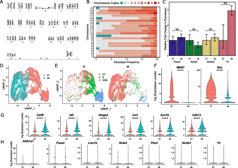
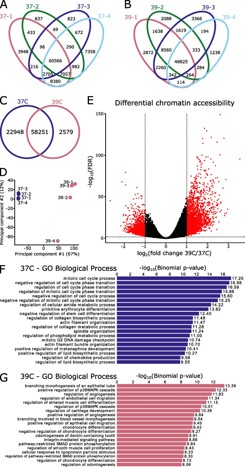
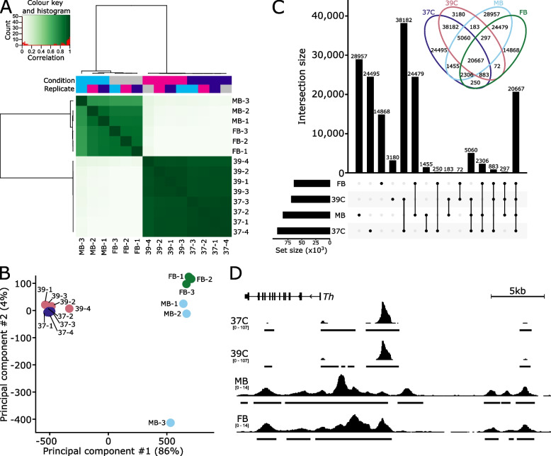
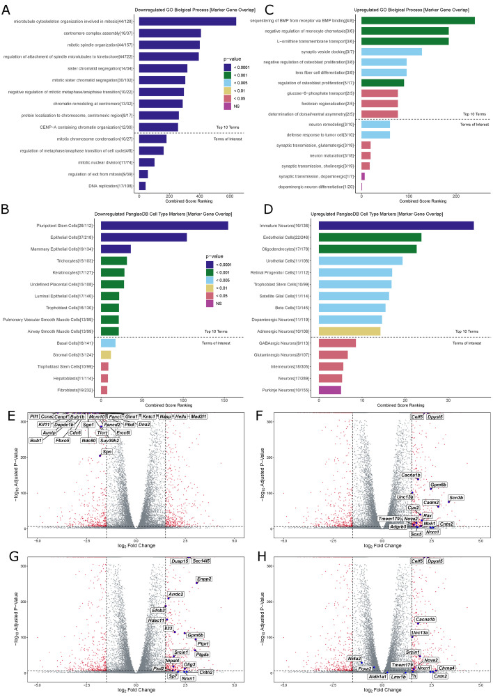
To overcome the ethical and technical limitations of in vivo human disease models, the broader scientific community frequently employs model organism-derived cell lines to investigate disease mechanisms, pathways, and therapeutic strategies. Despite the widespread use of certain in vitro models, many still lack contemporary genomic analysis supporting their use as a proxy for the affected human cells and tissues. Consequently, it is imperative to determine how accurately and effectively any proposed biological surrogate may reflect the biological processes it is assumed to model. One such cellular surrogate of human disease is the established mouse neural precursor cell line, SN4741, which has been used to elucidate mechanisms of neurotoxicity in Parkinson disease for over 25 years. Here, we are using a combination of classic and contemporary genomic techniques - karyotyping, RT-qPCR, single cell RNA-seq, bulk RNA-seq, and ATAC-seq - to characterize the transcriptional landscape, chromatin landscape, and genomic architecture of this cell line, and evaluate its suitability as a proxy for midbrain dopaminergic neurons in the study of Parkinson disease. We find that SN4741 cells possess an unstable triploidy and consistently exhibits low expression of dopaminergic neuron markers across assays, even when the cell line is shifted to the non-permissive temperature that drives differentiation. The transcriptional signatures of SN4741 cells suggest that they are maintained in an undifferentiated state at the permissive temperature and differentiate into immature neurons at the non-permissive temperature; however, they may not be dopaminergic neuron precursors, as previously suggested. Additionally, the chromatin landscapes of SN4741 cells, in both the differentiated and undifferentiated states, are not concordant with the open chromatin profiles of ex vivo, mouse E15.5 forebrain- or midbrain-derived dopaminergic neurons. Overall, our data suggest that SN4741 cells may reflect early aspects of neuronal differentiation but are likely not a suitable proxy for dopaminergic neurons as previously thought. The implications of this study extend broadly, illuminating the need for robust biological and genomic rationale underpinning the use of in vitro models of molecular processes.

Keywords: ATAC-seq; Chromatin accessibility; Disease-relevant model systems; Genomic characterization; Immortalized cell lines; Mouse-derived cell lines; Parkinson disease; RNA-seq; scRNA-seq.


Conflict of interest statement

The authors declare no competing interests.

Figures

Fig. 1
Fig. 1
Characterizing the genomic stability and differentiation consistency of the temperature sensitive SN4741 cell line. A A representative karyogram of SN4741 cells, indicating structural instability (M; marker chromosomes) and unstable triploidy. B A stacked bar plot summarizing the aneuploidy frequency of each chromosome over 20 SN4741 karyotypes. C Assaying expression of dopaminergic neuron markers by RT-qPCR indicates that Foxa2, Nr4a2,Slc6a3, and Th remain at similar expression levels when SN4741 cells are shifted from the permissive temperature (37 °C) to the non-permissive temperature (39 °C). D UMAP plot of scRNA-seq at the permissive and non-permissive temperatures indicates that cells at each temperature are transcriptionally distinct. E Analysis of scRNA-seq data demonstrates that shifting the cells to the non-permissive temperature is accompanied by a shift in cell cycle stage from G2M and S phases to primarily G1 phase. F Violin plots generated with scRNA-seq data show that Mki67, a marker of cellular proliferation, and Nes, a neural stem cell marker, are both expressed at the permissive temperature (37 °C), with little to no expression at the non-permissive temperature (39 °C). G Violin plots generated with scRNA-seq data show that transcripts associated with immature neurons are upregulated when SN4741 cells are shifted to the non-permissive temperature. H Violin plots generated with scRNA-seq data show that expression of DA neural markers, Aldh1a1, Foxa2, Lmx1b, Nr4a2, Pitx3, Slc6a3, and Th, remain at similar levels when SN4741 cells are shifted to the non-permissive temperature. (NS = Not significant, * = p < 0.05, ** = p < 0.01, *** = p < 0.001, **** = p < 0.0001)
Fig. 2
Fig. 2
Changes in chromatin accessibility suggest a reduction in potency at the non-permissive temperature. A, B Replicates are highly similar within temperature conditions, with the majority of peaks present in all four replicates. C The two temperatures share 58,251 regions of open chromatin but do not overlap completely. D Principal component analysis resolves the two temperatures on the first principal component. E Differential accessibility analysis identifies 5,055 differentially accessible regions, with 2,654 preferentially open in the permissive temperature (37 °C) and 2,401 preferentially accessible at the non-permissive temperature (39 °C). F Gene ontology (GO) of genes adjacent regions that are preferentially open at the permissive temperature are associated with regulation of the cell cycle and negative regulation of differentiation, as is appropriate for this temperature. G Gene ontology of genes adjacent regions that are preferentially open at the non-permissive temperature are associated with a variety of differentiation fates (blood vessels, muscle cells, cartilage/chondrocytes). Additionally, two of the top gene ontology terms relate to the p38 MAPK cascade, which has been found to be activated as a cellular response to heat stress
Fig. 3
Fig. 3
Chromatin accessibility of SN4741 cells do not resemble ex vivo dopaminergic neurons. A SN4741 samples are highly correlated with each other but very poorly correlate with the open chromatin landscape of either midbrain (MB) or forebrain (FB) embryonic mouse dopaminergic neurons. B Principal component analysis shows a clear separation between the ex vivo and in vitro samples along PC1, representing 86% of the variance. C An upset plot and associated Venn diagram quantify the overlap of peaks between the four conditions and show the poor relationship between the SN4741 cells and the ex vivo mouse dopaminergic neurons. Most peaks are specific to a single cell type/temperature or are restricted to either the ex vivo or in vitro samples. Few peaks are specifically shared between the non-permissive temperature and the ex vivo samples; for example, there are just 183 peaks that are shared exclusively by the MB dopaminergic neurons and the SN4741 cells at the non-permissive temperature. D A genome track showing the normalized read pile up and called consensus peaks in each of the cell types/temperatures at the key dopaminergic neuron specification gene, Th. The chromatin accessibility is largely similar within ex vivo or in vitro cells but bear little resemblance to each other
Fig. 4
Fig. 4
Gene Ontology and Differential Expression Analysis of Bulk RNA-seq Data: A Top 10 GO terms for downregulated DE genes in SN4741 cells at the non-permissive temperature, followed by GO terms of interest (below dotted line). Terms were evaluated using Combined Score Ranking = (p-value computed using the Fisher exact test)*(z-score computed by assessing the deviation from the expected rank), based on enrichment of DE genes that overlap with Enrichr input genes for each term (the end of each bar). B Top 10 predicted cell types based on downregulated DE genes in SN4741 cells at the non-permissive temperature, followed by predicted cell types of interest (below dotted line). Terms were evaluated using Combined Score Ranking = (p-value computed using the Fisher exact test)*(z-score computed by assessing the deviation from the expected rank), based on enrichment of DE genes that overlap with PanglaoDB input genes for each term (the end of each bar). C Top 10 GO terms for downregulated DE genes in SN4741 cells at the non-permissive temperature. D Top 10 predicted cell types based on upregulated DE genes in SN4741 cells at the non-permissive temperature. E Volcano plot of –log10 adjusted p-value versus log2 fold change with DESeq2 after lfc shrinkage, contrasting the fold change in expression of SN4741 cells at 39 °C, using SN4741 cells at 37 °C as reference. Red points = genes that are statistically differentially expressed (adjusted p-value < 0.01, |log2FC|> 1.5). Blue points = Overlapping immature neuron marker genes. F Blue points = Overlapping immature neuron marker genes. G Blue points = Overlapping oligodendrocyte marker genes. H Blue points = Overlapping DA neuron marker genes
Fig. 5
Fig. 5
Validation of gene ontology and comparison to bulk-RNA-seq from ex vivo DA neurons. A A bar chart showing normalized bulk RNA-seq read counts from genes upregulated at 39 °C that overlap the “immature neurons” predicted cell type. B A bar chart showing normalized bulk RNA-seq read counts from genes upregulated at 39 °C that overlap the “oligodendrocyte” predicted cell type. C A bar chart showing normalized bulk RNA-seq read counts from genes downregulated at 39 °C that overlap the “pluripotent stem cell” predicted cell type. D A Pearson correlation heatmap comparing the transcriptomes of SN4741 cells at 37 °C and 39 °C to midbrain (MB) or forebrain (FB) embryonic mouse dopaminergic neurons. (NS = Not significant, * = p < 0.05, ** = p < 0.01, *** = p < 0.001, **** = p < 0.0001)

Update of

References

    1. Ormond KE, Mortlock DP, Scholes DT, Bombard Y, Brody LC, Faucett WA, et al. Human germline genome editing. Am J Hum Genet. 2017;101(2):167–176. - PMC - PubMed
    1. Barbosa DJ, Capela JP, de Lourdes BM, Carvalho F. In vitro models for neurotoxicology research. Toxicol Res. 2015;4(4):801–842.
    1. Hirsch C, Schildknecht S. In vitro research reproducibility: keeping up high standards. Front Pharmacol. 2019;10:1484. - PMC - PubMed
    1. Fisher S, Grice EA, Vinton RM, Bessling SL, Urasaki A, Kawakami K, et al. Evaluating the biological relevance of putative enhancers using Tol2 transposon-mediated transgenesis in zebrafish. Nat Protoc. 2006;1(3):1297–1305. - PubMed
    1. Gorkin DU, Lee D, Reed X, Fletez-Brant C, Bessling SL, Loftus SK, et al. Integration of ChIP-seq and machine learning reveals enhancers and a predictive regulatory sequence vocabulary in melanocytes. Genome Res. 2012;22(11):2290–2301. - PMC - PubMed