. 2023 Jun 7;18(1):98.
doi: 10.1186/s13014-023-02291-8.

Chromosomal radiosensitivity in oncological and non-oncological patients with rheumatoid arthritis and connective tissue diseases

Affiliations

Chromosomal radiosensitivity in oncological and non-oncological patients with rheumatoid arthritis and connective tissue diseases

Dinah Rzepka et al. Radiat Oncol. .

Abstract

Background: The risk of developing late radiotoxicity after radiotherapy in patients with high chromosomal radiosensitivity after radiotherapy could potentially be higher compared to the risk in patients with average radiosensitivity. In case of extremely high radiosensitivity, dose reduction may be appropriate. Some rheumatic diseases (RhD), including connective tissue diseases (CTDs) appear to be associated with higher radiosensitivity. The question arises as to whether patients with rheumatoid arthritis (RA) also generally have a higher radiosensitivity and whether certain parameters could indicate clues to high radiosensitivity in RA patients which would then need to be further assessed before radiotherapy.

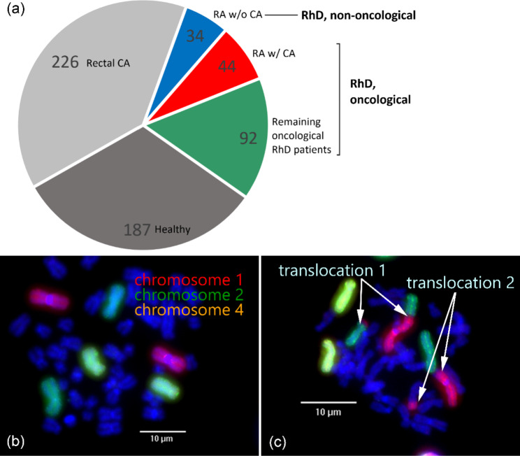
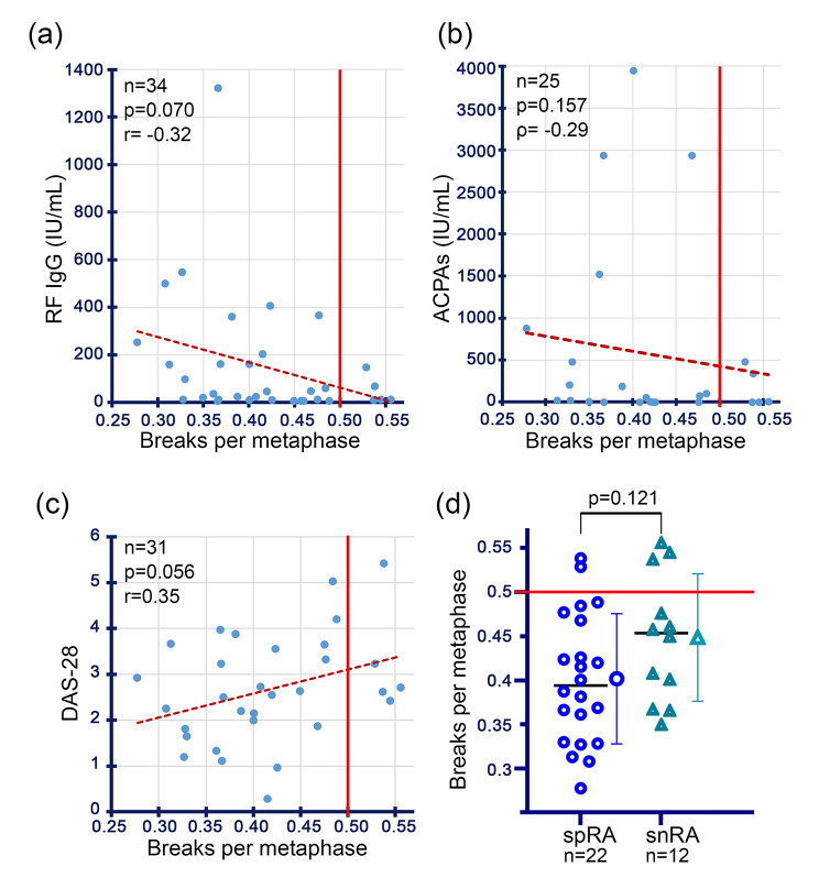
Methods: Radiosensitivity was determined in 136 oncological patients with RhD, 44 of whom were RA patients, and additionally in 34 non-oncological RA patients by three-colour fluorescence in situ hybridization (FiSH), in which lymphocyte chromosomes isolated from peripheral blood are analysed for their chromosomal aberrations of an unirradiated and an with 2 Gy irradiated blood sample. The chromosomal radiosensitivity was determined by the average number of breaks per metaphase. In addition, correlations between certain RA- or RhD-relevant disease parameters or clinical features such as the disease activity score 28 and radiosensitivity were assessed.

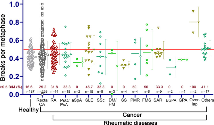
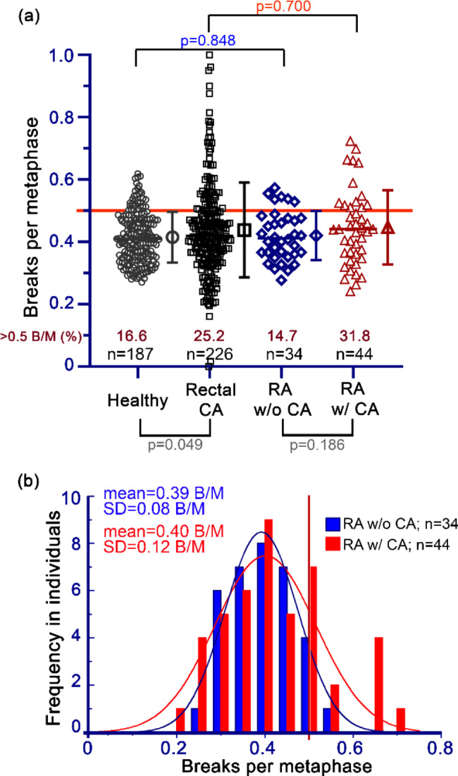
Results: Some oncological patients with RhD, especially those with connective tissue diseases have significantly higher radiosensitivity compared with oncology patients without RhD. In contrast, the mean radiosensitivity of the oncological patients with RA and other RhD and the non-oncological RA did not differ. 14 of the 44 examined oncological RA-patients (31.8%) had a high radiosensitivity which is defined as ≥ 0.5 breaks per metaphase. No correlation of laboratory parameters with radiosensitivity could be established.

Conclusions: It would be recommended to perform radiosensitivity testing in patients with connective tissue diseases in general. We did not find a higher radiosensitivity in RA patients. In the group of RA patients with an oncological disease, a higher percentage of patients showed higher radiosensitivity, although the average radiosensitivity was not high.

Keywords: Chromosomal aberrations; Collagen vascular disease; Connective tissue disease; Fluorescence in situ hybridisation; Radiation sensitivity; Radiosensitivity; Radiotherapy; Radiotoxicity; Rheumatism; Rheumatoid arthritis.


Conflict of interest statement

The authors declare that they have no competing interests.

Figures

Fig. 1
Fig. 1
G0 three-colour fluorescence in situ hybridization assay (FiSH) as radiosensitivity assay. (a) The pie chart gives an overview of the patient groups that were used and the number of individuals in each of the groups. In addition to the healthy and rectal CA (rectal cancer) cohorts [50], there is also a cohort of patients with RhD (rheumatic disease), which can be subdivided into non-oncological (RA w/o CA - non-oncological patients with rheumatoid arthritis) and oncological patients. The latter includes the RA w/ CA cohort (oncological patients with rheumatoid arthritis). (b) Metaphase without chromosomal aberrations. (c) Metaphase with chromosomal aberrations. Chromosome #1 is translocated to chromosome #2-each chromosome is broken once (translocation 1). Chromosome #1 is translocated to another one coloured in blue-both are broken once each (translocation 2). In total, four breaks are counted in this metaphase
Fig. 2
Fig. 2
Radiosensitivity in healthy individuals, rectal CA patients [50] and oncological patients with rheumatic diseases. Each symbol represents the individual’s radiosensitivity measured with the three-colour FiSH. The horizontal red line at 0.5 B/M marks the values above which the radiosensitivity (breaks per metaphase, B/M) is considered higher. The horizontal line of the respective cohort represents the median, and the symbol to the right of it in the same colour marks the mean from which the standard deviation (SD) is displayed. Note the percentage of patients with ≥ 0.5 B/M. Legend: RA = rheumatoid arthritis, PsO/PsA = psoriasis/psoriatic arthritis, aSpA = axial spondyloarthropathy, SLE = systemic lupus erythematosus, SSc = systemic sclerosis/scleroderma, DM/PM = dermatomyositis/polymyositis, SS = Sjögren’s syndrome, PMR = polymyalgia rheumatica, FMS = fibromyalgia syndrome, SAR = sarcoidosis, EGPA = eosinophilic granulomatosis with polyangiitis, GPA = granulomatosis with polyangiitis, Overlap = overlap syndrome, Others = rheumatic symptoms or undifferentiated connective tissue disease or “rheumatism” as diagnosis.
Fig. 3
Fig. 3
Radiosensitivity in non-oncological and oncological patients with rheumatoid arthritis. (a) Each symbol represents the radiosensitivity (breaks per metaphase, B/M) with the 3-colour FiSH. Comparison of the measured radiosensitivity of the patients with RA w/ CA (oncological patients with rheumatoid arthritis) and RA w/o CA (non-oncological patients with rheumatoid arthritis) cohorts with those of healthy individuals and with rectal cancer (rectal CA) patients [50]. In addition, the two comparison cohorts (healthy and rectal CA) and the two cohorts RA with CA and RA without CA were compared. Note the percentage of patients with ≥ 0.5 B/M. The horizontal line of the respective cohort represents the median, the symbol to the right of it in the same colour marks the mean from which the standard deviation (SD) is displayed. (b) The mean and the SD of the Gaussian normal distribution are calculated from the values of RA w/o and w/ CA. A vertical line at 0.5 B/M separates high and average radiosensitivity
Fig. 4
Fig. 4
Correlation of laboratory values and disease activity in non-oncological patients with rheumatoid arthritis with their radiosensitivity. (a) Correlation of radiosensitivity (breaks per metaphase, B/M) and IgG rheumatoid factor (RF, highest value). (b) Correlation of B/M and anti-citrullinated protein antibodies (ACPAs, highest value). (c). Correlation of B/M and disease activity score 28 (DAS-28, most recent value). (d) Comparison of B/M of patients with seropositive rheumatoid arthritis (spRA) with those with seronegative rheumatoid arthritis (snRA).

References

    1. Hara T, Iwadate M, Tachibana K, Waguri S, Takenoshita S, Hamada N. Metastasis of breast cancer cells to the bone, lung, and lymph nodes promotes resistance to ionizing radiation. Strahlenther Onkol. 2017;193:848–55. doi: 10.1007/s00066-017-1165-2. - DOI - PubMed
    1. Huang A, Xiao Y, Peng C, Liu T, Lin Z, Yang Q, Zhang T, Liu J, Ma H. 53BP1 expression and immunoscore are associated with the efficacy of neoadjuvant chemoradiotherapy for rectal cancer. Strahlenther Onkol. 2020;196:465–73. doi: 10.1007/s00066-019-01559-x. - DOI - PubMed
    1. Distel LV, Neubauer S, Keller U, Sprung CN, Sauer R, Grabenbauer GG. Individual differences in chromosomal aberrations after in vitro irradiation of cells from healthy individuals, cancer and cancer susceptibility syndrome patients. Radiother Oncol. 2006;81:257–63. doi: 10.1016/j.radonc.2006.10.012. - DOI - PubMed
    1. De Courcy L, Bezak E, Marcu LG. Gender-dependent radiotherapy: the next step in personalised medicine? Crit Rev Oncol Hematol. 2020;147:102881. doi: 10.1016/j.critrevonc.2020.102881. - DOI - PubMed
    1. Sicklick JK, Kato S, Okamura R, Schwaederle M, Hahn ME, Williams CB, De P, Krie A, Piccioni DE, Miller VA, et al. Molecular profiling of cancer patients enables personalized combination therapy: the I-PREDICT study. Nat Med. 2019;25:744–50. doi: 10.1038/s41591-019-0407-5. - DOI - PMC - PubMed

MeSH terms