. 2023 Jun 7;25(1):96.
doi: 10.1186/s13075-023-03081-z.

Role of high-temperature requirement serine protease A 2 in rheumatoid inflammation

Affiliations

Role of high-temperature requirement serine protease A 2 in rheumatoid inflammation

Gi Heon Jeong et al. Arthritis Res Ther. .

Abstract

Background: High-temperature requirement serine protease A 2 (HtrA2) is known to be involved in growth, unfolded protein response to stress, apoptosis, and autophagy. However, whether HtrA2 controls inflammation and immune response remains elusive.

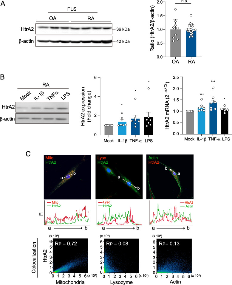
Methods: Expression of HtrA2 in the synovial tissue of patients was examined using immunohistochemistry and immunofluorescence staining. Enzyme-linked immunosorbent assay was used to determine the concentrations of HtrA2, interleukin-6 (IL-6), interleukin-8 (IL-8), chemokine (C-C motif) ligand 2 (CCL2), and tumor necrosis factor α (TNFα). Synoviocyte survival was assessed by MTT assay. For the downregulation of HtrA2 transcripts, cells were transfected with HtrA2 siRNA.

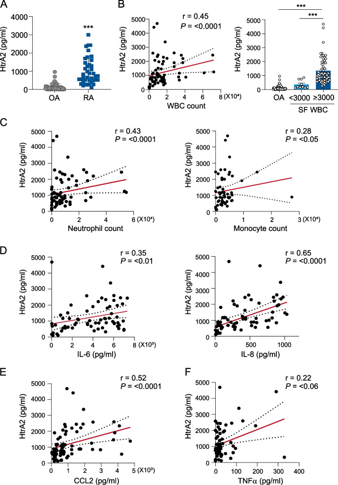
Results: We found that the concentration of HtrA2 was elevated in rheumatoid arthritis (RA) synovial fluid (SF) than in osteoarthritis (OA) SF, and its concentrations were correlated with the number of immune cells in the RA SF. Interestingly, HtrA2 levels in the SF of RA patients were elevated in proportion to synovitis severity and correlated with the expression of proinflammation cytokines and chemokines, such as IL-6, IL-8, and CCL2. In addition, HtrA2 was highly expressed in RA synovium and primary synoviocytes. RA synoviocytes released HtrA2 when stimulated with ER stress inducers. Knockdown of HtrA2 inhibited the IL1β-, TNFα-, and LPS-induced release of proinflammatory cytokines and chemokines by RA synoviocytes.

Conclusion: HtrA2 is a novel inflammatory mediator and a potential target for the development of an anti-inflammation therapy for RA.

Keywords: Fibroblast-like synoviocytes; High-temperature requirement serine protease A; Inflammation; Rheumatoid arthritis.


Conflict of interest statement

The authors declare no competing interests.

Figures

Fig. 1
Fig. 1
Correlation between HtrA2 and inflammatory cytokine levels. A HtrA2 concentrations in synovial fluids (SFs) of individuals with RA (n=72) or OA (n=61). B, C Fluid concentrations of HtrA2 depending on the count of white blood cells (WBCs, p < 0.0001), neutrophils (p < 0.0001), or monocytes (p < 0.05). D, E Correlation of HtrA2 levels with IL-6 (p < 0.01), IL-8 (p < 0.0001), CCL2 (p < 0.0001), and TNFα (p < 0.06) levels in the SF of patients with RA. Data present the mean ± s.e.m. ***p < 0.001. P values were determined by Mann-Whitney U test (A), Spearman correlation (CF and left panel of B), or one-way ANOVA (right panel of B)
Fig. 2
Fig. 2
Expression of HtrA2 in RA synovial fluids and the synovium. A HtrA2 levels in the SF of RA patients with considerable synovial hypertrophy (grayscale US, GSUS 2 and 3) compared with those with little or mild synovial hypertrophy (GSUS 0 and 1). B HtrA2 concentrations in RA patients with and without enhanced vascularity (power-Doppler US, PDUS 1–3). (PDUS 0). C HtrA2 concentrations in RA patients with and without active synovitis. Active synovitis was classified as GSUS ≥2 or PDUS ≥1. D Anti-HtrA2 antibody or isotype control antibody immunohistochemical staining of rheumatoid arthritis (RA, n=4) and osteoarthritis (OA, n=4) synovium. HtrA2 positivity was found in the cell lining layer (arrow), as well as in infiltrating leukocytes in the sublining layer (arrowhead). Scale bar, 20 μm. E Summary of the percentage of cells in each layer. Data are the mean ± s.e.m. *p < 0.05, **p < 0.01, ***p < 0.001. P values were determined by one-way ANOVA test (AC) or two-way ANOVA (E)
Fig. 3
Fig. 3
Increase in HtrA2 expression in FLSs by proinflammatory stimuli. A HtrA2 protein expression in RA-FLSs (n=20) and OA-FLSs (n=9) was assessed by western blot analysis. B RA-FLSs were stimulated with TNFα (10 ng/ml), IL1β (1 ng/ml), or LPS (1 μg/ml) in 1% FBS DMEM for 6 h or 12h. The mRNA and protein expression levels of HtrA2 in synoviocytes were determined by western blot analysis (left panel, n=5) and RT-PCR (right panel, n=7). Data are the mean ± s.e.m. *p < 0.05, **p < 0.01, ***p < 0.001. P values were determined by one-way ANOVA test. C Double immunofluorescence staining of synoviocytes. Cells were stained with Ab for HtrA2, mitochondria marker (RFP), F-actin marker (GFP), and lysosome marker (RFP) in RA-FLSs. Nuclei were stained with DAPI (blue). The fluorescence signal intensity (FI) was quantified along the white arrow. Scale bar, 20 μm. The colocalization study was carried out utilizing raw and deconvolved images, as well as Imaris, and ZEN software. The Rp indicates Pearson’s colocalization coefficient
Fig. 4
Fig. 4
The release of HtrA2 into the extracellular space. A Tunicamycin and thapsigargin induce HtrA2 release from the mitochondria into the cytosol. RA-FLSs were treated with 20 μg/ml tunicamycin or 20 μM thapsigargin for 16 h, after immunostaining with HtrA2 (green) and Tom20 (Mito, red). The fluorescence signal intensity (FI) was quantified along the white arrow. Scale bar, 20 μm. 3D images prepared with Imaris software. B HtrA2 expression was significantly elevated in the supernatant of RA-FLSs following ER stress-triggered apoptosis. RA FLSs (2×10⁴ cells) were cultured for 24h with 1% FBS DMEM in the presence of tunicamycin (n=7) or thapsigargin (n=4). Concentrations of HtrA2 were determined by ELISA. C FLSs treated with 100 μg/ml tunicamycin or 50 μM thapsigargin became spherical, shrunken, and detached from the bottom of the culture plates under phase-contrast microscopy, whereas untreated cells remained bipolar (upper panel). The nuclei were stained with DAPI (blue). Cell viability was determined by MTT assay (lower panel, n=4). Data presented as mean ± s.e.m. *p < 0.05, ***, p < 0.001. P values were determined by one-way ANOVA test (B and C)
Fig. 5
Fig. 5
The effect of HtrA2 siRNA on cytokine production. A Transfection efficiency of HtrA2 siRNA was measured by RT-qPCR (left panel, n=4) including western blotting (right panel). Twelve hours after transfection with siRNA for HtrA2, mRNA expression levels were determined by RT-qPCR analysis. B Fifty-eight hours after transfection with siRNA for HtrA2, cell viability was measured by an MTT assay (n=3). C Effect of HtrA2 knockdown on IL1β-, TNFα-, or LPS-induced increases of IL-6 and IL-8 production in RA synoviocytes. At 24 h after HtrA2 siRNA transfection, RA FLSs (n=9) were treated with IL1β (1 ng/ml), TNFα (10 ng/ml), or LPS (1 μg/ml) in 1% FBS DMEM for 24 h. Cytokine production was quantified by ELISA. D ELISA revealed CCL2 production by synoviocytes (n=7). Data represent the mean ± s.e.m. *p < 0.05, **p < 0.01, ***p < 0.001 versus siCon treated cells. P values were determined by Mann-Whitney U test (A) or Two-way ANOVA test (C, D)

References

    1. Feldmann M, Brennan FM, Maini RN. Rheumatoid arthritis. Cell. 1996;85:307–310. doi: 10.1016/S0092-8674(00)81109-5. - DOI - PubMed
    1. Firestein GS. Evolving concepts of rheumatoid arthritis. Nature. 2003;423:356–361. doi: 10.1038/nature01661. - DOI - PubMed
    1. Bottini N, Firestein GS. Duality of fibroblast-like synoviocytes in RA: passive responders and imprinted aggressors. Nat Rev Rheumatol. 2013;9:24–33. doi: 10.1038/nrrheum.2012.190. - DOI - PMC - PubMed
    1. Yamanishi Y, Firestein GS. Pathogenesis of rheumatoid arthritis: the role of synoviocytes. Rheum Dis Clin North Am. 2001;27:355–371. doi: 10.1016/S0889-857X(05)70206-4. - DOI - PubMed
    1. Feldmann M, Brennan FM, Maini RN. Role of cytokines in rheumatoid arthritis. Annu Rev Immunol. 1996;14:397–440. doi: 10.1146/annurev.immunol.14.1.397. - DOI - PubMed

Publication types