. 2023 Jun 7;14(1):159.
doi: 10.1186/s13287-023-03387-4.

Endometrium-derived mesenchymal stem cells suppress progression of endometrial cancer via the DKK1-Wnt/β-catenin signaling pathway

Affiliations

Endometrium-derived mesenchymal stem cells suppress progression of endometrial cancer via the DKK1-Wnt/β-catenin signaling pathway

Yuhui Xu et al. Stem Cell Res Ther. .

Abstract

Background: Mesenchymal stem cell (MSC) therapy is an attractive treatment option for various cancers. Whether MSCs can be used to treat well-differentiated endometrial cancer (EC) remains unclear. The aim of this study is to explore the potential therapeutic effects of MSCs on EC and the underlying mechanisms.

Methods: The effects of adipose-derived MSCs (AD-MSCs), umbilical-cord-derived MSCs (UC-MSCs), and endometrium-derived MSCs (eMSCs) on the malignant behaviors of EC cells were explored via in vitro and in vivo experiments. Three EC models, including patient-derived EC organoid lines, EC cell lines, and EC xenograft model in female BALB/C nude mice, were used for this study. The effects of MSCs on EC cell proliferation, apoptosis, migration, and the growth of xenograft tumors were evaluated. The potential mechanisms by which eMSCs inhibit EC cell proliferation and stemness were explored by regulating DKK1 expression in eMSCs or Wnt signaling in EC cells.

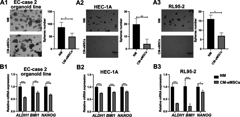
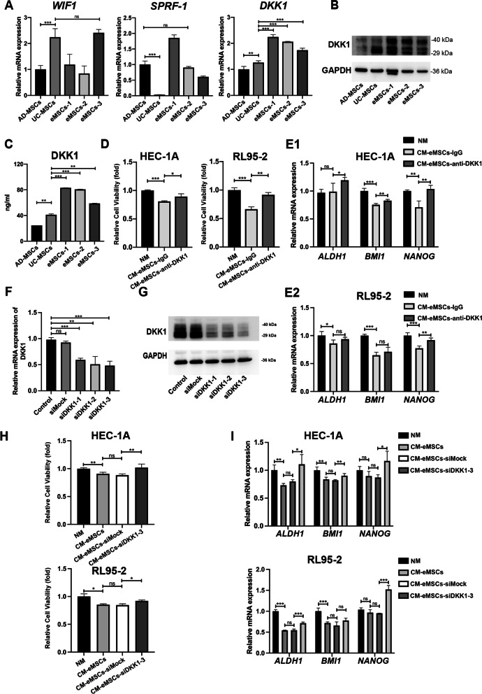
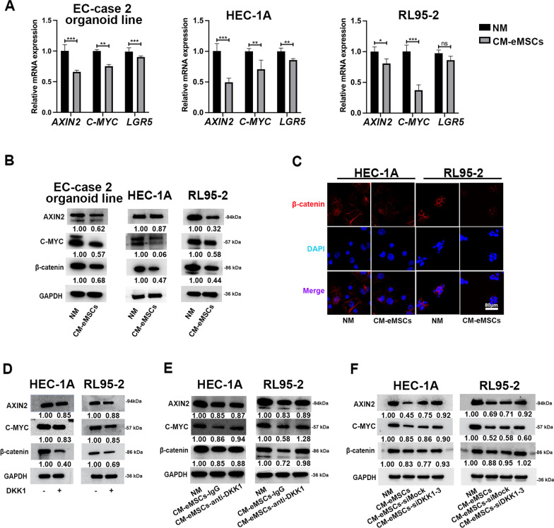
Results: Our results showed that eMSCs had the highest inhibitory effect on EC cell viability, and EC xenograft tumor growth in mice compared to AD-MSCs and UC-MSCs. Conditioned medium (CM) obtained from eMSCs significantly suppressed the sphere-forming ability and stemness-related gene expression of EC cells. In comparison to AD-MSCs and UC-MSCs, eMSCs had the highest level of Dickkopf-related protein 1 (DKK1) secretion. Mechanistically, eMSCs inhibited Wnt/β-catenin signaling in EC cells via secretion of DKK1, and eMSCs suppressed EC cell viability and stemness through DKK1-Wnt/β-catenin signaling. Additionally, the combination of eMSCs and medroxyprogesterone acetate (MPA) significantly inhibited the viability of EC organoids and EC cells compared with eMSCs or MPA alone.

Conclusions: The eMSCs, but not AD-MSCs or UC-MSCs, could suppress the malignant behaviors of EC both in vivo and in vitro via inhibiting the Wnt/β-catenin signaling pathway by secreting DKK1. The combination of eMSCs and MPA effectively inhibited EC growth, indicating that eMSCs may potentially be a new therapeutic strategy for young EC patients desiring for fertility preservation.

Keywords: DKK1; Endometrial cancer; Wnt/β-catenin signaling; eMSCs.


Conflict of interest statement

The authors declare no potential conflict of interest.

Figures

Fig. 1
Fig. 1
eMSCs inhibited the viability of EC patient-derived organoids. A Bright field images of EC organoids were taken from day 1 to day 6 after establishment. Original magnification, 4 × ; Scale bar, 400 μm. B Bright field images of EC organoids at Passage 1, Passage 5, and Passage 10. Original magnification, 4 × ; Scale bar, 400 μm. C Immunofluorescence staining images for DAPI, Ki67, and cytokeratin 7 (CK7) in EC organoids. Original magnification, 20 × ; Scale bar, 80 μm. D H&E and IHC staining images for Ki67, ERα, and PR in EC tissues and organoids. Original magnification, 20 × ; Scale bar, 80 μm. E eMSCs had the strongest inhibitory effect on the viability of EC organoids. EC organoids were treated with NM or CM derived from AD-MSCs, UC-MSCs, or eMSCs respectively for 96 h before three-dimensional cell viability assay. Three organoid lines derived from three patients with well-differentiated endometrial endometrioid cancer were used. Each EC organoid line was tested independently and was repeated three times in the same condition with independent analysis. NM, normal medium; CM, conditioned medium; Data were analyzed by ratio t-test (E). ns, not significant; *P < 0.05; **P < 0.01; ***P < 0.001
Fig. 2
Fig. 2
eMSCs suppressed the malignant behaviors of EC cells. A CM derived from eMSCs showed the most significant anti-proliferative effect on EC cells, compared to AD-MSCs and UC-MSCs. RL95-2 and HEC-1A cells were treated with NM or CM derived from MSCs for 48 h before Cell Viability Assay. B CM derived from eMSCs markedly increased BAX expression and BAX/BCL2 ratio and decreased BCL2 expression in EC cells, compared to AD-MSCs and UC-MSCs. RL95-2 and HEC-1A cells were treated with NM or CM derived from MSCs for 48 h before western blotting. BAX/BCL2 ratio was calculated from densitometry by ImageJ. C RL95-2 and HEC-1A cells seeded in 24‐well chambers were co-cultured with or without MSCs, as shown in the pattern diagram for 24 h before cell migration assay. D eMSCs did not affect EC cell migration, while AD-MSCs promoted EC cell migration. UC-MSCs slightly induced migration of HEC-1A cells but had no effect on RL95-2 cells. Bright field images were taken (Left), and the numbers of migratory cells per bright field image were calculated (Right). Original magnification 4 × ; Scale bar, 400 μm. E–G eMSCs had the highest inhibitory effect on EC xenograft tumor growth, compared to AD-MSCs and UC-MSCs. RL952 cells alone, or RL95-2 cells and MSCs (5:1) were subcutaneously injected in female nude mice. Mice were sacrificed after 28 days (E), tumor tissues were photographed (F), and tumor weight was then measured (G). H-I H&E and IHC staining images for Ki67 in xenograft tumors. NM, normal medium; CM, conditioned medium; The blots of BAX, BCL2, and GAPDH were all cropped (B) and full-length blots were presented in Additional file 7: Fig. S6. Data were analyzed by ratio t-test (A) and unpaired t-test (B, D, G, and I). ns, not significant; *P < 0.05; **P < 0.01; ***P < 0.001
Fig. 3
Fig. 3
eMSCs suppressed the tumor-related stemness of EC cells. A1-A3 eMSCs inhibited sphere-forming ability of EC organoids and cell lines. Single cell suspensions of EC cells were seeded in Matrigel and treated with NM or CM from eMSCs for 15 days. The numbers of visible spheres were calculated. Original magnification, 4 × ; Scale bar, 400 μm. B1-B3 eMSCs inhibited the transcriptional levels of EC stemness-associated genes (ALDH1, BMI1, and NANOG) both in EC organoids and cell lines. CM derived from eMSCs was used to treat EC organoids for 96 h or EC cell lines for 48 h. EC cells were then harvested for qRT-PCR analysis. NM, normal medium; CM, conditioned medium; Data were analyzed by unpaired t-test (A-B). *P < 0.05; **P < 0.01; ***P < 0.001
Fig. 4
Fig. 4
eMSCs inhibited EC cell proliferation and stemness through paracrine secretion of DKK1. A eMSCs showed the highest level of DKK1 mRNA, compared to AD-MSCs and UC-MSCs, while the mRNA levels of SFRP1 and WIF1 were not significantly elevated in eMSCs, compared to other MSCs. B eMSCs showed the highest level of DKK1 in eMSCs cell, compared to AD-MSCs and UC-MSCs, as detected by western blotting. C eMSCs showed the highest level of DKK1 secretion compared to AD-MSCs and UC-MSCs. CM obtained from the three MSCs was collected under the same condition as shown in Materials and Methods and the DKK1 levels were evaluated using an ELISA kit. D-E DKK1-neutralizing antibody compromised the inhibitory effect of eMSCs on proliferation (D) and stemness (E) in EC cells. CM obtained from eMSCs was pre-treated with DKK1-neutralizing antibody (anti-DKK1) or isotype control (IgG) overnight and then used to treat EC cells for 48 h. EC cells were harvested for Cell Viability Assay (D) and qRT-PCR (E) respectively. F–G. The mRNA and protein levels of DKK1 were reduced in eMSCs by transfection of DKK1 siRNAs. eMSCs were transfected with three different siRNAs (siDKK1-1, -2, and -3) targeting DKK1 or siMock for 6 h. At 48 h after transfection, eMSCs were harvested for examination of mRNA and protein levels of DKK1. siDKK1-3 was used for further investigation. H-I. Silencing DKK1 in eMSCs abrogated the anti-proliferation and anti-stemness effects of eMSCs on EC cells. The CM of eMSCs was collected after 24 h of siDKK1-3 transfection. EC cells were treated with the indicated CM of eMSCs for 48 h and then the mRNA levels of stemness-associated genes, ALDH1, BMI1, and NANOG, were measured by qRT-PCR. NM, normal medium; CM, conditioned medium; The blots of DKK1 and GAPDH were all cropped (B and G) and full-length blots were presented in Additional file 7: Figure S6. Data were analyzed by unpaired t-test (A, C, E, F, I) and ratio t-test (D and H). ns, not significant; *P < 0.05; **P < 0.01; ***P < 0.001
Fig. 5
Fig. 5
eMSCs inhibited Wnt/β-catenin signaling in EC cells through DKK1. A, B eMSCs inhibited expression of β-catenin protein and Wnt target genes in EC organoids and cell lines. CM from eMSCs was used to treat EC organoids for 96 h or EC cell lines for 48 h. EC cells were then harvested for qRT-PCR and western blotting. EC-case 2 organoid line was used in Fig. 5A. C Expression of β-catenin protein was reduced in cytoplasm and nuclei of EC cells after treatment of CM derived from eMSCs. Original magnification, 40 × ; Scale bar, 80 μm. D DKK1 inhibited expression of β-catenin and Wnt target proteins (AXIN2 and C-MYC) in EC cell lines. EC cells were treated with DKK1 (100 ng/ml) for 48 h and then harvested for western blotting. E DKK1-neutralizing antibody compromised the inhibitory effect of eMSCs on Wnt signaling in EC cells. CM obtained from eMSCs was pre-treated with DKK1-neutralizing antibody (anti-DKK1) or isotype control (IgG) overnight and then used to treat EC cells for 48 h. EC cells were harvested for western blotting analysis. F Silencing DKK1 by siDKK1-3 rescued Wnt signaling, which was inhibited by eMSCs in EC cells. EC cells were treated with the indicated CM of eMSCs for 48 h. EC cells were harvested for western blotting analysis. NM, normal medium; CM, conditioned medium. The blots of AXIN2, C-MYC, β-catenin, and GAPDH were all cropped (B, D-F) and full-length blots were presented in Additional file 7: Figure S6. Data were analyzed by unpaired t-test (A). ns, not significant; *P < 0.05; **P < 0.01; ***P < 0.001
Fig. 6
Fig. 6
eMSCs inhibited EC cell proliferation and stemness through DKK1-Wnt/β-catenin signaling. A The inhibitory effect of Wnt inhibitor Foscenvivint on EC cell sphere-forming ability. EC cells were treated with or without Foscenvivint (0.5 μM) for 15 days. The numbers of visible spheres were calculated and compared. Original magnification, 4 × ; Scale bar, 400 μm. B The inhibitory effect of Foscenvivint on expression of stemness-related proteins in EC cells. EC cells were treated with or without Foscenvivint (2 μM) for 48 h and then were harvested to analyze β-catenin and stemness-related proteins by western blotting. C The inhibitory effect of CM derived from eMSCs on sphere-forming ability was reversed after activating Wnt signaling. EC cells were treated with or without Wnt activator CHIR-99021 (5 nM) in the presence or absence of CM derived from eMSCs for 15 days. The numbers of visible spheres were calculated and compared. Original magnification, 4 × ; Scale bar, 400 μm. D The inhibitory effect of CM derived from eMSCs on Wnt/β-catenin signaling and stemness-related proteins were reversed after activating Wnt signaling by CHIR-99021. EC cells were treated with or without Wnt activator CHIR-99021 (5 nM) in the presence or absence of CM derived from eMSCs for 48 h and then were harvested to analyze β-catenin and Wnt target proteins by western blotting. E CHIR-99021 treatment rescued DKK1-induced inhibition of Wnt/β-catenin signaling and stemness-related proteins. EC cells were treated with or without Wnt activator CHIR-99021 (5 nM) in the presence or absence DKK1 (DKK1) for 48 h and then were harvested to analyze β-catenin and stemness-related proteins by western blotting. CM, conditioned medium; The blots of BMI1, Nanog, β-catenin, and GAPDH were all cropped (B, D, and E) and full-length blots were presented in Additional file 7: Figure S6. Data were analyzed by unpaired t-test (A, C). ***P < 0.001
Fig. 7
Fig. 7
eMSCs combined with MPA had the highest inhibitory effect on EC growth. A CM obtained from eMSCs combined with MPA had the highest proliferation-inhibiting effect on EC cells. RL95-2 and HEC-1A cells were treated with 10 μM MPA with or without CM of eMSCs for 48 h before Cell Viability Assay. EC organoids were treated with 10 μM MPA with or without CM of eMSCs for 96 h before CCK-8 assay. B CM obtained from eMSCs combined with 20 μM MPA effectively inhibited viability of EC organoids, compared to using CM or MPA alone. Three-dimensional cell viability assay was used to assess the viability of EC organoids treated with MPA and/or CM of eMSCs for 96 h. C DKK1 combined with MPA had highest anti-proliferative effect on EC organoids compared with DKK1 or MPA alone. EC organoids were treated with or without MPA (20 μM) in the presence of DKK1 (100 ng/mL) or not for 96 h to assess cell viability by three-dimensional cell viability assay. NM, normal medium; CM, conditioned medium; Data were analyzed by ratio t-test (A-C). ns, not significant; *P < 0.05; **P < 0.01; ***P < 0.001

Similar articles

Cited by

References

    1. Siegel RL, Miller KD, Fuchs HE, Jemal A. Cancer statistics. CA Cancer J Clin. 2021;71(2021):7–33. doi: 10.3322/caac.21654. - DOI - PubMed
    1. Washington CR, Haggerty A, Ronner W, Neff PM, Ko EM. Knowledge of endometrial cancer risk factors in a general gynecologic population. Gynecol Oncol. 2020;158:137–142. doi: 10.1016/j.ygyno.2020.03.032. - DOI - PubMed
    1. Shen L, Xie L, Li R, Shan B, Liang S, Tian W, Wang H, Ren Y. A preoperative prediction model for predicting coexisting adnexa malignancy of patients with G1/G2 endometrioid endometrial cancer. Gynecol Oncol. 2020;159:402–408. doi: 10.1016/j.ygyno.2020.08.017. - DOI - PubMed
    1. Soliman PT, Oh JC, Schmeler KM, Sun CC, Slomovitz BM, Gershenson DM, Burke TW, Lu KH. Risk factors for young premenopausal women with endometrial cancer. Obstet Gynecol. 2005;105:575–580. doi: 10.1097/01.AOG.0000154151.14516.f7. - DOI - PubMed
    1. Brooks RA, Fleming GF, Lastra RR, Lee NK, Moroney JW, Son CH, Tatebe K, Veneris JL. Current recommendations and recent progress in endometrial cancer. CA Cancer J Clin. 2019;69:258–279. - PubMed

Publication types

Substances