

Spinal meningiomas
- PMID: 37287574
- PMCID: PMC10243846
- DOI: 10.1093/noajnl/vdad013
Spinal meningiomas
Abstract
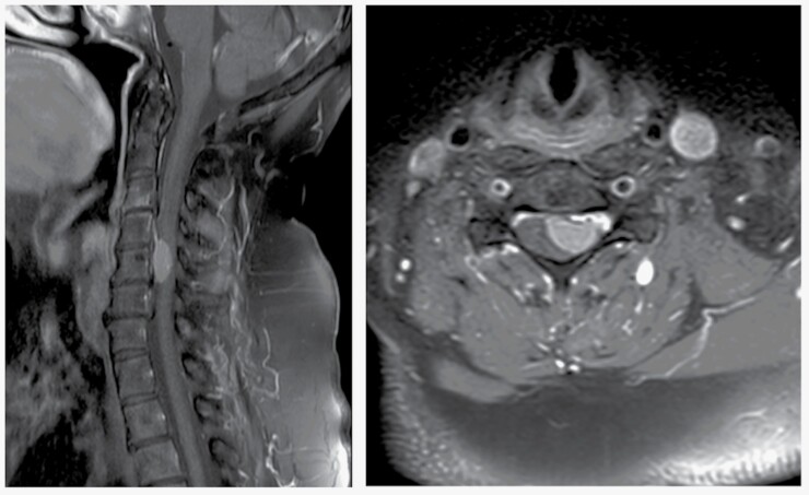
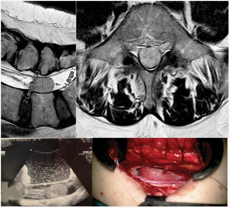
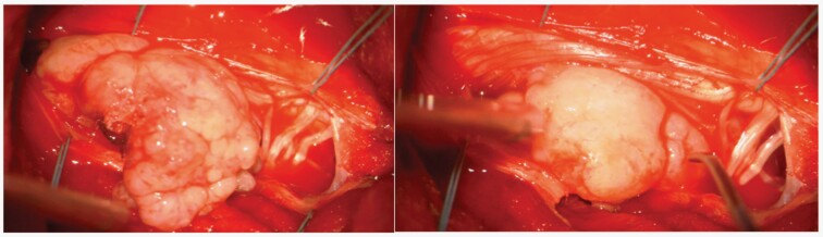
Spinal meningiomas (SM) are lesions with a mostly favorable oncological and surgical prognosis and a low incidence of tumor recurrence. SM account for approximately 1.2-12.7% of all meningiomas and 25% of all spinal cord tumors. Typically, SM are located in the intradural extramedullary space. SM grow slowly and spread laterally into the subarachnoid space, stretching and sometimes incorporating the surrounding arachnoid but rarely the pia. Standard treatment is surgery with the primary aims of achieving complete tumor resection as well as improving and recovering neurologic function. Radiotherapy may be considered in case of tumor recurrence, for challenging surgical cases, and for patients with higher-grade lesions (World Health Organization grade 2 or 3); however, radiotherapy is mostly used as an adjuvant therapy for SM. New molecular and genetic profiling increases the understanding of SM and may uncover additional treatment options.
Keywords: molecular and genetic targets; recurrence rate; surgical therapy.
© The Author(s) 2023. Published by Oxford University Press, the Society for Neuro-Oncology and the European Association of Neuro-Oncology.
Figures
References
-
- Jamilson Araújo Pereira B, Nogueira de Almeida A, Silva Paiva W, Henrique Pires de Aguiar P, Jacobsen Teixeira M, Kazue Nagahashi Marie S.. Neuro-oncological features of spinal meningiomas: Systematic review. Neurochirurgie. 2020;66:41–44.http://www.ncbi.nlm.nih.gov/pubmed/31672597.Accessed February 5, 2022. - PubMed
-
- Raco A, Pesce A, Toccaceli G, et al. Factors leading to a poor functional outcome in spinal meningioma surgery: remarks on 173 cases. Neurosurgery. 2017;80:602–609. http://www.ncbi.nlm.nih.gov/pubmed/28362922. Accessed 2019 April 5. - PubMed
-
- Helseth A, Mørk SJ.. Primary intraspinal neoplasms in Norway, 1955 to 1986. A population-based survey of 467 patients. J Neurosurg. 1989;71:842–845.http://www.ncbi.nlm.nih.gov/pubmed/2585075. Accessed Febraury 12, 2022. - PubMed
-
- Gottfried ON, Gluf W, Quinones-Hinojosa A, Kan P, Schmidt MH.. Spinal meningiomas: surgical management and outcome. Neurosurg Focus. 2003;14:1e2–1e7..http://www.ncbi.nlm.nih.gov/pubmed/15669787.Accessed April 5, 2019. - PubMed
-
- Nakamura M, Tsuji O, Fujiyoshi K, et al. Long-Term Surgical Outcomes of Spinal Meningiomas. Spine. 2012;37:E617–E623.http://content.wkhealth.com/linkback/openurl?sid=WKPTLP:landingpage&an=0...Accessed April 5, 2019. - PubMed
LinkOut - more resources
Full Text Sources