A very rare cause of leukoencephalopathy: Lymphomatosis cerebri
- PMID: 37287655
- PMCID: PMC10242392
- DOI: 10.5582/irdr.2022.01134
A very rare cause of leukoencephalopathy: Lymphomatosis cerebri
Abstract
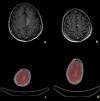
Leukoencephalopathy is a common finding on Magnetic Resonance Imaging (MRI), particularly in the elderly. A differential diagnosis may represent a very bet for clinicians when clear elements for diagnosis are lacking. Diffuse infiltrative "non mass like" leukoencephalopathy on MRI may represent the presentation of a very rare aggressive condition known as lymphomatosis cerebri (LC). The lack of orienting data, such as contrast enhancement on MRI or specific findings on examination of Cerebrospinal Fluid (CSF) or blood tests, may even far more complicate such a difficult diagnosis and orientate toward a less aggressive but time-losing mimic. A 69-old man initially presented to the Emergency Department (ED) complaining the recent appearance of unsteady walking, limitation of down and upgaze palsy, and hypophonia. Brain MRI revealed the presence of multiple, confluent hyperintense lesions on T2/Flair Attenuated Imaging Recovery (FLAIR) sequences involving either the withe matter of the semi-oval centres, juxtacortical structures, basal ganglia, or bilateral dentate nuclei. DWI sequences showed a wide restriction signal in the same brain regions but without any sign of contrast enhancement. Initial 18F-labeled fluoro-2-deoxyglucose positron emission tomography (FDG PET) and CSF studies were not relevant. Brain MRI revealed a high choline-signal, abnormal Choline/ N-Acetyl-Aspartate (NAA), and Choline/Creatine (Cr) ratios, as well as reduced NAA levels. Finally, a brain biopsy revealed the presence of diffuse large B-cell lymphomatosis cerebri. The diagnosis of lymphomatosis cerebri remains elusive. The valorisation of brain imaging may induce clinicians to suspect such a difficult diagnosis and go through the diagnostic algorithm.
Keywords: 1H-MRS; MRI; Primary Central Nervous System lymphoma (PNCSL); leukoencephalopathy; lymphomatosis cerebri.
2023, International Research and Cooperation Association for Bio & Socio - Sciences Advancement.
Conflict of interest statement
The authors have no conflicts of interest to disclose.
Figures


References
-
- Sugie M, Ishihara K, Kato H, Nakano I, Kawamura M. Primary central nervous system lymphoma initially mimicking lymphomatosis cerebri: An autopsy case report. Neuropathology. 2009; 29:704-707. - PubMed

