Case Reports
. 2023 May 29;18(8):2653-2658.
doi: 10.1016/j.radcr.2023.04.060. eCollection 2023 Aug.

A rare radiological presentation of pulmonary metastases from malignant melanoma

Affiliations
Case Reports

A rare radiological presentation of pulmonary metastases from malignant melanoma

Nanditha Guruvaiah Sridhara et al. Radiol Case Rep. .

Abstract

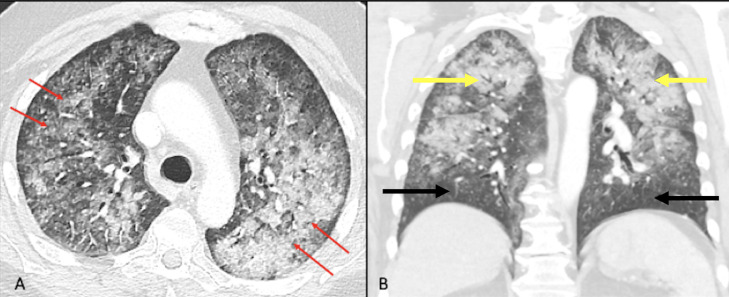
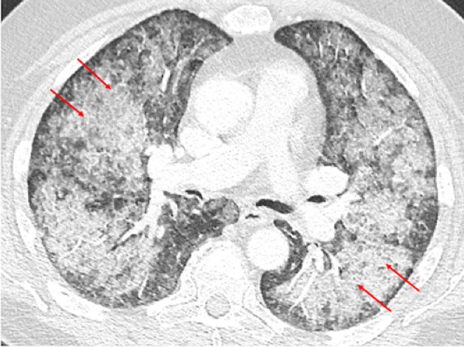
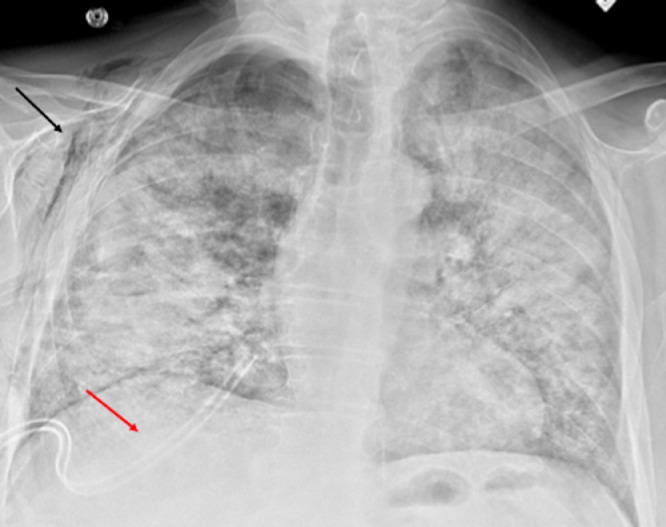
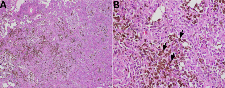
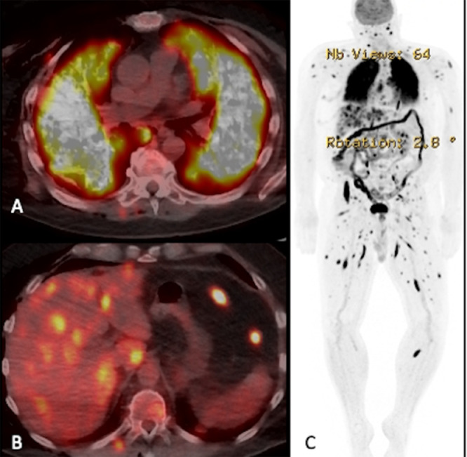
Malignant melanoma is a highly aggressive cancer with metastatic potential to various locations such as the lymph nodes, lungs, liver, brain, and bone. After the lymph nodes, the lungs are the most common site of malignant melanoma metastases. Pulmonary metastases from malignant melanoma commonly presents as solitary or multiple solid nodules, sub-solid nodules or miliary opacities on CT chest. We present a case of pulmonary metastases from malignant melanoma in a 74-year-old man which presented unusually on CT chest as a combination of patterns like "crazy paving," upper lobe predominance with subpleural sparing, and centrilobular micronodules. Video-assisted thoracoscopic surgery, wedge resection and tissue analysis were performed, which confirmed the diagnosis of malignant melanoma metastases, and the patient further underwent PET-CT for staging and surveillance. Patients with pulmonary metastases from malignant melanoma can have atypical imaging findings, therefore radiologists should be aware of these unconventional presentations to avoid any misdiagnoses.

Keywords: Atypical presentation; Crazy paving; Malignant melanoma; Oncology; Pulmonary metastases.


Figures

Fig 1:
Fig. 1
Axial CT chest image lung window (A) demonstrates bilateral diffuse consolidation and ground glass opacities with superimposed septal thickening, giving a “crazy-paving” pattern (red arrows). There is relative subpleural sparing. Coronal reformatted lung window (B) shows airspace disease predominantly involving upper lobes of lungs (yellow arrows) with relative sparing of bilateral lower lobes (black arrows).
Fig 2:
Fig. 2
Axial CT chest image lung window performed 4 weeks after baseline CT demonstrates interval worsening of bilateral diffuse airspace disease with crazy-paving pattern (red arrows).
Fig 3:
Fig. 3
Postoperative chest radiograph after wedge resections of right lung demonstrates bilateral diffuse airspace disease with expected postsurgical right subcutaneous emphysema (black arrow) and trace right pneumothorax. Right chest drainage tube is noted in the lung base (red arrow).
Fig 4:
Fig. 4
Representative H&E-stained slides showing the tumor histology. (A) The low-power picture shows diffuse growth of tumor containing dark-brown pigmentation. (B) At higher power magnification, the tumor cells show malignant nuclear features with melanin production (black arrows), consistent with metastatic melanoma.
Fig 5:
Fig. 5
FDG PET-CT axial section at the level of lungs (A) demonstrates avid uptake of FDG in bilateral diffuse airspace disease, consistent with pathology proven metastatic melanoma. FDG PET-CT axial section at the level of liver (B) demonstrates multiple avid FDG uptake involving both lobes of the liver, posterior chest wall and peritoneum, favoring metastatic disease. FDG PET image (C) demonstrates extensive FDG avid metastatic disease involving whole body.
Fig 6:
Fig. 6
Axial section post contrast T1 weighted images (A and B) demonstrates enhancing intraparenchymal lesions concerning for metastases in the ventral pontomedullary junction of the brainstem (red arrow in Fig. A) and in the right cerebral cortex (red arrow in Fig. B).

Similar articles

References

    1. Wang JY, Wang EB, Swetter SM. What is melanoma? JAMA. 2023;329(11):948. doi: 10.1001/jama.2022.24888. - DOI - PubMed
    1. Gershenwald JE, Scolyer RA, Hess KR, Sondak VK, Long GV., Ross MI, et al. Melanoma staging: evidence-based changes in the American Joint Committee on Cancer eighth edition cancer staging manual. CA Cancer J Clin. 2017;67(6):472–492. doi: 10.3322/caac.21409. - DOI - PMC - PubMed
    1. Pidhorecky I, Lee RJ, Proulx G, Kollmorgan DR, Jia C, Driscoll DL, et al. Risk factors for nodal recurrence after lymphadenectomy for melanoma. Ann Surg Oncol. 2001;8(2):109–115. doi: 10.1007/s10434-001-0109-2. - DOI - PubMed
    1. Younes R, Abrao FC, Gross J. Pulmonary metastasectomy for malignant melanoma: prognostic factors for long-term survival. Melanoma Res. 2013;23(4):307–311. doi: 10.1097/CMR.0b013e3283632cbe. - DOI - PubMed
    1. Detterbeck FC, Grodzki T, Gleeson F, Robert JH. Imaging requirements in the practice of pulmonary metastasectomy. J Thorac Oncol. 2010;5(6):134–139. doi: 10.1097/JTO.0b013e3181dcf64d. - DOI - PubMed

Publication types

LinkOut - more resources