. 2023 May 23:14:1156493.
doi: 10.3389/fimmu.2023.1156493. eCollection 2023.

Transcriptomic signatures reveal a shift towards an anti-inflammatory gene expression profile but also the induction of type I and type II interferon signaling networks through aryl hydrocarbon receptor activation in murine macrophages

Affiliations

Transcriptomic signatures reveal a shift towards an anti-inflammatory gene expression profile but also the induction of type I and type II interferon signaling networks through aryl hydrocarbon receptor activation in murine macrophages

Johannes R Schmidt et al. Front Immunol. .

Abstract

Introduction: The aryl hydrocarbon receptor (AhR) is a ligand-activated transcription factor that regulates a broad range of target genes involved in the xenobiotic response, cell cycle control and circadian rhythm. AhR is constitutively expressed in macrophages (Mϕ), acting as key regulator of cytokine production. While proinflammatory cytokines, i.e., IL-1β, IL-6, IL-12, are suppressed through AhR activation, anti-inflammatory IL-10 is induced. However, the underlying mechanisms of those effects and the importance of the specific ligand structure are not yet completely understood.

Methods: Therefore, we have compared the global gene expression pattern in activated murine bone marrow-derived macrophages (BMMs) subsequently to exposure with either benzo[a]pyrene (BaP) or indole-3-carbinol (I3C), representing high-affinity vs. low-affinity AhR ligands, respectively, by means of mRNA sequencing. AhR dependency of observed effects was proved using BMMs from AhR-knockout (Ahr-/-) mice.

Results and discussion: In total, more than 1,000 differentially expressed genes (DEGs) could be mapped, covering a plethora of AhR-modulated effects on basal cellular processes, i.e., transcription and translation, but also immune functions, i.e., antigen presentation, cytokine production, and phagocytosis. Among DEGs were genes that are already known to be regulated by AhR, i.e., Irf1, Ido2, and Cd84. However, we identified DEGs not yet described to be AhR-regulated in Mϕ so far, i.e., Slpi, Il12rb1, and Il21r. All six genes likely contribute to shifting the Mϕ phenotype from proinflammatory to anti-inflammatory. The majority of DEGs induced through BaP were not affected through I3C exposure, probably due to higher AhR affinity of BaP in comparison to I3C. Mapping of known aryl hydrocarbon response element (AHRE) sequence motifs in identified DEGs revealed more than 200 genes not possessing any AHRE, and therefore being not eligible for canonical regulation. Bioinformatic approaches modeled a central role of type I and type II interferons in the regulation of those genes. Additionally, RT-qPCR and ELISA confirmed a AhR-dependent expressional induction and AhR-dependent secretion of IFN-γ in response to BaP exposure, suggesting an auto- or paracrine activation pathway of Mϕ.

Keywords: aryl hydrocarbon receptor; benzo[a]pyrene; immunomodulation; indol-3-carbinol; innate immunity; macrophage activation; transcriptomics; type I/II interferons.


Conflict of interest statement

The authors declare that the research was conducted in the absence of any commercial or financial relationships that could be construed as a potential conflict of interest.

Figures

Figure 1
Figure 1
Effects of AhR activation on gene expression profiles during PAMP-induced BMM activation. Murine Ahr+/+ BMMs were exposed to AhR ligands (BaP or I3C) or treated with vehicle control (DMSO) for 6 h (n = 4). Subsequently, cells were activated with hk S.E. for 3 h or 20 h (p.a. – post administration) for PAMP activation. Whole cellular RNA was extracted and analyzed by means of RNA sequencing. Relative quantitative whole gene expression profiles of ligand-exposed vs. DMSO-treated cells retrieved from DESeq2 analysis were inspected via gene set enrichment analysis against biological process category of gene ontology and canonical pathways of databases REACTOME and Wiki Pathways. NES of gene sets were used for Euclidean Clustering. Gene sets were selected to cover innate immunity and representative basal cellular processes and annotated in die figure. Complete results are provided as Supplementary Table 2 .
Figure 2
Figure 2
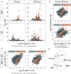
Identification of AhR-dependently differentially expressed genes. Murine Ahr+/+ BMMs were exposed to AhR ligands (BaP or I3C) or treated with vehicle control (DMSO) for 6 h (n = 4). Subsequently, cells were activated with hk S.E. for 3 h or 20 h (p.a. – post administration) for PAMP activation. Whole cellular RNA was extracted and analyzed by means of RNA sequencing. Mapped read counts were analyzed by DESeq2 to identify DEGs (n = 4). (A) Volcano plots representing log2-transformed fold changes (FCs) of gene expression after ligand exposure compared to DMSO-treated BMMs and −log10-transformed adjusted p values (FDR). (B) Correlation of gene expression 3 h to 20 h post administration (p.a.) of hk S.E. for BaP (upper panel) and I3C exposure (lower panel) represented by log2-FCs. (C) Correlation of gene expression after BaP (x-axis) to I3C (y-axis) exposure after 3 h (left panel) and 20 h p.a. of hk S.E. (right panel) represented by log2-FCs. Linear regression is represented by correlation of all genes (blue) and AhR-dependently DEGs (AhR-dep, orange). Corresponding Pearson’s correlation coefficients (ρ) are indicated in the plots. Complete statistics of correlation analyses are provided as Supplementary Table 3 . (D) Venn-Euler diagrams of total AhR-dependent DEGs comparing 3 h and 20 h p.a. of hk S.E. (upper panel) or I3C and BaP exposure (lower panel).
Figure 3
Figure 3
Changes in relative Il10 (A) and Il1b (B) expression following exposure to different concentrations of BaP or I3C. BMMs from Ahr+/+ mice were treated with indicated concentrations of BaP or I3C or vehicle control (DMSO) for 6 h. Subsequently, cells were activated with hk S.E. for 20 h (p.a. – post administration) for PAMP activation. Changes in gene transcription were assessed by real-time RT-PCR. Data represent the mean of the relative cytokine gene expression ± SEM after normalization with housekeeping genes Alas1 and Hprt (n=4). *p ≤ 0.05 indicates significant differences between treated and untreated cells.
Figure 4
Figure 4
Putative mechanisms in AhR-dependent gene expression. Murine Ahr+/+ BMMs were exposed to BaP or treated with vehicle control (DMSO) for 6 h. Subsequently, cells were activated with hk S.E. for 20 h for PAMP activation. Whole cellular RNA was extracted and analyzed by means of RNA sequencing. Mapped read counts were analyzed by DESeq2 to identify DEGs (n = 4). (A) Potential upstream regulators were predicted from AhR-dependent gene expression to identify 24 regulator elements (upper panel) that target 14 non-AHRE motif-possessing genes (lower panel). Circle plot illustrating functional interactions of upstream regulators to downstream targets. Outer grid color represents predicted activation z-score (for upstream regulators) and measured log2-fold change (for downstream targets). (B) Upstream regulator interaction network modified from IPA output ( Supplementary Figure 2 ). Nodes represent measured or predicted upstream regulator activities and interactions based on experimental results from BaP-exposed BMMs activated for 20 h with hk S.E. (C) BMMs from Ahr+/+ and Ahr-/- mice were exposed to BaP (8 nM or 800 nM) or DMSO (vehicle control) for 6 h. Changes in gene transcription of Ifng were assessed by quantitative real-time RT-PCR after activation of BMMs with hk S.E. for 20 h. Data represent the mean of the relative expression ± SEM after normalization with housekeeping genes Alas1 and Hprt (n = 3). *p ≤ 0.05 indicates significant differences between treated and untreated cells.
Figure 5
Figure 5
Proposed model for the modulation of PAMP-induced Mϕ activation through AhR ligand exposure. After entering the cytoplasm, AhR ligands such as BaP or I3C bind to a complex of several proteins, containing AhR, HSP90, XAP2, P23, and Src. AhR ligand binding induces a conformational change in this protein complex resulting in translocation of the whole complex into the nucleus where it is degraded to release the AhR molecule. Inside the nucleus, AhR associates with the Aryl hydrocarbon nuclear translocator (ARNT) and binds to the Aryl hydrocarbon response element (AHRE) in the promotor region of target genes, i.e., Cyp1a1, Cyp1a2, Cyp1b1, inducing their transcriptional activity (➪ canonical, genomic targets). This study confirms or suggest the expressional activation of several key immune genes, i.e., Il21r, Irf1, but also Spint1, Slpi, and Ido2 as putative AHRE motif-possessing canonical targets. In the cytoplasm, the AhR complex included src tyrosine kinase phosphorylates STAT3 leading to its dimerization and nuclear translocation. In the nucleus STAT3 binds to a response element in the Il10 promotor, and thus inducing IL-10 production (81). This is the most prominent non-canonical AhR-dependent pathway. If IL-10 is secreted, it exerts autocrine and paracrine anti-inflammatory effects, leading to the suppression of gene expression of proinflammatory cytokines, i.e., Il1b, Tnfa, Il12a/b. Further, this study suggests the AhR-dependent expressional induction of key immune genes for Mϕ activation, such as Il12rb1, Fcgr1, Cd84, that do possess an AHRE motif and thus, are regulated by non-canonical or secondary mechanisms. Of note, from several expressional profiles it is anticipated that the transcriptional activation of type I (IFN-α, IFN-β) and type II (IFN-γ) interferons is regulated by AhR via so far unknown molecular mechanisms possibly including both canonical and non-canonical pathways. Gene expressional profiles and bioinformatic approaches suggest an autocrine/paracrine effect of secreted interferons partly explaining the here presented results. Solid lines ⟹ known mechanisms covered in the figure; dashed lines ⟹ known mechanisms not covered in the figure; dotted line ⟹ unkown mechanisms. Boxes indicate genes expressionally induced in the study.

References

    1. Gu YZ, Hogenesch JB, Bradfield CA. The PAS superfamily: sensors of environmental and developmental signals. Annu Rev Pharmacol Toxicol (2000) 40:519–61. doi: 10.1146/annurev.pharmtox.40.1.519 - DOI - PubMed
    1. Denison MS, Soshilov AA, He G, DeGroot DE, Zhao B. Exactly the same but different: promiscuity and diversity in the molecular mechanisms of action of the aryl hydrocarbon (dioxin) receptor. Toxicol Sci (2011) 124(1):1–22. doi: 10.1093/toxsci/kfr218 - DOI - PMC - PubMed
    1. Ge NL, Elferink CJ. A direct interaction between the aryl hydrocarbon receptor and retinoblastoma protein. Linking dioxin Signaling to Cell Cycle J Biol Chem (1998) 273(35):22708–13. doi: 10.1074/jbc.273.35.22708 - DOI - PubMed
    1. Gutiérrez-Vázquez C, Quintana FJ. Regulation of the immune response by the aryl hydrocarbon receptor. Immunity (2018) 48(1):19–33. doi: 10.1016/j.immuni.2017.12.012 - DOI - PMC - PubMed
    1. Naseri-Nosar P, Nogalski MT, Shenk T. The aryl hydrocarbon receptor facilitates the human cytomegalovirus-mediated G1/S block to cell cycle progression. Proc Natl Acad Sci USA (2021) 118(12):e2026336118. doi: 10.1073/pnas.2026336118 - DOI - PMC - PubMed

Publication types