Review
. 2023 May 23:10:1206138.
doi: 10.3389/fcvm.2023.1206138. eCollection 2023.

Advanced imaging of fetal cardiac function

Affiliations
Review

Advanced imaging of fetal cardiac function

Henriette Kühle et al. Front Cardiovasc Med. .

Abstract

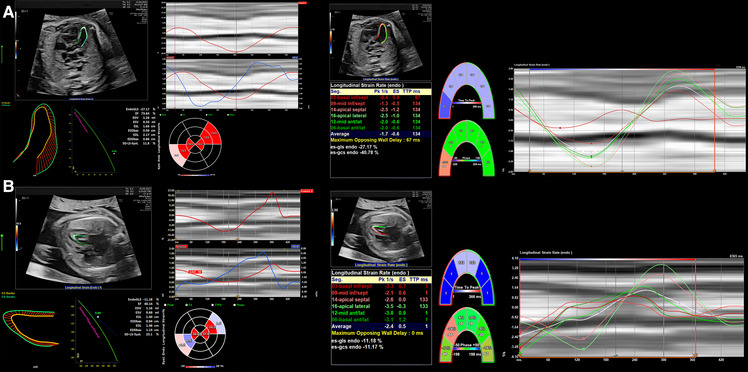
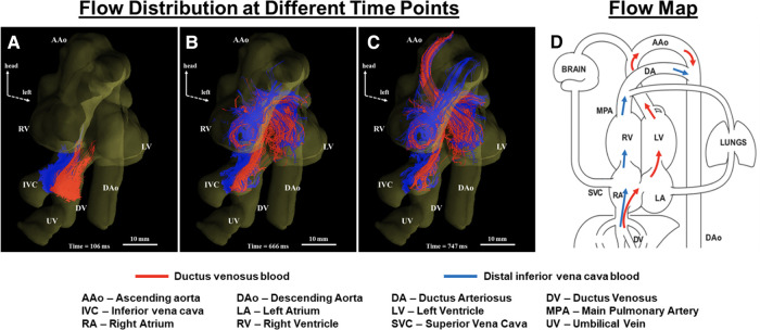
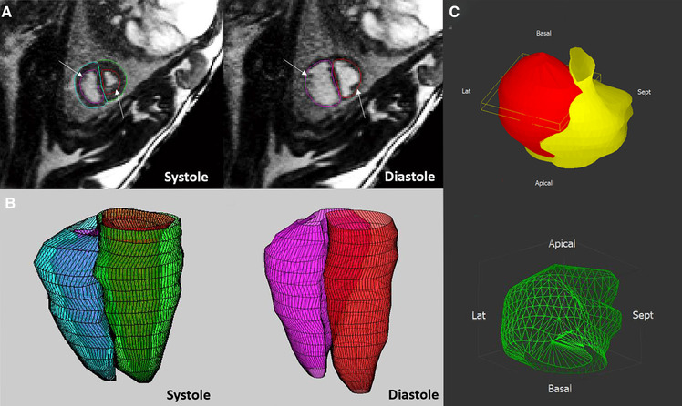
Over recent decades, a variety of advanced imaging techniques for assessing cardiovascular physiology and cardiac function in adults and children have been applied in the fetus. In many cases, technical development has been required to allow feasibility in the fetus, while an appreciation of the unique physiology of the fetal circulation is required for proper interpretation of the findings. This review will focus on recent advances in fetal echocardiography and cardiovascular magnetic resonance (CMR), providing examples of their application in research and clinical settings. We will also consider future directions for these technologies, including their ongoing technical development and potential clinical value.

Keywords: echocardiography; fetal cardiac function; magnetic resonance imaging; speckle tracking; strain.


Conflict of interest statement

The authors declare that the research was conducted in the absence of any commercial or financial relationships that could be construed as a potential conflict of interest.

Figures

Figure 1
Figure 1
Cardiovascular profile score (CVPS) to assess fetal heart function, adapted from (89).
Figure 2
Figure 2
The assessment of fetal cardiac function and cardiovascular profile in Ebstein anomaly showing severe cardiomegaly (cardiothoracic ratio: 0.69) with severely dilated right atrium and right ventricle and compressed lungs and mild to moderately reduced RV function, fetal hydrops with a small pericardial effusion, significant ascites and scalp edema. The Dopplers reveal intermittent a-wave reversal in the ductus venosus and umbilical vein notching suggestive of elevated cardiac filling pressures and intermittently absent diastolic flow in the umbilical artery consistent with a systemic steal. (A) Four chamber view suggesting severe cardiomegaly (cardiothoracic ratio: 0.69) and dilated right heart. (B) Color Doppler showing tricuspid regurgitation. (C) Doppler of tricuspid valve showing biphasic flow with high velocity. (D) Intermittently absent diastolic flow in the umbilical artery. (E) Umbilical vein notching. (F) The intermittent a-wave reversal in the DV.
Figure 3
Figure 3
2D and M-mode assessment of left ventricular function in (A) a fetus with a family history of hypertrophic cardiomyopathy at 30 + 1 weeks and (B) a fetus with severe aortic stenosis and left ventricular endocardial fibroelastosis at 26 + 1 week.
Figure 4
Figure 4
2D strain in LV and M-mode analysis from 4CV in an artificial placenta piglet model by echocardiography obtained using a voluson S6 (GE healthcare ultrasound, WI, USA) and post-processing software by TOMTEC (TOMTEC imaging systems GmbH, Germany). A gated M-Mode loop of two cardiac cycles was generated using a four-chamber cine loop, allowing for automatic LV strain analysis. Compared to the original M-mode measurements, the post-processing technique seems feasible and generated similar values for ejection fraction, with good repeatability between serial measurements. (A) Strain Analysis. (B) M-mode. (C) Segmental endocardial strain. (D) Segmental endocardial strain rate. Superimposed anatomical M-Mode behind graphs, endocardial border detection.
Figure 5
Figure 5
2D strain on LV analysis from 4 chamber view in (A) a patient with a family history of hypertrophic cardiomyopathy resulting in increased CVO and overall elevated strain measurements and (B) a patient with aortic stenosis resulting in reduced CVO and deviating strain measurements between the ventricular septum due to indirect movement of the hypoplastic and dysfunctional LV by the adjacent RV.
Figure 6
Figure 6
Tracing blood from inferior vena cava (IVC) and ductus venosus (DV) in a human fetal heart (A–C). Coronal view showing blood from the IVC (blue) and DV (red) at different cardiac phases over two heartbeats. Streams from IVC and DV with limited mixing enter the right atrium (A), with blood from the DV (oxygenated) being mainly directed into the left ventricle (B) to supply the coronaries and upper fetal body (C). (D) Corresponding flow map. Adapted from (33).
Figure 7
Figure 7
The application of ventricular volumetry in late gestation fetal sheep (A,B) and human fetus (C). (A,B) In fetal sheep, manual endocardial contours were applied on the stack of short-axis cine acquisitions in systole and diastole (A) to generate a 3D reconstruction of the right and left ventricles (B). (C) Morphological and quantitative models (green, mass 40 mm3) of a human fetal heart with a cardiac rhabdomyoma (red), and the compressed left ventricle (yellow). Adapted from (31) (A,B) and (40) (C).
Figure 8
Figure 8
Gadolinium imaging (left) and SSFP short-axis cine feature tracking analysis (right) in a fetal sheep model of myocardial infarction on the day of surgery and 6 days post-surgery in both injured and sham twins suggesting good correlation between regional myocardial dysfunction in the apical left ventricular lateral wall and evidence of myocardial infarction in the respective segments in early and late gadolinium enhancement imaging. Anterolateral ischemia was noted at day of surgery from apex to near-mid ventricle (top row; early gadolinium enhancement; white arrow) and there was regional myocardial dysfunction in apical segments as measured by decreased circumferential strain by feature tracking (top row; right). 6 days after the myocardial infarction surgery, injury was noted in similar regions (bottom row; late gadolinium enhancement; white arrow) and there was regional myocardial dysfunction in the same regions, overall showing good correlation between injury and regional dysfunction measured by regional circumferential strain by feature tracking. In both scans, the internal sham control (fetus B) remained asymptomatic and did not demonstrate any regional wall motion abnormalities.

References

    1. Donofrio MT, Moon-Grady AJ, Hornberger LK, Copel JA, Sklansky MS, Abuhamad A, et al. Diagnosis and treatment of fetal cardiac disease: a scientific statement from the American heart association. Circulation. (2014) 129(21):2183–242. 10.1161/01.cir.0000437597.44550.5d - DOI - PubMed
    1. Sepulveda W, Wong AE, Sepulveda F, Martinez-Ten P, Ximenes R. Fetal magnetic resonance imaging and three-dimensional ultrasound in clinical practice: general aspects. Best Pract Res Clin Obstet Gynaecol. (2012) 26(5):575–91. 10.1016/j.bpobgyn.2012.05.010 - DOI - PubMed
    1. Crispi F, Valenzuela-Alcaraz B, Cruz-Lemini M, Gratacós E. Ultrasound assessment of fetal cardiac function. Australas J Ultrasound Med. (2013) 16(4):158–67. 10.1002/j.2205-0140.2013.tb00242.x - DOI - PMC - PubMed
    1. Di Salvo G, Russo MG, Paladini D, Felicetti M, Castaldi B, Tartaglione A, et al. Two-dimensional strain to assess regional left and right ventricular longitudinal function in 100 normal foetuses. Eur J Echocardiogr. (2008) 9(6):754–6. 10.1093/ejechocard/jen134 - DOI - PubMed
    1. Abuhamad A, Chaoui R. A practical guide to fetal echocardiography: Normal and abnormal hearts. 4 edn. Philadelphia: Lippincott Williams & Wilkins; (2022).

LinkOut - more resources