Review
. 2023 May 23:14:1189007.
doi: 10.3389/fendo.2023.1189007. eCollection 2023.

The versatile role of Serpina3c in physiological and pathological processes: a review of recent studies

Affiliations
Review

The versatile role of Serpina3c in physiological and pathological processes: a review of recent studies

Yang Li et al. Front Endocrinol (Lausanne). .

Abstract

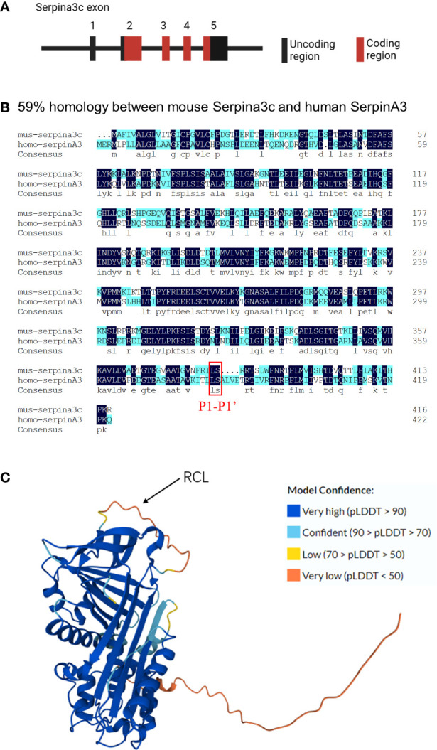
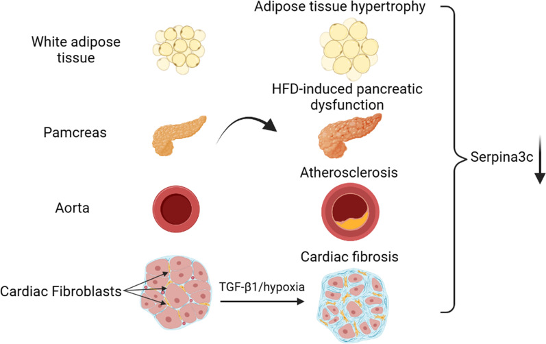
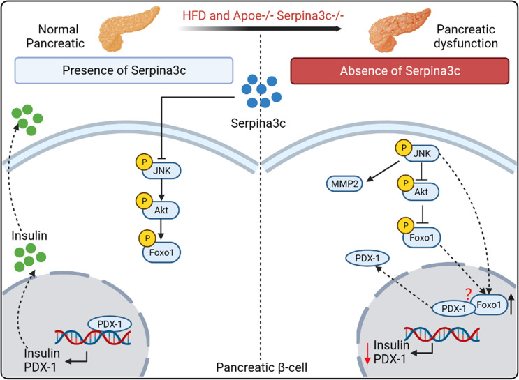
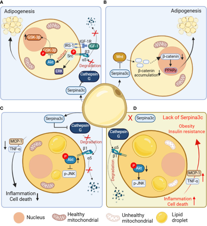
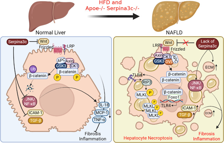
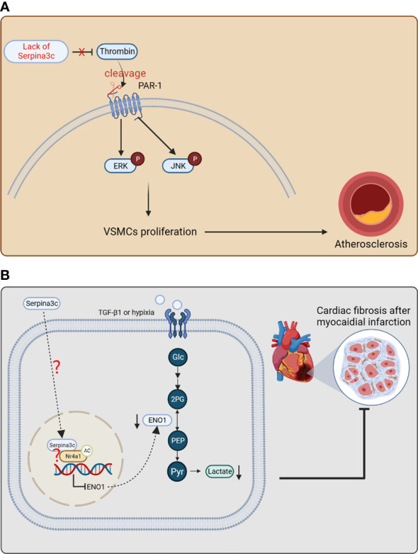
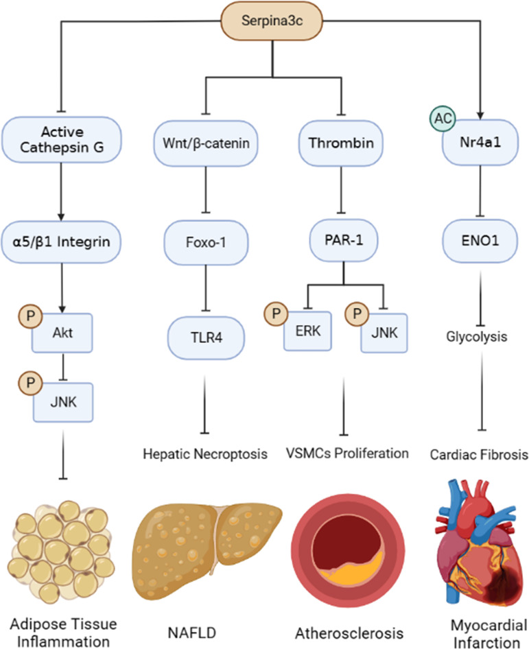
Murine Serpina3c belongs to the family of serine protease inhibitors (Serpins), clade "A" and its human homologue is SerpinA3. Serpina3c is involved in some physiological processes, including insulin secretion and adipogenesis. In the pathophysiological process, the deletion of Serpina3c leads to more severe metabolic disorders, such as aggravated non-alcoholic fatty liver disease (NAFLD), insulin resistance and obesity. In addition, Serpina3c can improve atherosclerosis and regulate cardiac remodeling after myocardial infarction. Many of these processes are directly or indirectly mediated by its inhibition of serine protease activity. Although its function has not been fully revealed, recent studies have shown its potential research value. Here, we aimed to summarize recent studies to provide a clearer view of the biological roles and the underlying mechanisms of Serpina3c.

Keywords: Cardiometabolic diseases; NAFLD; cathepsin G; inflammation; insulin resistance; obesity; serine protease inhibitors; thrombin.


Conflict of interest statement

The authors declare that the research was conducted in the absence of any commercial or financial relationships that could be construed as a potential conflict of interest.

Figures

Figure 1
Figure 1
Molecular features of Serpina3c (A) The Serpina3c gene contains five exons, The first exon of the Serpina3c gene does not encode amino acid residues, and the second, third, fourth, and fifth exons of the Serpina3c gene encode 212, 92, 50 and 63 amino acid residues, respectively. (B) Comparison and alignment of murine Serpina3c protein sequences with human SerpinA3 protein. Two identical residues are represented in dark blue. A homology of 59% is detected between the murine Serpina3c protein and human SerpinA3 protein. The red box represents the P1-P1’ bond, which is conserved between Serpina3c and SerpinA3. (C) Prediction 3D structure of Serpina3c protein. The amino acid sequences were derived from UniProt (https://www.uniprot.org/uniprotkb/P29621), and the 3D structure was predicted by AlphaFold (https://alphafold.ebi.ac.uk/entry/P29621). AlphaFold produces a per-residue confidence score (pLDDT) between 0 and 100. Some regions below 50 pLDDT may be unstructured in isolation.
Figure 2
Figure 2
Serpina3c expression change in different tissues under cardiometabolic stress. Serpina3c protein expression is down-regulated in white adipose tissue (WAT) of obese mice induced by high-fat diet (HFD) feeding. Serpina3c protein levels were reduced in the pancreas of HFD-fed or Apoe-/- mice compared with normal mice. Compared with normal mice, the mRNA and protein levels of Serpina3c in the aorta of mice in the Apoe-/- group were down-regulated; and the mRNA and protein levels of Serpina3c were further down-regulated in Apoe-/- mice fed with HFD compared with the ones fed with chow diet (CD). Serpina3c protein expression is down-regulated in primary mouse cardiac fibroblasts treated with transforming growth factor-β1(TGF-β1) or hypoxia for 24 h. (Created with BioRender.com).
Figure 3
Figure 3
The role and mechanism of Serpina3c in the pancreatic β cells. Serpina3c can protect against pancreatic dysfunction induced by HFD feeding. Under HFD feeding in Apoe-/- mice, the expression of phosphorylated JNK in the pancreas increased, which inhibited the phosphorylation of AKT, thus reducing the inhibition of phosphorylation of forkhead box protein O1 (Foxo1) and increasing the entry of Foxo1 into the nucleus. Overexpression of JNK can also promote the entry of Foxo1 into the nucleus. Foxo1 inhibits the activity of pancreatic transcription factor pancreatic and duodenal homeobox 1 (PDX-1) and the transcription of insulin and PDX-1 genes in the nucleus. And Foxo-1 is involved in the nucleocytoplasmic translocation of PDX-1, which promotes the transfer of PDX-1 from the nucleus to cytoplasm. However, the mechanism of Foxo-1-mediated nucleocytoplasmic translocation of PDX-1 needs to be further studied. Serpina3c can facilitate insulin secretion by inhibiting JNK/AKT/Foxo1/PDX-1 signal pathway in the pancreatic β cells of HFD-fed Apoe-/- mice. (Created with BioRender.com).
Figure 4
Figure 4
The role and mechanism of Serpina3c in preadipocytes and adipose tissue. (A) In preadipocytes, Serpina3c inhibits Cathepsin G, reduces the degradation of integrin α5, and promotes complex formation between insulin-like growth factors-1 receptor (IGF-1R) and integrin α5/β3 heterodimer. In the case of ligand binding, integrin β3 phosphorylates and binds to Src to activate extracellular regulated protein kinases (ERK). Insulin receptor substrate-1 (IRS-1) can instantly induce protein kinase B (AKT) signal transduction, reduce the phosphorylation of glycogen synthase kinase-3β (GSK-3β) to promote its nuclei entry, thereby facilitating adipogenesis. (B) In preadipocytes, Serpina3c can promote adipogenesis by inhibiting Wnt/β-catenin signal pathway, reducing the accumulation of β-catenin in the cytoplasm and its nuclei entry, and reducing the β-catenin-mediated transcriptional inhibition of peroxisome proliferators-activated receptor γ(PPARγ). (C) In adipocytes, Serpina3c maintained the integrity of integrin α5/β1 by inhibiting the activity of Cathepsin G and reducing the degradation of integrin α5. The increased integrity of integrin α5/β1 activates AKT, thereby reducing c-Jun N-terminal kinase (JNK) phosphorylation. This will lead to reduced release of inflammatory factors tumor necrosis factor-α (TNF-α) and macrophage chemoattractant protein-1 (MCP-1) from adipocytes, thereby inhibiting the inflammation of adipose tissue and cell death of adipocytes. (D) When Serpina3c is lacking in white adipose tissue, it cannot inhibit the degradation of integrin α5 by cathepsin G, resulting in a decrease in intracellular AKT phosphorylation and a failure to inhibit JNK phosphorylation, thereby promoting the release of inflammatory factors TNF-α and MCP-1 in adipose tissue, resulting in increased cell death. This eventually leads to insulin resistance and obesity in mice. (Created with BioRender.com).
Figure 5
Figure 5
The beneficial roles of Serpina3c in NAFLD. In normal liver, Serpina3c can inhibit Wnt/β-catenin signal pathway, promotes ubiquitination-mediated degradation of β-catenin, inhibits JNK/NF-κB signal pathway, reduces the expression of inflammatory cytokines interleukin 18 (IL-18), MCP-1, TNF- α, and decreases the expression level of intercellular adhesion molecule 1(ICAM-1) and TGF-β. These will reduce liver inflammation and fibrosis. However, in the NAFLD livers of Apoe and Serpina3c double knockout mice fed with HFD, due to the lack of Serpina3c, Wnt/β-catenin signal pathway could not be inhibited, making the entry of β-catenin into the nuclei, which interacts with Foxo-1 to activate TLR4 transcription. Increased expression of TLR4, and activation of downstream receptor interaction protein 3 (RIP3) and phosphorylation mixed lineage kinase domain-like pseudokinase (MLKL) will then induce hepatocyte necroptosis. The deletion of Serpina3c leads to the activation of JNK/NF-κB signal pathway and the increase of the expression of ICAM-1 and TGF-β. The content of extracellular matrix (ECM) will be increased. Together, these lead to more severe inflammation and fibrosis in the liver. (Created with BioRender.com).
Figure 6
Figure 6
The protective effects of Serpina3c against atherosclerosis and myocardial infarction. (A) Serpina3c deficiency leads to the failure to inhibit the cleavage and activation of protease-activated receptor-1 (PAR-1) by thrombin. PAR-1 subsequently increases the phosphorylation of ERK and JNK, which promotes the excessive proliferation of arterial smooth muscle cells and leads to atherosclerosis. VSMCs, vascular smooth muscle cells. (B) In fibroblasts, Serpina3c enters the nuclei of mouse cardiac fibroblasts and interacts with Nr4a1 to promote Nr4a1 acetylation, which inhibits ENO1 transcription to prevent excessive activation of glycolysis. These will inhibit fibroblasts proliferation and differentiation, thus alleviating fibrosis after myocardial infarction. However, as a secretory protein, how serpina3c enters the nuclei and how it leads to acetylation of Nr4a1 remains to be studied further. (Glc, glucose; 2PG, 2-phosphoglycerate; PEP, phosphoenolpyruvate; Pyr, enolpyruvic acid.) (Created with BioRender.com).
Figure 7
Figure 7
Summary of the protective roles and mechanisms of Serpina3c in pathophysiological processes. Serpina3c inhibits adipose tissue inflammation by inhibiting Cathepsin G, maintaining integrin α5/β1 integrity, activating AKT and inhibiting JNK phosphorylation. Serpina3c can inhibit non-alcoholic fatty liver disease (NAFLD) by inhibiting Wnt/β-catenin signal pathway in hepatocyte, reducing the transcription of Toll-like receptor 4 (TLR4) by Foxo-1, and inhibiting hepatic necroptosis. Seprina3c can bind and inhibit thrombin activity, prevent its cleavage and activation of protease-activated receptor-1 (PAR-1), weaken its downstream ERK and JNK phosphorylation, and inhibit the excessive proliferation of vascular smooth muscle cells (VSMCs), thus inhibiting atherosclerosis. Serpina3c can bind to nuclear receptor subfamily 4 group A member 1 (Nr4a1) in the mice cardiac fibroblasts nuclei and promote its acetylation, inhibits the transcription of enolase 1 (ENO1) and suppresses the excessive activation of glycolysis, thus alleviating cardiac fibrosis after myocardial infarction. (Created with BioRender.com).

Similar articles

Cited by

References

    1. Forsyth S, Horvath A, Coughlin P. A review and comparison of the murine alpha1-antitrypsin and alpha1-antichymotrypsin multigene clusters with the human clade a serpins. Genomics (2003) 81(3):336–45. doi: 10.1016/S0888-7543(02)00041-1 - DOI - PubMed
    1. Inglis JD, Hill RE. The murine spi-2 proteinase inhibitor locus: a multigene family with a hypervariable reactive site domain. EMBO J (1991) 10(2):255–61. doi: 10.1002/j.1460-2075.1991.tb07945.x - DOI - PMC - PubMed
    1. Li BY, Guo YY, Xiao G, Guo L, Tang QQ. SERPINA3C ameliorates adipose tissue inflammation through the cathepsin G/Integrin/AKT pathway. Mol Metab (2022) 61:101500. doi: 10.1016/j.molmet.2022.101500 - DOI - PMC - PubMed
    1. Choi Y, Choi H, Yoon BK, Lee H, Seok JW, Kim HJ, et al. . Serpina3c regulates adipogenesis by modulating insulin growth factor 1 and integrin signaling. iScience (2020) 23(3):100961. doi: 10.1016/j.isci.2020.100961 - DOI - PMC - PubMed
    1. Ji JJ, Qian LL, Zhu Y, Wu YP, Guo JQ, Ma GS, et al. . Serpina3c protects against high-fat diet-induced pancreatic dysfunction through the JNK-related pathway. Cell Signal (2020) 75:109745. doi: 10.1016/j.cellsig.2020.109745 - DOI - PubMed

Publication types