Review
. 2023 Jul;39(1-3):19-39.
doi: 10.1089/ars.2023.0352. Epub 2023 Jul 10.

Emerging Chemical Biology of Protein Persulfidation

Affiliations
Review

Emerging Chemical Biology of Protein Persulfidation

Thibaut Vignane et al. Antioxid Redox Signal. 2023 Jul.

Erratum in

Abstract

Significance: Protein persulfidation (the formation of RSSH), an evolutionarily conserved oxidative posttranslational modification in which thiol groups in cysteine residues are converted into persulfides, has emerged as one of the main mechanisms through which hydrogen sulfide (H2S) conveys its signaling. Recent Advances: New methodological advances in persulfide labeling started unraveling the chemical biology of this modification and its role in (patho)physiology. Some of the key metabolic enzymes are regulated by persulfidation. RSSH levels are important for the cellular defense against oxidative injury, and they decrease with aging, leaving proteins vulnerable to oxidative damage. Persulfidation is dysregulated in many diseases. Critical Issues: A relatively new field of signaling by protein persulfidation still has many unanswered questions: the mechanism(s) of persulfide formation and transpersulfidation and the identification of "protein persulfidases," the improvement of methods to monitor RSSH changes and identify protein targets, and understanding the mechanisms through which this modification controls important (patho)physiological functions. Future Directions: Deep mechanistic studies using more selective and sensitive RSSH labeling techniques will provide high-resolution structural, functional, quantitative, and spatiotemporal information on RSSH dynamics and help with better understanding how H2S-derived protein persulfidation affects protein structure and function in health and disease. This knowledge could pave the way for targeted drug design for a wide variety of pathologies. Antioxid. Redox Signal. 39, 19-39.

Keywords: aging; cysteine; hydrogen sulfide; posttranslational modifications; protein persulfidation; redox signaling.


Conflict of interest statement

No competing financial interests exist.

Figures

FIG. 1.
FIG. 1.
Biosynthesis and oxidation of H2S. α-KB, α-ketobutyrate; α-KG, α-ketoglutarate; 3-MP, 3-mercaptopyruvate; CAT, cysteine aminotransferase; CBS, cystathionine β-synthase; CSE, cystathionine γ-lyase; ETHE1, persulfide dioxygenase; GSH, glutathione; H2S, hydrogen sulfide; MPST, mercaptopyruvate sulfur transferase; SBP1 (alternatively SELENBP1), methanethiol oxidase; SO, sulfite oxidase; SQR, sulfide:quinone oxidoreductase; TST, thiosulfate sulfur transferase (alternatively rhodanese).
FIG. 2.
FIG. 2.
Electrophilic nature of organic persulfides and polysulfides. (A) Nucleophilic attack on electrophilic sulfur (red circle) of RSSH could occur on either of the two sulfur atoms with reaction path a being favored most of the time. (B) Instability of organic trisulfides is illustrated by their amine-induced or thiol-induced decomposition.
FIG. 3.
FIG. 3.
Antiferroptotic effects of RSSH. RSSH are excellent H-atom donors (upper reaction scheme) preventing formation and radical amplification of lipid radicals (L), peroxyl radical (ROO), and alkoxyl radical (LO), which are the main drivers of membrane damage and ferroptosis (lower reaction scheme). GPX4, glutathione peroxidase 4.
FIG. 4.
FIG. 4.
Reaction of protein persulfides with two-electron oxidants. DTT, dithiothreitol; RNS, reactive nitrogen species; ROS, reactive oxygen species; Trx, thioredoxin.
FIG. 5.
FIG. 5.
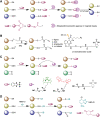
Methods for persulfide detection. (A) Biotin thiol assay and its variants. (B) Low-ph quantitative thiol reactivity profiling method. (C) Tag-switch method. (D) Dimedone-switch method.
FIG. 6.
FIG. 6.
Metalloprotein-catalyzed RSSH formation. (A) Cytochrome c oxidizes H2S to HS, which can react with protein thiols to form RSSH. Reduced cytochrome c is then reoxidized by complex IV of mitochondrial respiratory chain, establishing a pseudocatalytic cycle for RSSH formation. (B) Zinc center in metalloproteins could play a general role in catalyzing protein persulfide formation by: (i) shifting the redox potential of O2 to a more positive value favoring superoxide formation, (ii) lowering the pKa of H2S (akin to Zn binding to OH in carbonic anhydrase) favoring formation of HS, and (iii) acting as a template to bring O2 and H2S in a close proximity enabling efficient electron shuttling.
FIG. 7.
FIG. 7.
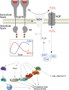
Regulation of EGFR signaling by RSOH to RSSH switching. Upon the receptor stimulation with EGF, NOX produces H2O2, which is transported into the cell via aquaporins, and H2O2 modifies C797 of EGFR to RSOH, increasing its kinase activity. H2O2 also stimulates expression of H2S producing enzymes. H2S reacts with RSOH to form RSSH on EGFR, which inhibits the phosphorylation of EGFR and downstream signaling. The temporal phase shifted waves of RSOH and RSSH caused by EGFR activation modulate many of the downstream targets involved in cytoskeleton regulation and cell motility. EGFR, epidermal growth factor receptor; H2O2, hydrogen peroxide; NOX, NADPH oxidase.
FIG. 8.
FIG. 8.
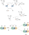
Transpersulfidation steps in RNA thiolation and iron–sulfur cluster assembly. Cysteine desulfurase (IscS in prokaryotes, NFS1 in eukaryotes) converts cysteine to alanine forming the intermediate persulfide (sulfur originating from the cysteine is marked red). Through transpersulfidation, this sulfur is transferred to ThiI and then to tRNA (upper figure), or transferred to ISCU. Alternatively, NFS1 persulfide could transfer sulfur to LMW RSH. FXN, frataxin, a protein involved in iron–sulfur cluster assembly; ISCU, iron-sulfur cluster assembly scaffold protein; LMW, low molecular weight; ThiI, tRNA sulfur transferase.
FIG. 9.
FIG. 9.
Protein transpersulfidation. (A) Cysteine persulfide of TSTD1 (left) is more exposed (yellow arrow) and accessible to engage in transpersulfidation reactions than cysteine of TST (right). (B) Energy diagram of RSSH tautomerization to thiosulfoxide (sulfane sulfur is marked red). TSTD1, thiosulfate sulfur transferase-like domain-containing 1 protein.
FIG. 10.
FIG. 10.
Enzyme-catalyzed protein depersulfidation. (A) Trx catalyzes protein depersulfidation via two different mechanisms. Oxidized Trx is then reduced by TrxR. (B) Grx catalyzes protein depersulfidation oxidizing GSH, which is reduced back by GR. GR, glutathione reductase; Grx, glutaredoxin; TrxR, thioredoxin reductase.
FIG. 11.
FIG. 11.
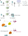
Persulfidation affects enzyme activity. (A) Persulfidation of parkin stimulates its E3 ligase activity leading to higher ubiquitination of target proteins and their proteosomal degradation. In Parkinson's disease, H2S production declines and nitrosation of thiols occurs, leading to inactivation of parkin. (B) Tau interacts with CSE stimulating its H2S producing activity, which results in persulfidation of GSK3β and inhibition of its activity. With aging and in AD, there is a decline in CSE levels. GSK3β becomes more active leading to hyperphosphorylation of tau and its aggregation. AD, Alzheimer's disease; GSK3β, glycogen synthase kinase 3β.
FIG. 12.
FIG. 12.
Hypothetic scheme for antiaging effects of RSSH. During aging or age-induced diseases, restoring protein persulfidation can serve as a mechanism to reduce ROS-oxidized cysteine residues, preventing their irreversible overoxidation.

References

    1. Abe K, Kimura H. The possible role of hydrogen sulfide as an endogenous neuromodulator. J Neurosci 1996;16(3):1066–1071. - PMC - PubMed
    1. Abiko Y, Yoshida E, Ishii I, et al. . Involvement of reactive persulfides in biological bismethylmercury sulfide formation. Chem Res Toxicol 2015;28(6):1301–1306; doi: 10.1021/acs.chemrestox.5b00101 - DOI - PubMed
    1. Akaike T, Ida T, Wei FY, et al. . Cysteinyl-TRNA synthetase governs cysteine polysulfidation and mitochondrial bioenergetics. Nat Commun 2017;8(1):1177; doi: 10.1038/s41467-017-01311-y - DOI - PMC - PubMed
    1. Akter S, Fu L, Jung Y, et al. . Chemical proteomics reveals new targets of cysteine sulfinic acid reductase. Nat Chem Biol 2018;14(11):995–1004; doi: 10.1038/s41589-018-0116-2 - DOI - PMC - PubMed
    1. Alberti S, Hyman AA. Biomolecular condensates at the nexus of cellular stress, protein aggregation disease and ageing. Nat Rev Mol Cell Biol 2021;22(3):196–213; doi: 10.1038/s41580-020-00326-6 - DOI - PubMed

Publication types

LinkOut - more resources