. 2023 Jun 29;97(6):e0043423.
doi: 10.1128/jvi.00434-23. Epub 2023 Jun 8.

Characterization of an Emergent Chicken H3N8 Influenza Virus in Southern China: a Potential Threat to Public Health

Affiliations

Characterization of an Emergent Chicken H3N8 Influenza Virus in Southern China: a Potential Threat to Public Health

Peiwen Chen et al. J Virol. .

Abstract

Although influenza A viruses of several subtypes have occasionally infected humans, to date only those of the H1, H2, and H3 subtypes have led to pandemics and become established in humans. The detection of two human infections by avian H3N8 viruses in April and May of 2022 raised pandemic concerns. Recent studies have shown the H3N8 viruses were introduced into humans from poultry, although their genesis, prevalence, and transmissibility in mammals have not been fully elucidated. Findings generated from our systematic influenza surveillance showed that this H3N8 influenza virus was first detected in chickens in July 2021 and then disseminated and became established in chickens over wider regions of China. Phylogenetic analyses revealed that the H3 HA and N8 NA were derived from avian viruses prevalent in domestic ducks in the Guangxi-Guangdong region, while all internal genes were from enzootic poultry H9N2 viruses. The novel H3N8 viruses form independent lineages in the glycoprotein gene trees, but their internal genes are mixed with those of H9N2 viruses, indicating continuous gene exchange among these viruses. Experimental infection of ferrets with three chicken H3N8 viruses showed transmission through direct contact and inefficient transmission by airborne exposure. Examination of contemporary human sera detected only very limited antibody cross-reaction to these viruses. The continuing evolution of these viruses in poultry could pose an ongoing pandemic threat. IMPORTANCE A novel H3N8 virus with demonstrated zoonotic potential has emerged and disseminated in chickens in China. It was generated by reassortment between avian H3 and N8 virus(es) and long-term enzootic H9N2 viruses present in southern China. This H3N8 virus has maintained independent H3 and N8 gene lineages but continues to exchange internal genes with other H9N2 viruses to form novel variants. Our experimental studies showed that these H3N8 viruses were transmissible in ferrets, and serological data suggest that the human population lacks effective immunological protection against it. With its wide geographical distribution and continuing evolution in chickens, other spillovers to humans can be expected and might lead to more efficient transmission in humans.

Keywords: H3N8; chicken; ferret; infectivity; influenza; pathogenesis; transmissibility; zoonosis.


Conflict of interest statement

The authors declare no conflict of interest.

Figures

FIG 1
FIG 1
Distribution of H3N8 influenza virus and isolation of H3 subtype viruses in Guangdong and Jiangxi. (A) The location of the cities in China that reported the two H3N8 human infection cases are highlighted by a red human figure (13, 17). Blue shading and gray shading indicate the provinces that were positive and negative, respectively, for H3N8 viruses (data from this work and the previous studies of Yang et al. [15] and Sit et al. [16]). Black dots denote cities where our sampling occurred. The four cities surveyed in Guangdong province were Guangzhou, Dongguan, Huizhou, and Shantou, from left to right. Shape file for the China boundary was downloaded from the Stanford Digital Repository. (B) Monthly isolation rates of H3 influenza viruses in Guangdong and Jiangxi. NS, not sampled.
FIG 2
FIG 2
Maximum likelihood phylogenies of selected H3 hemagglutinin (n = 60) (A), N8 neuraminidase (n = 50) (B), and PB2 (n = 92) (C) genes. Full trees are given in Fig. S1 and S2. The recent human H3N8 viruses are labeled in red, and the chicken viruses isolated in this study are in blue. Viruses used in the animal experiments are indicated by red circles. Hong Kong/1/1968, the prototype human H3N2 pandemic virus, has a blue background. Lineage sources of the six internal gene segments are indicated by the colors of the boxes (left to right: PB2, PB1, PA, NP, M, and NS) to the right of the tree, and dots in the NS box indicate allele B (A). Viruses of the chicken H3N8 lineage are highlighted with a green background and their subtree is magnified by ×10 relative to the rest of the tree (A and B). Gray backgrounds indicate viruses from the avian gene pool, with Eurasian viruses in darker gray and North American viruses in light gray. Orange boxes in the PB2 phylogeny (C) highlight viruses from enzootic H9N2 lineages in China. Numbers at nodes indicate topological support as assessed by 1,000 bootstrap replicates. The length of the scale bar corresponds to 0.01 nucleotide substitution per site. (Host species: Ck, chicken; SCk, silkie chicken; Dk, duck; Qa, quail; Pa, partridge; Gs, goose; W.f. Gs, white-fronted goose; N. pintail, Northern pintail; MDk, muscovy duck). Geography: Chinese province: JX, Jiangx; GD, Guangdong; cities in Guangdong province: ST, Shantou; DG, Dongguan; GZ, Guangzhou; HZ, Huizhou.
FIG 3
FIG 3
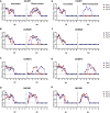
Transmissibility of H3N8 and H3N2 viruses in ferrets. Donor ferrets (n = 3) were inoculated with 106 TCID50 of Ck/481 (A and E), Ck/8520 (B and F), Ck/6673 (C and G), and HK1/68 (D and H). Naïve ferrets were introduced at 1 dpi. The efficiency of direct-contact (A to D) and airborne (E to H) transmission was confirmed by detecting viruses in nasal washes. Nasal washes were collected daily (for 14 days) from each ferret and titrated in MDCK cells. In each subfigure, the left set of graphs is from inoculated ferrets and the right set is from naive ferrets. The detection limit is 2.02 log10 TCID50/mL; dpc, days postcontact.
FIG 4
FIG 4
Viral distribution of H3N8 and H3N2 virus in ferrets. Ferrets were euthanized at 2, 4 and 7 dpi. The viral titers of each tissue are expressed as log10 TCID50/mL. Arithmetic mean of transformed titers with standard derivation (SD) is shown. The dashed lines indicate the detection limit at 1.52 log10TCID50/mL. (Tissues: NT, nasal turbinate; Tr, trachea; Lu, lung; DC, diencephalon; CC, cerebral cortex; CB, cerebellum; BS, brain stem; OB, olfactory bulb; SC, spinal cord; Duo, duodenum; Jej, jejunum; Rec, rectum; EB, eyeball; Coj, conjunctiva; Spl, spleen; Kid, kidney; Liv, liver; Hrt, heart).
FIG 5
FIG 5
Histopathological examination of respiratory tracts and brains of ferrets infected with avian H3N8 virus. The tissues were collected at 2, 4, and 7 dpi. The pathological changes and NP expressions were similar among three H3N8 viruses. The NP expression and pathological change in above tissues of ferrets infected with Ck/8520 are shown. (A to C) NP-positive cells in nasal turbinate at 2 dpi (A), 4 dpi (B), and 7 dpi (C). (D) The NP antigen was absent in olfactory bulb at any time point (data of 4 dpi was shown). (E to G) The NP-positive cells were detected in submucosal glands and epithelial cells in the bronchus of ferrets at 4 dpi (F) but could not be detected at 2 dpi (E) and 7 dpi (G). (H) The NP antigen was absent in brains at any time point (data of cerebral cortex at 4 dpi was shown). (I to K) H&E staining of nasal turbinate at 2 dpi (I), 4 dpi (J), and 7 dpi (K). (L) No pathological change was observed in olfactory bulb at any time point (H&E staining at 4 dpi was shown). (M to O) H&E staining of lungs at 2 dpi (M), 4 dpi (N), and 7 dpi (O). (P) No pathological change was observed in brain at any time points (H&E staining of cerebral cortex at 4 dpi was shown). The pictures of the same time point were collected from the same animal. NP-positive cells are indicated by black arrows. All scale bars = 100 μm.

References

    1. Smith GJ, Vijaykrishna D, Bahl J, Lycett SJ, Worobey M, Pybus OG, Ma SK, Cheung CL, Raghwani J, Bhatt S, Peiris JS, Guan Y, Rambaut A. 2009. Origins and evolutionary genomics of the 2009 swine-origin H1N1 influenza A epidemic. Nature 459:1122–1125. doi:10.1038/nature08182. - DOI - PubMed
    1. Garten RJ, Davis CT, Russell CA, Shu B, Lindstrom S, Balish A, Sessions WM, Xu X, Skepner E, Deyde V, Okomo-Adhiambo M, Gubareva L, Barnes J, Smith CB, Emery SL, Hillman MJ, Rivailler P, Smagala J, de Graaf M, Burke DF, Fouchier RA, Pappas C, Alpuche-Aranda CM, Lopez-Gatell H, Olivera H, Lopez I, Myers CA, Faix D, Blair PJ, Yu C, Keene KM, Dotson PD, Jr, Boxrud D, Sambol AR, Abid SH, St George K, Bannerman T, Moore AL, Stringer DJ, Blevins P, Demmler-Harrison GJ, Ginsberg M, Kriner P, Waterman S, Smole S, Guevara HF, Belongia EA, Clark PA, Beatrice ST, Donis R, et al. 2009. Antigenic and genetic characteristics of swine-origin 2009 A(H1N1) influenza viruses circulating in humans. Science 325:197–201. doi:10.1126/science.1176225. - DOI - PMC - PubMed
    1. Guan Y, Peiris JS, Lipatov AS, Ellis TM, Dyrting KC, Krauss S, Zhang LJ, Webster RG, Shortridge KF. 2002. Emergence of multiple genotypes of H5N1 avian influenza viruses in Hong Kong SAR. Proc Natl Acad Sci USA 99:8950–8955. doi:10.1073/pnas.132268999. - DOI - PMC - PubMed
    1. Guo YJ, Krauss S, Senne DA, Mo IP, Lo KS, Xiong XP, Norwood M, Shortridge KF, Webster RG, Guan Y. 2000. Characterization of the pathogenicity of members of the newly established H9N2 influenza virus lineages in Asia. Virology 267:279–288. doi:10.1006/viro.1999.0115. - DOI - PubMed
    1. Lam TT, Wang J, Shen Y, Zhou B, Duan L, Cheung CL, Ma C, Lycett SJ, Leung CY, Chen X, Li L, Hong W, Chai Y, Zhou L, Liang H, Ou Z, Liu Y, Farooqui A, Kelvin DJ, Poon LL, Smith DK, Pybus OG, Leung GM, Shu Y, Webster RG, Webby RJ, Peiris JS, Rambaut A, Zhu H, Guan Y. 2013. The genesis and source of the H7N9 influenza viruses causing human infections in China. Nature 502:241–244. doi:10.1038/nature12515. - DOI - PMC - PubMed

Publication types