. 2023 Jun 29;97(6):e0049523.
doi: 10.1128/jvi.00495-23. Epub 2023 Jun 8.

Interaction of Teleost Fish TRPV4 with DEAD Box RNA Helicase 1 Regulates Iridovirus Replication

Affiliations

Interaction of Teleost Fish TRPV4 with DEAD Box RNA Helicase 1 Regulates Iridovirus Replication

Zhiyong Luo et al. J Virol. .

Abstract

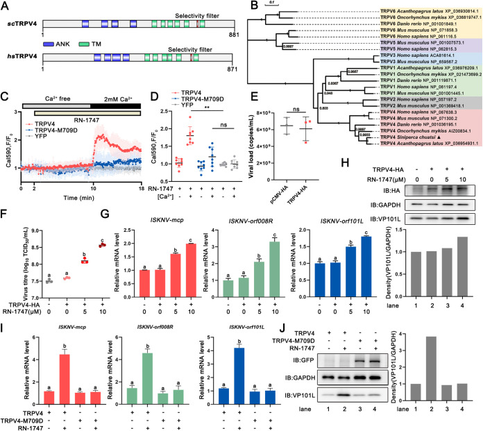
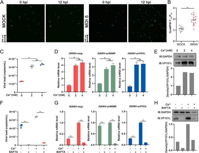
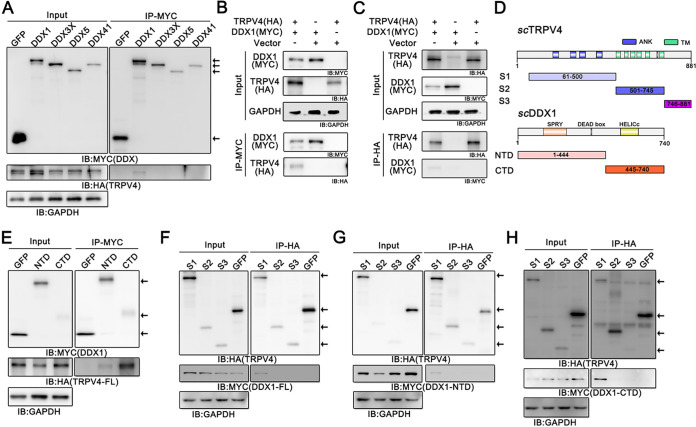
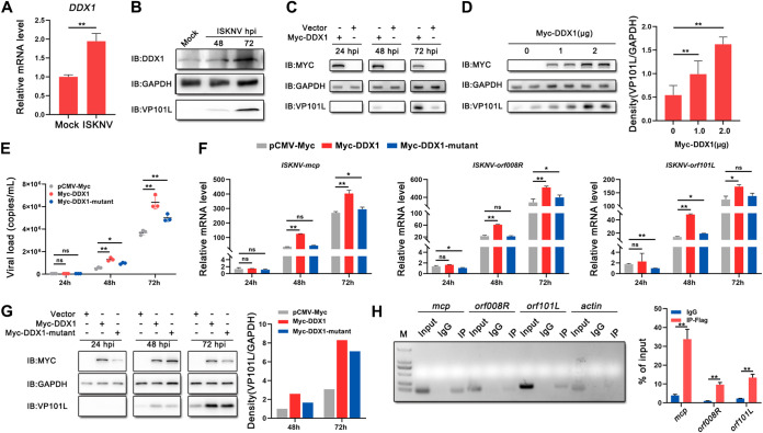
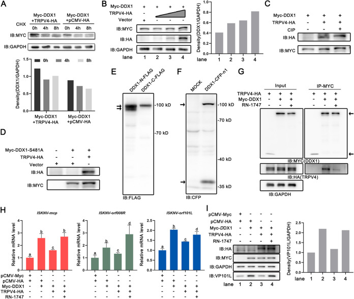
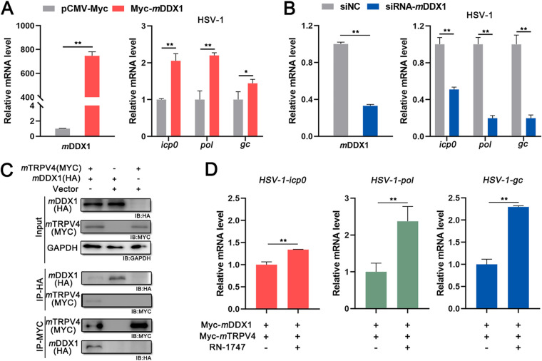
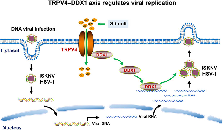
Viral diseases are a significant risk to the aquaculture industry. Transient receptor potential vanilloid 4 (TRPV4) has been reported to be involved in regulating viral activity in mammals, but its regulatory effect on viruses in teleost fish remains unknown. Here, the role of the TRPV4-DEAD box RNA helicase 1 (DDX1) axis in viral infection was investigated in mandarin fish (Siniperca chuatsi). Our results showed that TRPV4 activation mediates Ca2+ influx and facilitates infectious spleen and kidney necrosis virus (ISKNV) replication, whereas this promotion was nearly eliminated by an M709D mutation in TRPV4, a channel Ca2+ permeability mutant. The concentration of cellular Ca2+ increased during ISKNV infection, and Ca2+ was critical for viral replication. TRPV4 interacted with DDX1, and the interaction was mediated primarily by the N-terminal domain (NTD) of TRPV4 and the C-terminal domain (CTD) of DDX1. This interaction was attenuated by TRPV4 activation, thereby enhancing ISKNV replication. DDX1 could bind to viral mRNAs and facilitate ISKNV replication, which required the ATPase/helicase activity of DDX1. Furthermore, the TRPV4-DDX1 axis was verified to regulate herpes simplex virus 1 replication in mammalian cells. These results suggested that the TRPV4-DDX1 axis plays an important role in viral replication. Our work provides a novel molecular mechanism for host involvement in viral regulation, which would be of benefit for new insights into the prevention and control of aquaculture diseases. IMPORTANCE In 2020, global aquaculture production reached a record of 122.6 million tons, with a total value of $281.5 billion. Meanwhile, frequent outbreaks of viral diseases have occurred in aquaculture, and about 10% of farmed aquatic animal production has been lost to infectious diseases, resulting in more than $10 billion in economic losses every year. Therefore, an understanding of the potential molecular mechanism of how aquatic organisms respond to and regulate viral replication is of great significance. Our study suggested that TRPV4 enables Ca2+ influx and interactions with DDX1 to collectively promote ISKNV replication, providing novel insights into the roles of the TRPV4-DDX1 axis in regulating the proviral effect of DDX1. This advances our understanding of viral disease outbreaks and would be of benefit for studies on preventing aquatic viral diseases.

Keywords: Ca2+; DDX1; HSV-1; ISKNV; TRPV4.


Conflict of interest statement

The authors declare no conflict of interest.

Figures

FIG 1
FIG 1
Activated TRPV4 facilitates viral replication. (A) Protein domains of S. chuatsi TRPV4 (scTRPV4) predicted by the SMART program. hsTRPV4, Homo sapiens TRPV4. (B) Phylogenetic tree based on multiple alignments of S. chuatsi TRPV4 and TRPV proteins from the other species. The neighbor-joining tree was generated using MEGA X, and a 1,000-replicate bootstrap analysis was performed. (C) Intracellular Ca2+ signals (Calbryte-590) obtained from MFF-1 cells transfected with peYFP-N1 (n = 8) (gray line), TRPV4-eYFP-N1 (n = 8) (red line), and TRPV4-M709D-eYFP-N1 (n = 8) (blue line). Cells were exposed to 10 μM RN-1747 in the absence and presence of extracellular Ca2+, as indicated. (D) Quantitative data for the Calbryte-590 fluorescence intensity (mean ± SD) from panel C. (E) Absolute quantification of the viral load (genomic copies) of ISKNV in MFF-1 cells transfected with pCMV-HA or TRPV4-HA and then infected with ISKNV at an MOI of 1 was performed by qPCR. ns, not significant. (F) MFF-1 cells were transfected with pCMV-HA or TRPV4-HA for 24 h, followed by infection with ISKNV and treatment with different concentrations of RN-1747 (0, 5, and 10 μM). Cells were collected, and the viral titers were then measured by a TCID50 assay. (G and H) ISKNV replication in MFF-1 cells transfected with pCMV-HA or TRPV4-HA. Cells were treated with different concentrations of RN-1747 (0, 5, and 10 μM) at the time of viral infection. (G) The relative mRNA levels of ISKNV-mcp, ISKNV-orf008R, and ISKNV-orf101L were determined via RT-qPCR. (H) The expression level of ISKNV-VP101L was determined by Western blotting. (I and J) ISKNV replication in MFF-1 cells transfected with TRPV4-eYFP-N1 or TRPV4-M709D-eYFP-N1. Cells were treated with 10 μM RN-1747 during viral infection. (I) The relative mRNA levels of ISKNV-mcp, ISKNV-orf008R, and ISKNV-orf101L were determined via RT-qPCR. (J) The expression level of ISKNV-VP101L was determined by Western blotting. All immunoblot (IB) data are representative of data from three independent experiments with similar results. Data are shown as means ± SD (n = 3). Statistical significance is indicated by lowercase letters. The different and same letters indicate the presence and absence of significant differences, respectively.
FIG 2
FIG 2
Ca2+ is critical for viral replication. (A) Representative images at 0 and 12 hpi of mock-infected (left) and ISKNV-infected (MOI = 5) (right) GCaMP6f cells. (B) Quantification of GCaMP6f fluorescence from panel A. (C to E) Buffering out extracellular calcium hinders ISKNV replication, significantly reducing the total viral load (C), the relative viral mRNA level (D), and the level of the viral protein VP101L (E), and excess extracellular Ca2+ (~4 mM) does not impact replication. (F to H) The stunted ISKNV replication in the presence of BAPTA-AM (20 μM) was detected via qPCR (F), RT-qPCR (G), and Western blot analysis (H). As a Ca2+ chelator, BAPTA-AM can significantly reduce the cytoplasmic Ca2+ concentration. Data are shown as means ± SD (n = 3). Statistical significance is indicated by asterisks (*, P < 0.05; **, P < 0.01).
FIG 3
FIG 3
TRPV4 interacts with DDX1. (A) Results of the detection of the interactions between TRPV4 and S. chuatsi DDX family proteins. (B and C) Co-IP of TRPV4-HA and Myc-DDX1 in FHM cells. (B) TRPV4 as a coprecipitant of DDX1; (C) the reverse result. (D) Schematic showing the domain structures of scTRPV4 and scDDX1. The amino acid residues of the TRPV4 and DDX1 fragments used in this study are indicated. S1, aa 61 to 500 of TRPV4; S2, aa 501 to 745 of TRPV4; S3, aa 746 to 881 of TRPV4; NTD, aa 1 to 444 of DDX1; CTD, aa 445 to 740 of DDX1. (E) Myc immunoprecipitates from lysates of FHM cells expressing the Myc-tagged NTD and CTD were immunoblotted for the detection of TRPV4. (F to H) HA immunoprecipitated from the lysates of FHM cells expressing HA-tagged S1, S2, and S3 of TRPV4 were immunoblotted for the detection of full-length DDX1 (F) and the NTD (G) and CTD (H) of DDX1.
FIG 4
FIG 4
DDX1 has a proviral effect, benefiting from its ATPase/helicase activity. (A and B) Endogenous DDX1 mRNA (A) and protein (B) levels after mock infection or ISKNV infection (MOI = 1). (C) Expression levels of ISKNV-VP101L determined by Western blotting in MFF-1 cells overexpressing Myc-DDX1 or pCMV-Myc 24, 48, and 72 h after ISKNV infection (MOI = 1). (D) Expression levels of ISKNV-VP101L determined by Western blotting in MFF-1 cells overexpressing 0, 1, or 2 μg of the Myc-DDX1 plasmid at 72 hpi (MOI = 1). Quantitative analysis of the data obtained from two replicates was performed using ImageJ software. (E to G) ISKNV replication in MFF-1 cells transfected with pCMV-Myc, Myc-DDX1, or the Myc-DDX1 mutant. Cells were infected with ISKNV (MOI = 1) at 24 hpt and collected at 24, 48, and 72 hpi. (E) The viral loads (genomic copies) of ISKNV were determined by qPCR. (F) The relative mRNA levels of ISKNV-mcp, ISKNV-orf008R, and ISKNV-orf101L were determined by RT-qPCR. (G) The expression level of ISKNV-VP101L was determined by Western blotting. (H) Interaction between FLAG-tagged DDX1 protein and ISKNV RNA analyzed by PCR (left) and RT-qPCR (right). The agarose gel electrophoresis schematic shows the amplification efficiency of PCR products derived from the lysates. The enrichment of ISKNV RNA pulled down with anti-FLAG antibody or IgG is indicated as a percentage of the input. All immunoblot data are representative of data from three independent experiments with similar results. Data are shown as means ± SD (n = 3). Statistical significance is indicated by asterisks (*, P < 0.05; **, P < 0.01). M, molecular weight marker.
FIG 5
FIG 5
The interplay of TRPV4-DDX1 regulates ISKNV replication. (A) Immunoblot analysis of cell lysates cotransfected with the Myc-DDX1 plasmid and an empty vector or the TRPV4-HA plasmid. Cells were treated with CHX for 0, 4, and 8 h before sampling. (B) Immunoblot analysis of cell lysates cotransfected with the Myc-DDX1 plasmid and different doses of the TRPV4-HA plasmid. (C) Immunoblot analysis of cell lysates cotransfected with the Myc-DDX1 plasmid and the TRPV4-HA plasmid or an empty vector. Protein dephosphorylation was conducted at 37°C for 45 min in 100-μL reaction mixtures made up of 100 μg of cell protein and 10 U of CIP. (D) Immunoblot analysis of cell lysates cotransfected with the Myc-DDX1-S481A plasmid and the TRPV4-HA plasmid or an empty vector. (E and F) Immunoblot analysis of cell lysates overexpressing the DDX1-N-FLAG, DDX1-C-FLAG, or DDX1-CFP-N1 plasmid. (G) Co-IP of TRPV4-HA and Myc-DDX1 in FHM cells. Cells were treated with RN-1747 or dimethyl sulfoxide (DMSO) (negative control) overnight before sampling. (H and I) ISKNV replication in MFF-1 cells transfected with different plasmids. Cells were treated with 10 μM RN-1747 or DMSO at the time of viral infection. (H) The relative mRNA levels of ISKNV-mcp, ISKNV-orf008R, and ISKNV-orf101L were determined via RT-qPCR. (I) The expression level of ISKNV-VP101L was determined by Western blotting. All immunoblot data are representative of data from three independent experiments with similar results. Data are shown as means ± SD (n = 3). Statistical significance is indicated by lowercase letters. The different and same letters indicate the presence and absence of significant differences, respectively.
FIG 6
FIG 6
The TRPV4-DDX1 axis regulates HSV-1 replication. (A) HSV-1 replication in BHK-21 cells transfected with pCMV-Myc or Myc-mDDX1. Cells were infected with HSV-1 (MOI = 1) at 24 hpt and collected at 48 hpi. The relative mRNA levels of mDDX1, HSV-1-icp0, HSV-1-pol, and HSV-1-gc were determined by RT-qPCR. (B) HSV-1 replication in BHK-21 cells transfected with siNC or siRNA-mDDX1. Cells were infected with HSV-1 (MOI = 1) at 24 hpt and collected at 48 hpi. The relative mRNA levels of mDDX1, HSV-1-icp0, HSV-1-pol, and HSV-1-gc were determined by RT-qPCR. (C) Co-IP of Myc-tagged mTRPV4 and HA-tagged mDDX1 in BHK-21 cells. (D) HSV-1 replication in BHK-21 cells transfected with Myc-mDDX1 and Myc-mTRPV4. Cells were treated with 10 μM RN-1747 or DMSO during viral infection. The relative mRNA levels of HSV-1-icp0, HSV-1-pol, and HSV-1-gc were determined via RT-qPCR. Data are shown as means ± SD (n = 3). Statistical significance is indicated by asterisks.
FIG 7
FIG 7
Model of the interplay between TRPV4 and DDX1 in the regulation of viral replication. TRPV4 anchors DDX1 in a resting state. Upon TRPV4 activation by stimuli (heat or RN-1747), the intracellular Ca2+ concentration increases, and DDX1 dissociates from TRPV4. Dissociated DDX1 is transported to bind to viral RNA and is hijacked by viruses to induce their translation. Altogether, the interaction between TRPV4 and DDX1 regulates viral replication.

Similar articles

Cited by

References

    1. Voets T, Talavera K, Owsianik G, Nilius B. 2005. Sensing with TRP channels. Nat Chem Biol 1:85–92. doi:10.1038/nchembio0705-85. - DOI - PubMed
    1. Strotmann R, Harteneck C, Nunnenmacher K, Schultz G, Plant TD. 2000. OTRPC4, a nonselective cation channel that confers sensitivity to extracellular osmolarity. Nat Cell Biol 2:695–702. doi:10.1038/35036318. - DOI - PubMed
    1. Galindo-Villegas J, Montalban-Arques A, Liarte S, de Oliveira S, Pardo-Pastor C, Rubio-Moscardo F, Meseguer J, Valverde MA, Mulero V. 2016. TRPV4-mediated detection of hyposmotic stress by skin keratinocytes activates developmental immunity. J Immunol 196:738–749. doi:10.4049/jimmunol.1501729. - DOI - PubMed
    1. Liedtke W, Choe Y, Renom MAM, Bell AM, Denis CS, Sali A, Hudspeth AJ, Friedman JM, Heller S. 2000. Vanilloid receptor-related osmotically activated channel (VR-OAC), a candidate vertebrate osmoreceptor. Cell 103:525–535. doi:10.1016/s0092-8674(00)00143-4. - DOI - PMC - PubMed
    1. Suzuki M, Watanabe Y, Oyama Y, Mizuno A, Kusano E, Hirao A, Ookawara S. 2003. Localization of mechanosensitive channel TRPV4 in mouse skin. Neurosci Lett 353:189–192. doi:10.1016/j.neulet.2003.09.041. - DOI - PubMed

Publication types

Substances

LinkOut - more resources