. 2023 Jun 8;21(6):e3002144.
doi: 10.1371/journal.pbio.3002144. eCollection 2023 Jun.

Host-specific sensing of coronaviruses and picornaviruses by the CARD8 inflammasome

Affiliations

Host-specific sensing of coronaviruses and picornaviruses by the CARD8 inflammasome

Brian V Tsu et al. PLoS Biol. .

Abstract

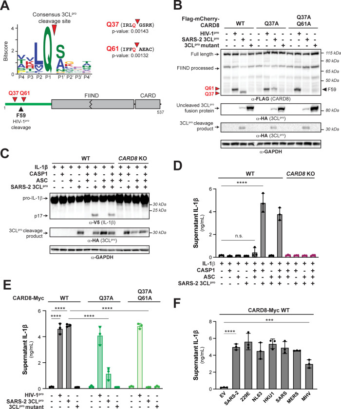
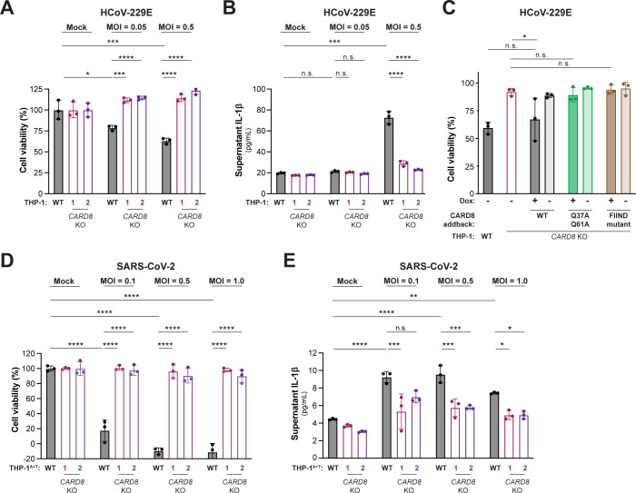
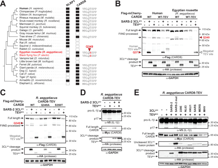
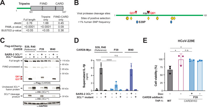
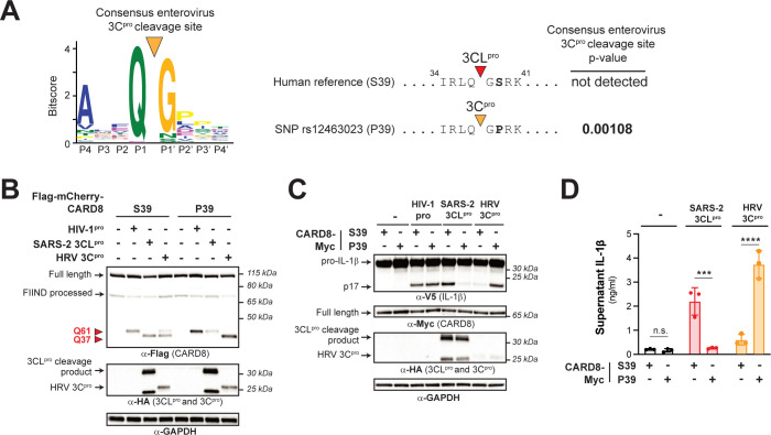
Hosts have evolved diverse strategies to respond to microbial infections, including the detection of pathogen-encoded proteases by inflammasome-forming sensors such as NLRP1 and CARD8. Here, we find that the 3CL protease (3CLpro) encoded by diverse coronaviruses, including Severe Acute Respiratory Syndrome Coronavirus 2 (SARS-CoV-2), cleaves a rapidly evolving region of human CARD8 and activates a robust inflammasome response. CARD8 is required for cell death and the release of pro-inflammatory cytokines during SARS-CoV-2 infection. We further find that natural variation alters CARD8 sensing of 3CLpro, including 3CLpro-mediated antagonism rather than activation of megabat CARD8. Likewise, we find that a single nucleotide polymorphism (SNP) in humans reduces CARD8's ability to sense coronavirus 3CLpros and, instead, enables sensing of 3C proteases (3Cpro) from select picornaviruses. Our findings demonstrate that CARD8 is a broad sensor of viral protease activities and suggests that CARD8 diversity contributes to inter- and intraspecies variation in inflammasome-mediated viral sensing and immunopathology.


Conflict of interest statement

The authors have declared that no competing interests exist.

Figures

Fig 1
Fig 1. 3CLpro from SARS-CoV-2 and other coronaviruses cleaves and activates the human CARD8 inflammasome.
(A) A consensus Beta-CoV 3CLpro cleavage motif (upper panel; S1 Fig and Materials and methods) was used to predict two 3CLpro cleavage sites (red triangles) within the disordered “tripwire” N-terminus of human CARD8 (lower panel, green) near the described site of HIV-1pro cleavage (black triangle). Flanking residues and p-values of prediction for each site (Q37 and Q61) are shown. (B) HEK293T cells were transfected with the indicated CARD8 construct in the presence (“+”) or absence (“−”) of indicated proteases. Active (SARS-2 3CLpro) or catalytically inactive (3CLpro mutant) protease from SARS-CoV-2 was expressed as an HA-tagged fusion construct (S2 Fig). HIV-1pro was expressed from an untagged gag-pol construct. Triangles are as described in (A). (C, D) WT or CARD8 KO HEK293T cells were transfected with (“+”) or without (“−”) indicated constructs. Inflammasome activation was monitored by immunoblotting for mature IL-1β (p17) (C) or measuring culture supernatant levels of bioactive IL-1β using IL1R-expressing reporter cells (D). (E, F) CARD8 KO HEK293T cells were cotransfected with the indicated CARD8 and protease constructs and supernatant levels of bioactive IL-1β were measured by IL1R reporter assay. 3CLpros from the following viruses were used: hCoV-229E (229E), hCoV-NL63 (NL63), hCoV-HKU1 (HKU1), SARS-CoV (SARS), MERS-CoV (MERS), and mouse hepatitis virus (MHV). (D-F) Individual values (n = 3), averages, and standard deviations shown are representative of experiments performed in triplicate. Data were analyzed using two-way ANOVA with Šidák’s post-test (D, E) or one-way ANOVA with Tukey’s post-test (F). *** = p < 0.001, **** = p < 0.0001, n.s. = not significant. Data for Fig 1A, 1D, 1E, and 1F can be found in S1 Data. Beta-CoV, betacoronavirus; HIV-1pro, HIV-1 protease; IL, interleukin; MHV, mouse hepatitis virus; SARS-CoV-2, Severe Acute Respiratory Syndrome Coronavirus 2; WT, wild-type; 3CLpro, 3CL protease.
Fig 2
Fig 2. Coronavirus infection activates the CARD8 inflammasome in THP-1 cells.
WT, CARD8 KO1, and CARD8 KO2 THP-1 cells (A, B), or THP-1 cells expressing ACE2 and TMPRSS2 (THP-1T+A) (D, E) were primed with 0.5 μg/mL Pam3CSK4 for 6 h, followed by infection with the coronaviruses hCoV-229E (A, B) or SARS-CoV-2 (SARS-2) (D, E) at the indicated MOI. (C) CARD8 KO1 THP-1 cells were complemented with a using a Dox-inducible lentiviral construct expressing the indicated CARD8 variant. WT, CARD8 KO1, or complemented cell lines were treated with Dox as indicated and infected with 200 PFUs hCoV-229E. After 48 h, cell viability (A, C, D) was measured using the Cell Titer Glo assay and IL-1β levels were measured using the IL1R reporter assay (B, E) as in Fig 1D. Data were analyzed using two-way ANOVA with Šidák’s post-test (A, B, D, E) or one-way ANOVA with Tukey’s post-test (C). * = p < 0.05, ** = p < 0.01, *** = p < 0.001, **** = p < 0.0001, n.s. = not significant. Data for Fig 2A, 2B, 2C, 2D and 2E can be found in S1 Data. Dox, doxycycline; IL, interleukin; MOI, multiplicity of infection; PFU, plaque-forming unit; SARS-CoV-2, Severe Acute Respiratory Syndrome Coronavirus 2; WT, wild-type.
Fig 3
Fig 3. Megabat CARD8 is antagonized rather than activated by coronavirus 3CLpro.
(A) Presence (filled rectangle) or absence (empty rectangle) of predicted NLRP1 or CARD8 orthologs in the indicated mammalian species. To the left is a species phylogeny. Megabat species are indicated in red. To the right is an alignment of a predicted 3CLpro cleavage site in Rousettus aegyptiacus CARD8 (red triangle indicates site and number indicates residue position). (B) Human CARD8 or R. aegyptiacus CARD8 was cotransfected with either SARS-CoV-2 (SARS-2) 3CLpro or protease from tobacco etch virus (TEVpro). For human or R. aegyptiacus CARD8 constructs labeled “WT-TEV,” a TEVpro site was introduced into the N-terminus. The red triangles and amino acid number indicate the sites of 3CLpro cleavage in human and R. aegyptiacus CARD8. The gray triangles indicate the sites of TEV protease cleavage within each CARD8 WT-TEV. (C) Mapping of the 3CLpro site within R. aegyptiacus CARD8 was performed by transfecting the indicated point mutants with SARS-CoV-2 (SARS-2) 3CLpro or TEVpro. 3CLpro and TEVpro sites are marked by triangles as in (B). (D) CARD8 KO HEK293T cells were cotransfected with R. aegyptiacus IL-1β and CASP1, along with the indicated CARD8 and protease constructs. Presence of mature IL-1 β (p17) upon TEVpro addition indicates successful reconstitution of the R. aegyptiacus CARD8 inflammasome, whereas absence of p17 upon SARS-2 3CLpro indicates antagonism of the R. aegyptiacus CARD8 inflammasome. (C) R. aegyptiacus CARD8 inflammasome activation assays were performed as in (D) with the indicated 3CLpro constructs. IL, interleukin; SARS-CoV-2, Severe Acute Respiratory Syndrome Coronavirus 2; TEV, tobacco etch virus; 3CLpro, 3CL protease.
Fig 4
Fig 4. Human polymorphism in CARD8 reduces sensing of coronavirus 3CLpro.
(A) Evolutionary analyses of positive selection were performed on full length CARD8 (encoding residues 1–537), the disordered N-terminal “tripwire” region (encoding residues 1–161), and the FIIND-CARD region (encoding residues 162–537). P values from PAML and BUSTED analyses are shown, along with the dN/dS value obtained from PAML. (B) Schematic of the CARD8 “tripwire” region. Red and black triangles and amino acid numbers indicate sites of 3CLpro and HIV-1pro cleavage, respectively. Yellow diamonds indicate codons predicted to be evolving under positive selection by at least one evolutionary analysis (S4 Table). Blue diamonds indicate high frequency (>1% allele frequency) nonsynonymous SNPs in humans (S5 and S6 Tables). The position of the S39P substitution that results from SNP rs12463023 is shown. (C, D) Reference human CARD8 (S39, R40) or human CARD8 variants (P39 or W40) were coexpressed with the indicated protease construct and assayed for 3CLpro-mediated cleavage (C) or CARD8 inflammasome activation (D). (E) The CARD8 variant P39 was complemented into CARD8 KO1 THP-1 cells using a Dox-inducible lentiviral construct and were infected along with WT and CARD8 KO1 THP-1 cells with hCoV-229E in the presence or absence of 100 ng/mL Dox. Cell viability was measured using the Cell Titer Glo assay, 48 h post-infection. Individual values (n = 3), averages, and standard deviations shown are representative of experiments performed at least twice. Data were analyzed using two-way ANOVA with Šidák’s post-test (D) or one-way ANOVA with Tukey’s post-test (E). * = p < 0.05, **** = p < 0.0001, n.s. = not significant. Data for Fig 4D and 4E can be found in S1 Data. Dox, doxycycline; SNP, single nucleotide polymorphism; WT, wild-type; 3CLpro, 3CL protease.
Fig 5
Fig 5. A human polymorphism dictates CARD8 sensing of coronavirus 3CLpro and human rhinovirus 3Cpro.
(A) A previously generated 3Cpro consensus cleavage motif for enteroviruses (a genus within picornaviruses) [14], in which a proline is the most common amino acid found at the P2’ position, is shown. A 3Cpro cleavage site is predicted in the CARD8 P39 variant (encoded by rs12463023) but not the CARD8 S39 variant (encoded by the reference allele). (B-D). Human CARD8 S39 or CARD8 P39 were transfected with the indicated proteases and assayed for 3CLpro-mediated cleavage (B), CARD8 inflammasome-mediated maturation of IL-1β (C), or the release of bioactive IL-1β (D), as in Fig 1. Individual values (n = 3), averages, and standard deviations shown are representative of experiments performed in triplicate. Data were analyzed using two-way ANOVA with Šidák’s post-test. *** = p < 0.001, **** = p < 0.0001, n.s. = not significant. Data for Fig 5A and 5D can be found in S1 Data. HRV 3Cpro, human rhinovirus 3Cpro; IL, interleukin; 3Cpro, 3C protease; 3CLpro, 3CL protease.

Update of

References

    1. Cui H, Tsuda K, Parker JE. Effector-triggered immunity: from pathogen perception to robust defense. Annu Rev Plant Biol 2015;66:487–511. Epub 2014/12/11. doi: 10.1146/annurev-arplant-050213-040012 . - DOI - PubMed
    1. Fischer NL, Naseer N, Shin S, Brodsky IE. Effector-triggered immunity and pathogen sensing in metazoans. Nat Microbiol 2020;5(3):528. Epub 2020/02/12. doi: 10.1038/s41564-020-0682-4 . - DOI - PMC - PubMed
    1. Jones JD, Vance RE, Dangl JL. Intracellular innate immune surveillance devices in plants and animals. Science. 2016;354(6316). Epub 2016/12/10. doi: 10.1126/science.aaf6395 . - DOI - PubMed
    1. Mitchell PS, Sandstrom A, Vance RE. The NLRP1 inflammasome: new mechanistic insights and unresolved mysteries. Curr Opin Immunol 2019;60:37–45. Epub 2019/05/24. doi: 10.1016/j.coi.2019.04.015 ; PubMed Central PMCID: PMC6800612. - DOI - PMC - PubMed
    1. Vance RE, Isberg RR, Portnoy DA. Patterns of pathogenesis: discrimination of pathogenic and nonpathogenic microbes by the innate immune system. Cell Host Microbe 2009;6(1):10–21. Epub 2009/07/21. doi: 10.1016/j.chom.2009.06.007 ; PubMed Central PMCID: PMC2777727. - DOI - PMC - PubMed

Publication types