. 2023 Oct;69(4):391-403.
doi: 10.1165/rcmb.2022-0217OC.

Targeting EB3-IP3R3 Interface with Cognate Peptide Protects from Acute Respiratory Distress Syndrome

Affiliations

Targeting EB3-IP3R3 Interface with Cognate Peptide Protects from Acute Respiratory Distress Syndrome

Man Long Kwok et al. Am J Respir Cell Mol Biol. 2023 Oct.

Abstract

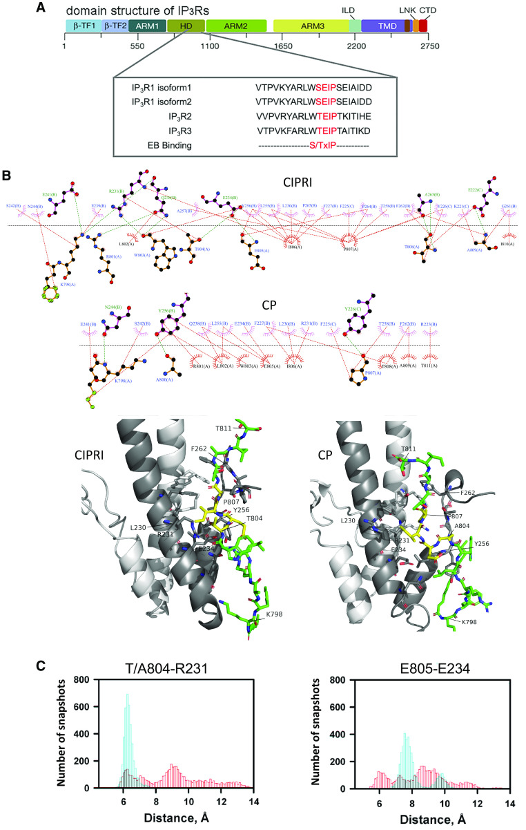
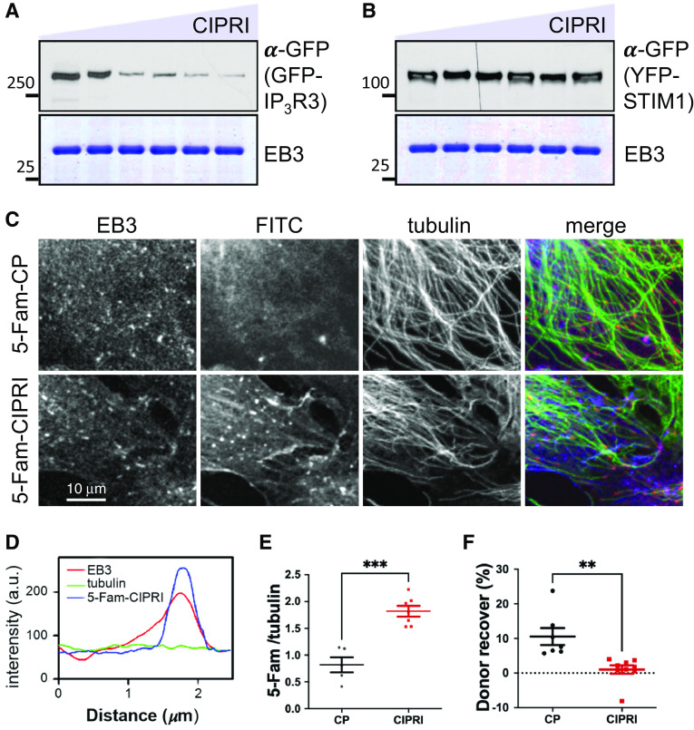
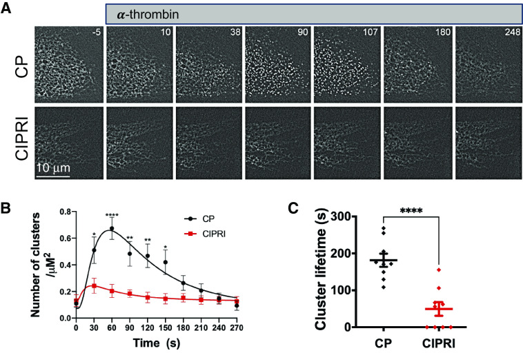
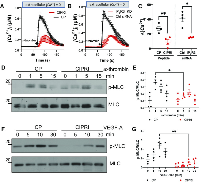
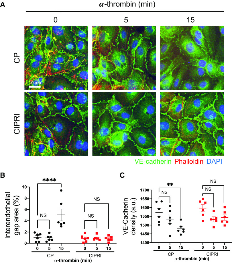
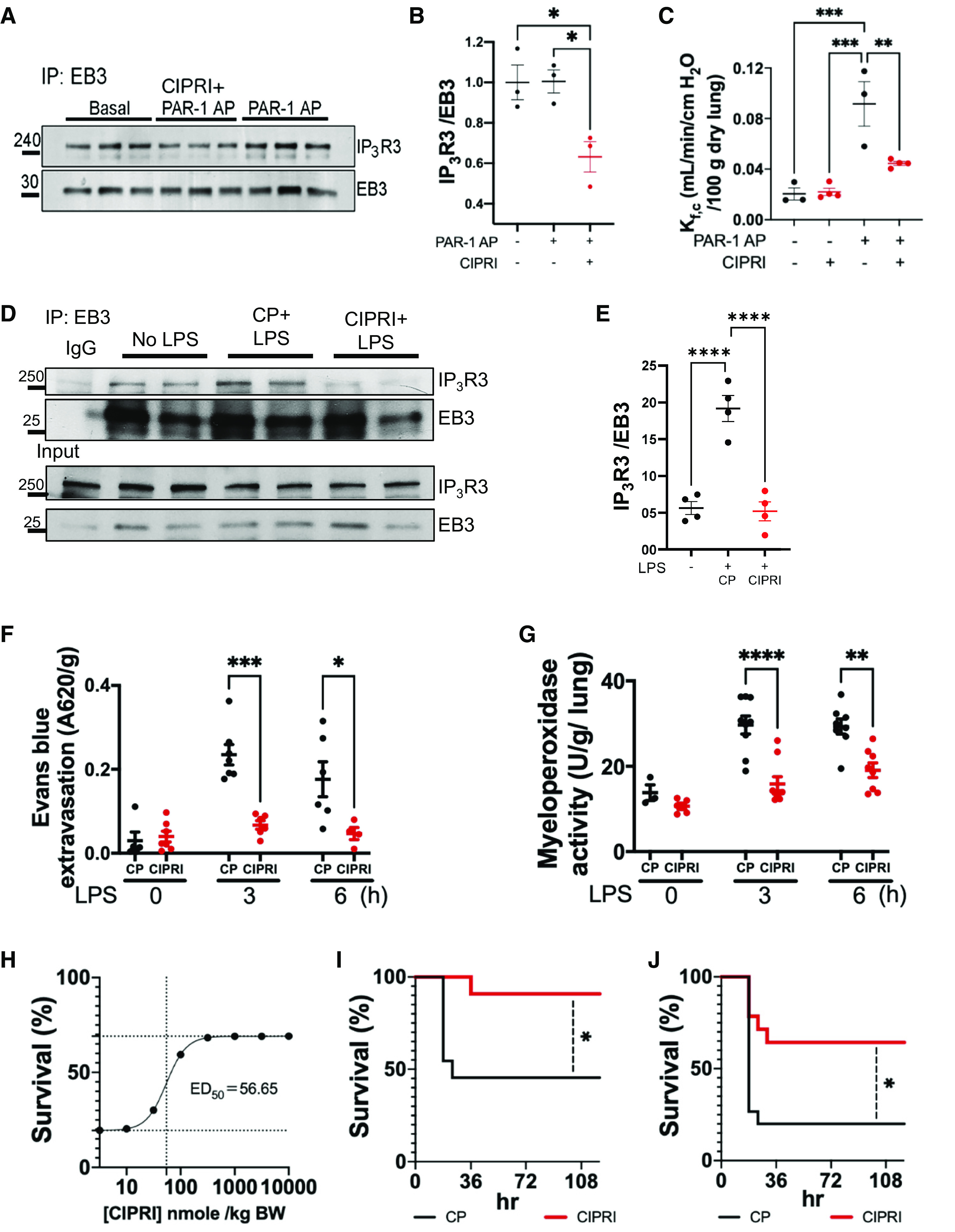
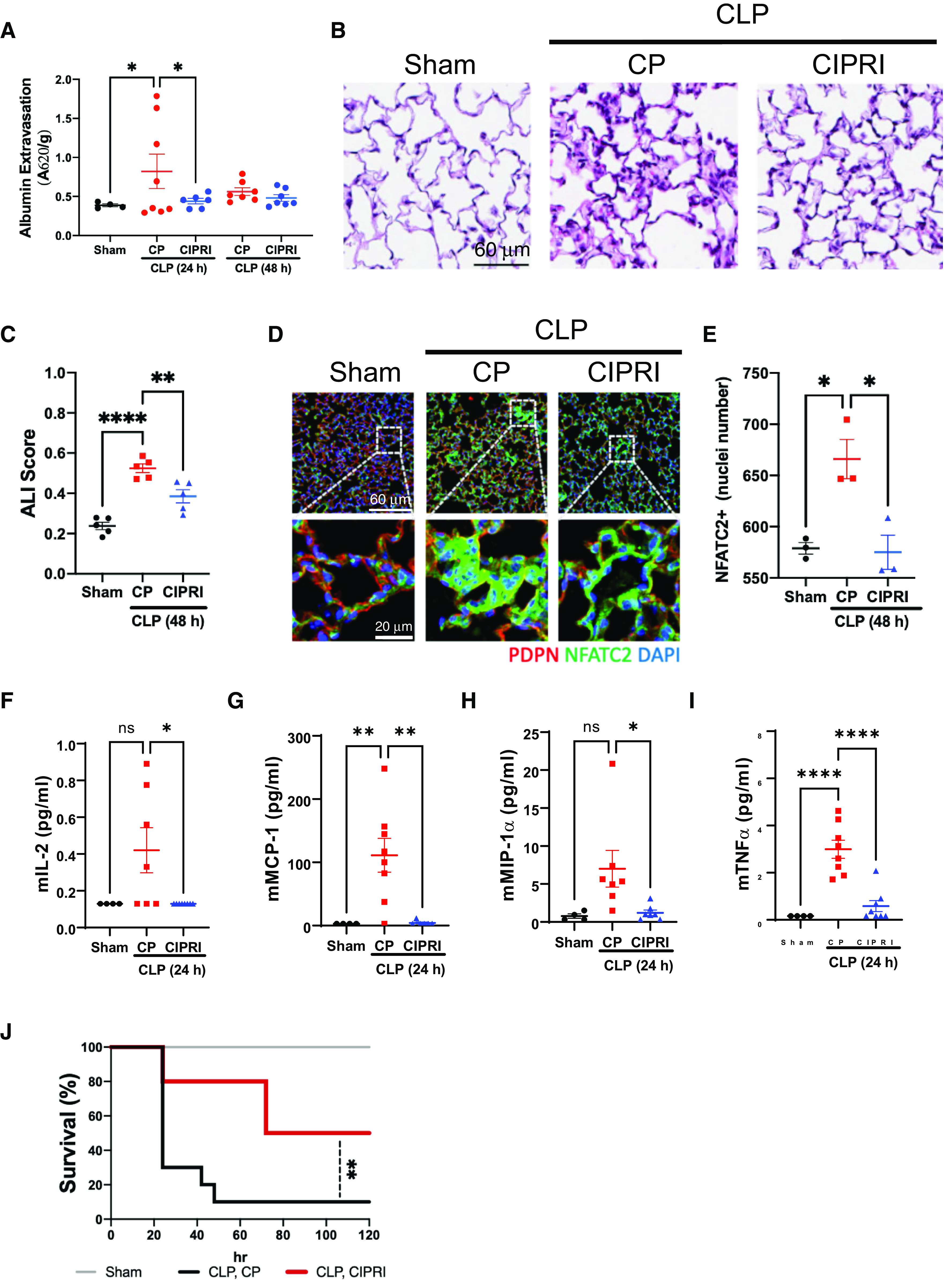
Acute respiratory distress syndrome (ARDS) is a lung disease characterized by acute onset of noncardiogenic pulmonary edema, hypoxemia, and respiratory insufficiency. The current treatment for ARDS is mainly supportive in nature, providing a critical need for targeted pharmacological management. We addressed this medical problem by developing a pharmacological treatment for pulmonary vascular leakage, a culprit of alveolar damage and lung inflammation. Our novel therapeutic target is the microtubule accessory factor EB3 (end binding protein 3), which contributes to pulmonary vascular leakage by amplifying pathological calcium signaling in endothelial cells in response to inflammatory stimuli. EB3 interacts with IP3R3 (inositol 1,4,5-trisphosphate receptor 3) and orchestrates calcium release from endoplasmic reticulum stores. Here, we designed and tested the therapeutic benefits of a 14-aa peptide named CIPRI (cognate IP3 receptor inhibitor), which disrupted EB3-IP3R3 interaction in vitro and in lungs of mice challenged with endotoxin. Treatment with CIPRI or depletion of IP3R3 in lung microvascular endothelial monolayers mitigated calcium release from endoplasmic reticulum stores and prevented a disassembly of vascular endothelial cadherin junctions in response to the proinflammatory mediator α-thrombin. Furthermore, intravenous administration of CIPRI in mice mitigated inflammation-induced lung injury, blocked pulmonary microvascular leakage, prevented activation of NFAT (nuclear factor of activated T cells) signaling, and reduced production of proinflammatory cytokines in the lung tissue. CIPRI also improved survival of mice from endotoxemia and polymicrobial sepsis. Together, these data demonstrate that targeting EB3-IP3R3 interaction with a cognate peptide is a promising strategy to address hyperpermeability of microvessels in inflammatory lung diseases.

Keywords: calcium signaling; endotoxemia; lung inflammation; sepsis; vascular leakage.


Figures

Figure 1.
Figure 1.
Analysis of the EB3 (end binding protein 3)–IP3R3 (inositol 1,4,5-trisphosphate receptor 3) interaction. (A) Sequence alignment of IP3R3 isoforms from different species (human, mouse, rat, bovine) with EB-targeting motif S/TxIP. (B) Representative snapshots of CIPRI (cognate IP3 receptor inhibitor) and the control peptide (CP) bound to EB3 dimer. Top: a LigPlot rendering of the interface residues in EB3 and CIPRI or CP: (A) CIPRI or CP, (B and C) two chains of EB3 homodimer; green dashed lines indicate hydrogen bonds; red dashed lines indicate hydrophobic interactions; arches indicate the approximate combined direction of the interfacing atoms or groups. Bottom: a PYMOL rendering of the three-dimensional structure of the EB3–CIPRI and EB3–CP complexes. The key residues are marked for reference. The amino acid sequence TEIP and AEIP in CIPRI and CP, respectively, are rendered as yellow sticks. The remaining portions of the peptides are rendered as green sticks. Note a pronounced difference in the number of interactions for CIPRI compared with CP. (C) A distribution analysis of the distances between Ile806 or Pro807 of CIPRI (cyan) and the CP (red) and Leu230 or Phe263 of EB3, respectively. Amino acids within CIPRI and CP are labeled based on human IP3R3 sequence. Each bar corresponds to a number of snapshots binned based on distance (10 per 1 Å). Note that, in the molecular-dynamic simulation with the CP, the distance distribution is dispersed and spread between a wider range of distances.
Figure 2.
Figure 2.
The CIPRI peptide competes with IP3R3 for binding to EB3. (A and B) Western blot analysis of EB3’s interaction with GFP–IP3R3 (A) and yellow fluorescent protein (YFP)–tagged STIM1 (Stromal interaction molecule-1) (B) in the presence of various concentrations of Myr-CIPRI (no peptide and 1:0.1, 1:0.5, 1:1, 1:2, and 1:5 molar EB3/CIPRI ratios) as indicated. Upper panels show Western blots for GFP to detect YFP-STIM1, and lower panels show Coomassie brilliant blue–stained gels loaded with 5% of the (6× His)-EB3 used for the pull-down. Results are representative of three independent experiments. The CIPRI peptide inhibits the interaction between EB3 and IP3R3 in a dose-dependent manner. (C) The Myr-5-Fam–labeled CIPRI colocalizes with EB3 at the growing microtubule (MT) plus ends. Human lung microvascular endothelial cells (HLMVECs) were pretreated with 1 μM 5-Fam-CIPRI or 5-Fam CP for 30 minutes and fixed with cold methanol. Cells were immunostained for EB3 (red) and tubulin (green); FITC is shown in blue. Scale bar, 10 μm. (D) Line-scan analysis demonstrating accumulation of Myr-5-Fam-CIPRI (blue) at MT plus ends (identified by EB3; red). (E) Accumulation of Myr-5-Fam-CIPRI and Myr-5-Fam–labeled CP at the growing MT tips. The 5-Fam fluorescence integrated intensity at the MT plus ends was normalized to tubulin intensity (***P < 0.001 using Student’s t test). (F) The Myr-CIPRI peptide inhibits the interaction between IP3R3 and EB3 in cells. Graph showing percentage recovery of the donor fluorescence (dequenching) after acceptor photobleaching in CHO-K1 cells treated with 1 μM Myr-CP or Myr-CIPRI peptides. Dequenching of donor fluorescence from GFP-IP3R3 was recorded while EB3–monomeric red fluorescent protein (mRFP) fluorescence was photobleached at the MT tip; n = 5–8 cells per group (**P < 0.01 using Student’s t test).
Figure 3.
Figure 3.
The CIPRI peptide inhibits clustering of IP3R3. (A) Time-lapse images of GFP–IP3R3 in HLMVECs treated with 1 μM Myr-CP or Myr-CIPRI peptides and stimulated with 50 nM α-thrombin at t = 0 seconds. Times (in seconds) are shown on each panel. Scale bar, 10 μm. (B) Time-dependent assembly of GFP–IP3R3 clusters in response to α-thrombin in A. The data are normalized to t = 0 (baseline clusters). Mean ± SEM from n = 8–11 cells per group (*P < 0.05, **P < 0.01, and ****P < 0.0001 using two-way ANOVA with Bonferroni’s multiple comparisons test). (C) The lifetime of the GFP–IP3R3 clusters of the data in A (see data supplement) (****P < 0.0001 using Student’s t test).
Figure 4.
Figure 4.
The CIPRI peptide inhibits calcium signaling in endothelial cells. (A and B) Representative tracing of endoplasmic reticulum (ER) calcium release measured as a function of Fura 2-AM cytosolic calcium increase in endothelial cells treated with 1 μM Myr-CP or the Myr-CIPRI peptides (A) or control or IP3R3 siRNA (B) following stimulation with α-thrombin at 60 seconds. The ER calcium release was measured in calcium-free Hanks’ balanced salt solution buffer. (C) Peak changes in [Ca2 + ]ER release evoked by α-thrombin (in absence of extracellular calcium) in A and B; n = 5–14 experiments (*P < 0.05 and **P < 0.01 using Student’s t test). (D and E) Time course of MLC (myosin light chain) phosphorylation in HLMVECs treated with 1 μM Myr-control or Myr-CIPRI peptides and stimulated with 50 nM α-thrombin at t = 0 (D) and quantification of data (E). Data are representative of four experiments (*P < 0.05 using two-way ANOVA with Bonferroni’s multiple comparisons test). (F and G) Time course of MLC phosphorylation in HLMVECs treated with 1 μM Myr-control or Myr-CIPRI peptides and stimulated with 100 ng/ml VEGF-A at t = 0 (F) and quantification of data (G). Data are representative of four experiments (**P < 0.01 using two-way ANOVA with Bonferroni’s multiple comparisons test).
Figure 5.
Figure 5.
The CIPRI peptide prevents disassembly of adherens junctions (AJs). (A) Immunofluorescent staining of HPAECs treated with 1 μM Myr-CP or the Myr-CIPRI peptides and stained for vascular endothelial (VE)-cadherin (green) and phalloidin (red) at indicated times after stimulation with α-thrombin. Scale bar, 10 μm. (B and C) The area of interendothelial gaps expressed as percentage of total area (B) and VE-cadherin density at AJs (C) of data in A; n = 6 filed per condition (**P < 0.01 and ****P < 0.0001 using two-way ANOVA with Dunnett’s test). NS = not significant.
Figure 6.
Figure 6.
The CIPRI peptide suppresses endotoxemia-induced transvascular leakage and inflammation in the lung by blocking the EB3–IP3R3 interaction. (A) The Myr-CIPRI peptide suppresses the interaction between EB3 and IP3R3 in lung challenged with PAR-1 agonist peptide. Lungs were left untreated or infused with 30 μM PAR-1 (protease-activated receptor-1) agonist peptide (AP; TFLLRN-NH2) with or without 10 μM Myr-CIPRI as indicated. EB3 was precipitated from lung homogenate using an antibody. Resulted precipitates were probed for IP3R3 and EB3; data derived from three independent experiments. (B) Quantification of data in A expressed as IP3R3–EB3 ratio (*P < 0.05 using one-way ANOVA with Tukey’s multiple comparisons test). (C) The CIPRI peptide suppresses permeability of endothelial vessel wall to fluids. Microvascular filtration coefficient Kf,c was assessed using ex vivo lung preparation. Lungs isolated from CD1 mice were infused with 10 μM Myr-control or Myr-CIPRI and 30 μM PAR-1 AP as indicated. Kf,c was calculated from the slope of the weight gain curve at 15–20 minutes; n = 3 lungs per group (**P < 0.01 and ***P < 0.01 using one-way ANOVA with Tukey’s multiple comparisons test). (D) The CIPRI peptide suppresses the EB3–IP3R3 interaction in the lung of mice challenged with the 50% lethal dose (LD50) (10 mg/kg body weight [BW]) of LPS for 3 hours. Mice received 1 μmol/kg BW Myr-CP or Myr-CIPRI. EB3 was precipitated from lung homogenates using an antibody and probed for IP3R3 and EB3 as in A; data derived from four mice per group from four independent experiments. (E) Quantification of data in D expressed as IP3R3–EB3 ratio (***P < 0.001 using one-way ANOVA with Tukey’s test). (F) Extravasation of Evans blue–labeled albumin (EBA) tracer in lungs of CD1 mice challenged with LPS. Mice received Myr-control or Myr-CIPRI (1 μmol/kg BW) and were challenged with LPS at LD50 (30 mg/kg BW). Extravasation of EBA was assessed before and 3 and 6 hours after LPS challenge (see Methods). n = 4–7 mice per group (*P < 0.05 and ***P < 0.001 using two-way ANOVA with Bonferroni’s test). (G) Treatment with Myr-CIPRI mitigates sequestration of neutrophils into the lung tissue as determined by myeloperoxidase activity. Mice treated as in D; n = 3–9 mice per group (**P < 0.01 and ****P < 0.0001 using two-way ANOVA with Bonferroni’s test). (H) The Myr-CIPRI peptide dose response on mice survival. CD-1 mice treated with the indicated doses of Myr-CIPRI peptide were intraperitoneally injected with 50 mg/kg BW LPS at the 80% lethal dose (LD80) and were monitored for 5 days (n = 10 mice per group). The 50% effective dose (ED50) calculated based on the dose–response studies is 56 nmol/kg BW. (I) The Myr-CIPRI peptide prevents lethality in LPS-induced sepsis. Mice treated with the Myr-CIPRI peptide demonstrate mortality reduction in LD50- (I) and LD80- (J) challenged control mice; n = 11–15 mice per group (*P < 0.05 using a log-rank test).
Figure 7.
Figure 7.
The CIPRI peptide protects from sepsis-induced lung injury and inflammation by blocking NFAT (nuclear factor of activated T cells) signaling. (A) Extravasation of EBA tracer in the lungs of CD1 mice at 24 and 48 hours after cecal ligation and puncture (CLP) surgery. Mice received 1 μmol/kg BW Myr-control or Myr-CIPRI 30 minutes before CLP surgery (see Methods); n = 4–8 mice per group (*P < 0.01 using one-way ANOVA with Fisher’s least significant difference test). (B and C) Representative images of the histopathological assessment of lung injury induced by CLP in mice with Myr-CP or Myr-CIPRI (B). Scale bar, 60 μm. Images of mouse lung sections were stained with hematoxylin and eosin and scored for acute lung injury (see Methods), quantified in C. Statistics: one-way ANOVA with Tukey post hoc test (**P < 0.01 and ****P < 0.0001). (D) Representative immunofluorescent-stained images of mice lungs that undergo CLP treated with Myr-CP or Myr-CIPRI for NFATC2. The dashed white box indicates the enlarged inset below to show nuclear accumulation of NFATC2. Epithelial marker podoplanin is shown in red, NFATC2 in green, and DAPI in blue. Scale bars: top image, 60 μm; bottom image, 20 μm. (E) Quantification of the number cells with NFATC2-positive nuclei in D. One-way ANOVA with Tukey post hoc test (*P < 0.05). (F–I) Analyses of cytokine levels in the BAL fluid of mice treated as in A at 24 hours after CLP surgery. Myr-CIPRI prevented an increase in IL-2 (F), MCP-1 (G), MIP-1α (H), and TNF-α (I) (*P < 0.05, **P < 0.01, and ****P < 0.0001 using one-way ANOVA with Dunnett’s test). (J) The Myr-CIPRI peptide prevents lethality in polymicrobial sepsis. CD-1 mice that received 1 μmol/kg BW Myr-control or the Myr-CIPRI peptide underwent CLP surgery using a 16-gauge needle to puncture the cecum. This surgery caused 90% lethality in mice treated with the Myr-CP. Mice treated with Myr-CIPRI showed marked improvement in survival rate; n = 10 mice per group (**P < 0.01 using a log-rank test).

Comment in

References

    1. Fishman AP. Shock lung: a distinctive nonentity. Circulation . 1973;47:921–923. - PubMed
    1. Wang D, Hu B, Hu C, Zhu F, Liu X, Zhang J, et al. Clinical characteristics of 138 hospitalized patients with 2019 novel coronavirus-infected pneumonia in Wuhan, China. JAMA . 2020;323:1061–1069. - PMC - PubMed
    1. Bernard GR, Artigas A, Brigham KL, Carlet J, Falke K, Hudson L, et al. The American-European Consensus Conference on ARDS. Definitions, mechanisms, relevant outcomes, and clinical trial coordination. Am J Respir Crit Care Med . 1994;149:818–824. - PubMed
    1. Brigham KL, Bowers R, Haynes J. Increased sheep lung vascular permeability caused by Escherichia coli endotoxin. Circ Res . 1979;45:292–297. - PubMed
    1. Steinberg KP, Milberg JA, Martin TR, Maunder RJ, Cockrill BA, Hudson LD. Evolution of bronchoalveolar cell populations in the adult respiratory distress syndrome. Am J Respir Crit Care Med . 1994;150:113–122. - PubMed

Publication types