. 2023 Jul 1;142(1):31-50.
doi: 10.1097/AOG.0000000000005229. Epub 2023 Jun 7.

Placenta Accreta Spectrum

Affiliations

Placenta Accreta Spectrum

Brett D Einerson et al. Obstet Gynecol. .

Abstract

Placenta accreta spectrum (PAS) is one of the most dangerous conditions in pregnancy and is increasing in frequency. The risk of life-threatening bleeding is present throughout pregnancy but is particularly high at the time of delivery. Although the exact cause is unknown, the result is clear: Severe PAS distorts the uterus and surrounding anatomy and transforms the pelvis into an extremely high-flow vascular state. Screening for risk factors and assessing placental location by antenatal ultrasonography are essential for timely diagnosis. Further evaluation and confirmation of PAS are best performed in referral centers with expertise in antenatal imaging and surgical management of PAS. In the United States, cesarean hysterectomy with the placenta left in situ after delivery of the fetus is the most common treatment for PAS, but even in experienced referral centers, this treatment is often morbid, resulting in prolonged surgery, intraoperative injury to the urinary tract, blood transfusion, and admission to the intensive care unit. Postsurgical complications include high rates of posttraumatic stress disorder, pelvic pain, decreased quality of life, and depression. Team-based, patient-centered, evidence-based care from diagnosis to full recovery is needed to optimally manage this potentially deadly disorder. In a field that has relied mainly on expert opinion, more research is needed to explore alternative treatments and adjunctive surgical approaches to reduce blood loss and postoperative complications.


Conflict of interest statement

Financial Disclosure Lisa C. Zuckerwise reports money was paid to her institution from Laborie Medical Technologies Corp for an ongoing clinical study unrelated to placenta accreta spectrum. She received payment from Gershon, Willoughby & Getz, LLC, and Huff, Powell & Bailey, LLC for legal expert consulting not related to placenta accreta spectrum. The other authors did not report any potential conflicts of interest.

Figures

Figure 1:
Figure 1:
Managing unexpected and intraoperative placenta accreta spectrum (PAS) cases outside of PAS specialty centers. Real-time discovery of previously undiagnosed PAS is possible at any center that provides obstetric care, presenting an urgent need for complex clinical decision-making. Critical stability assessment of the pregnant patient and fetus is required throughout resource mobilization and consideration for potential transfer. Opportunities for transfer to a PAS referral center may be present before delivery of the infant, after delivery of the infant but prior to placental delivery (particularly if the placenta was not disrupted at hysterotomy), and following attempted removal of the placenta but prior to initiation of hysterectomy.
Figure 2:
Figure 2:
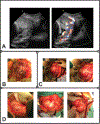
Appearance of severe placenta accreta spectrum (PAS). Visual correlation with patient outcomes at each stage of diagnosis and management is important for honing skills of PAS recognition, developing classification schemes predictive of clinical morbidity, and iterative learning for the multidisciplinary PAS care team. All images and photographs in this figure are from the same patient case. A. Representative antenatal ultrasound images (29 weeks of gestation) show complete placenta previa with placental bulging toward the bladder, loss of retroplacental clear zone, myometrial thinning, and extreme uterovesical hypervascularity with bridging vessels visible by color Doppler (right panel). B. Taken at the time of abdominal entry for scheduled delivery at 35 weeks of gestation, this photograph demonstrates obvious massive dehiscence of the previous cesarean scar with a large portion of the placental base visible through a thin translucent layer of uterine serosa. C. The placental edge was mapped with intraoperative ultrasonography to create a hysterotomy well away from the placental edge. The infant was delivered, avoiding placental disruption, and the hysterotomy was closed. Reduction in uterine volume with infant delivery allowed for full pelvic anatomy assessment, and a clear surgical plane could not be identified between the bladder and uterus. The patient was deemed a candidate for delayed interval hysterectomy. D. These photographs, taken at the time of completion laparotomy 40 days later, demonstrate the degree of tissue involution and reduction in vasculature that is the primary impetus for the delayed hysterectomy approach.
Figure 3:
Figure 3:
Endovascular approaches to reducing blood loss in placenta accreta spectrum (PAS). Endovascular adjunctive approaches, most commonly performed by interventional radiologists, can be used as salvage therapy in the case of active bleeding, or in a prophylactic manner to simplify dissection by “drying” the surgical field. In cases where the uterus is maintained in situ after delivery, these techniques are thought to hasten involution of the uterus, placenta, and pelvic vasculature. A. Diagrammatic representation of uteroplacental anatomy (left of dashed line) and major abdominopelvic vasculature (right of dashed line) in PAS after infant delivery. A hallmark of PAS disease, and the predominant source of its morbidity, is pervasive hypertrophied pelvic neovasculature, which can arise from any major artery in the lower abdomen and pelvis. B. Balloon occlusion approaches range from distal to proximal, with placement in uterine arteries (UA), internal iliac arteries (IIA), common iliac arteries (CIA), or the distal abdominal aorta (AA). More proximal placement can be faster and result in greater reduction in pelvic blood flow, but also carries greater risk for nontarget ischemic sequelae. C. Multivessel targeted embolization may require more specialized interventional radiology experience for optimization, but early data has demonstrated comparable blood loss reduction to abdominal aortic occlusion with dramatic reduction in nontarget vascular sequelae.

Similar articles

Cited by

References

    1. Leonard SA, Kennedy CJ, Carmichael SL, et al. An Expanded Obstetric Comorbidity Scoring System for Predicting Severe Maternal Morbidity. Obstet Gynecol 2020;136(3):440–49. doi: 10.1097/aog.0000000000004022 [published Online First: 2020/August/10] - DOI - PMC - PubMed
    1. Jauniaux E, Bunce C, Grønbeck L, et al. Prevalence and main outcomes of placenta accreta spectrum: a systematic review and meta-analysis. Am J Obstet Gynecol 2019;221(3):208–18. doi: 10.1016/j.ajog.2019.01.233 [published Online First: 2019/February/05] - DOI - PubMed
    1. Eller AG, Porter TF, Soisson P, et al. Optimal management strategies for placenta accreta. BJOG 2009;116(5):648–54. doi: 10.1111/j.1471-0528.2008.02037.x [published Online First: 2009/February/05] - DOI - PubMed
    1. Morlando M, Schwickert A, Stefanovic V, et al. Maternal and neonatal outcomes in planned versus emergency cesarean delivery for placenta accreta spectrum: A multinational database study. Acta Obstet Gynecol Scand 2021;100 Suppl 1:41–49. doi: 10.1111/aogs.14120 [published Online First: 2021/March/14] - DOI - PubMed
    1. Placenta accreta spectrum. Obstetric Care Consensus No. 7. American College of Obstetricians and Gynecologists. Obstetrics and Gynecology 2018;132:e259–75. - PubMed