. 2023 Sep:300:122185.
doi: 10.1016/j.biomaterials.2023.122185. Epub 2023 May 31.

Polymeric nanoparticle gel for intracellular mRNA delivery and immunological reprogramming of tumors

Affiliations

Polymeric nanoparticle gel for intracellular mRNA delivery and immunological reprogramming of tumors

Sarah Y Neshat et al. Biomaterials. 2023 Sep.

Abstract

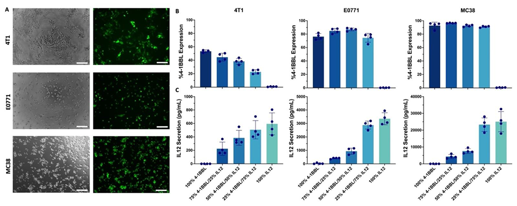
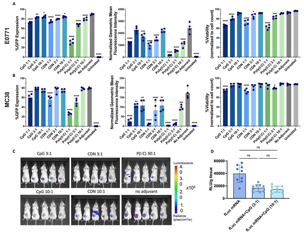
Immuno-oncology therapies have been of great interest with the goal of inducing sustained tumor regression, but clinical results have demonstrated the need for improved and widely applicable methods. An antigen-free method of cancer immunotherapy can stimulate the immune system to recruit lymphocytes and produce immunostimulatory factors without prior knowledge of neoantigens, while local delivery reduces the risk of systemic toxicity. To improve the interactions between tumor cells and cytotoxic lymphocytes, a gene delivery nanoparticle platform was engineered to reprogram the tumor microenvironment (TME) in situ to be more immunostimulatory by inducing tumor-associated antigen-presenting cells (tAPCs) to activate cytotoxic lymphocytes against the tumor. Biodegradable, lipophilic poly (beta-amino ester) (PBAE) nanoparticles were synthesized and used to co-deliver mRNA constructs encoding a signal 2 co-stimulatory molecule (4-1BBL) and a signal 3 immuno-stimulatory cytokine (IL-12), along with a nucleic acid-based immunomodulatory adjuvant. Nanoparticles are combined with a thermoresponsive block copolymer for gelation at the injection site for local NP retention at the tumor. The reprogramming nanoparticle gel synergizes with immune checkpoint blockade (ICB) to induce tumor regression and clearance in addition to resistance to tumor rechallenge at a distant site. In vitro and in vivo studies reveal increases in immunostimulatory cytokine production and recruitment of immune cells as a result of the nanoparticles. Intratumoral injection of nanoparticles encapsulating mRNA encoding immunostimulatory agents and adjuvants via an injectable thermoresponsive gel has great translational potential as an immuno-oncology therapy that can be accessible to a wide range of patients.

Keywords: Immuno-oncology; Immunoengineering; Immunotherapy; Nanoparticles; Nonviral gene delivery; mRNA.


Conflict of interest statement

Declaration of competing interest The authors declare the following financial interests/personal relationships which may be considered as potential competing interests:Stephany Y. Tzeng reports a relationship with OncoSwitch Therapeutics, LLC that includes: equity or stocks. Jordan J. Green reports a relationship with OncoSwitch Therapeutics, LLC that includes: equity or stocks. Sarah Y. Neshat, Stephany Y. Tzeng, Jordan J. Green has patent pending to Johns Hopkins University.

Figures

Figure 1.
Figure 1.. PBAE mRNA nanoparticles transfect three different tumor cell lines in vitro.
(A) Representative images of 4T1, E0771, and MC38 cells 24 h after transfection with 7-90,c12-63 80% NPs at 30 w/w. Brightfield and GFP filters are shown, scale bar: 200 nm). (B) Flow cytometry was used to measure surface presentation of 4-1BBL after transfecting 4T1, E0771, and MC38 with mRNA NPs with varying mRNA ratios (n=4 wells per group). (C) Sandwich ELISA was used to measure secretion of IL-12 after transfecting 4T1, E0771, and MC38 with mRNA NPs with varying mRNA ratios (n=4 wells per group). Mean ± SE is shown for all graphs.
Figure 2.
Figure 2.. Block co-polymer thermoresponsive gel demonstrates sequestered expression and release in vivo and in vitro.
(A) Block co-polymer structure shown (PLGA-PEG-PLGA LG 50:50 (w:w)). (B) The commercially available block co-polymers are engineered to be liquid and injectable at 4C but form a gel-like hydrogel network at body temperature (37°C). (C) Lead formulation PBAE fLuc mRNA NPs (30 w/w 1m% DMG-PEG2k 2.5 μg/tumor) combined with or without the block co-polymer is injected (single) 4T1 intratumorally and imaged by IVIS after 12 and 24 h to show biodistribution of expression (n=5 mice per group). (D) In vitro degradation and release of 1:1 block co-polymer and PBAE Cy5 labeled GFP mRNA nanoparticle formulated with 1 mass% DMG-PEG2k (n=5). Mean ± SE is shown.
Figure 3.
Figure 3.. Formulation optimization with various GFP mRNA:adjuvant ratios in vitro and fLuc mRNA:adjuvant ratios in vivo.
Percent GFP expression, normalized geometric mean of expression, and viability of NP transfection (30 w/w, 150ng/well) in (A) E0771 and (B) MC38 cells. (C) IVIS imaging at 12 h of PBAE fLuc mRNA NPs (30 w/w 1m% PEG 2.5 μg/tumor) + block co-polymer with varying adjuvants and mRNA:adjuvant ratios 4T1 tumors (n=5 mice per group). (D) Relative luminescence units (RLU)/g tissue readings of tumors ex vivo analysis comparing gel-embedded NPs carrying mRNA only or mRNA along with CpG at two ratios (n=9 mice per group). For A-B, statistically significant differences were measured by one-way ANOVA with Dunnett post-tests comparing to the control no adjuvant nanoparticle group. For D, statistically significant differences were measured by one-way ANOVA with Tukey’s post-tests comparing all groups to each other. All bar graphs show mean ± SE. Four (n=4) replicates were used for each formulation group. *P < 0.05; **P < 0.01; ***P < 0.001; ****P < 0.0001.
Figure 4.
Figure 4.. Cytokine secretion on d3 after co-culture of tumor cells and splenocytes/immune cells in vitro after NP administration.
Supernatant from E0771 and MC38 cells co-cultured with splenocytes analyzed using bead-based LEGENDplex immunoassay for IFN-γ, TNF-α, IL-27, and IFN-β. Statistically significant differences were measured by one-way ANOVA with Tukey’s post-tests comparing each group to each other. Only comparisons between 4-1BBL+IL-12 NPs and 4-1BBL+IL-12+CpG are shown here, full report can be found in Table S2. All bar graphs show mean ± SE. Four (n =4) well replicates were used per group. *=p<0.05, **=p<0.01, ***=p<0.001, ****=p<0.0001
Figure 5.
Figure 5.. Intratumoral injection of tAPC reprogramming NPs elicits a strong and long-term anti-tumor effect.
(A) Schematic of NP and anti-PD-1 (aPD1) dosing regimen. Tumor area measurements for (B) E0771 and (D) E0771 survivors rechallenge. Kaplan-Meier survival plots for (D) E0771 and (E) E0771 survivors rechallenge. For A, statistically significant differences in the growth rate were measured by two-way ANOVA with Dunnett’s post-test. For B and E, differences in survival were calculated by the Mantel-Cox log-rank test compared to controls Group 1 (fLuc NPs) and naïve, respectively, with Bonferroni p-value correction for multiple comparisons. All graphs show mean ± SE. Seven or eight (n=7-8) mice were used per group. *=p<0.05, **=p<0.01, ***=p<0.001, ****=p<0.0001
Figure 6.
Figure 6.. Ex vivo analysis of therapeutic study for immune analysis in E0771 tumors treated with mRNA NPs and anti-PD-1.
(A) Serum analysis collected 72 h after NP injections analyzed using bead-based LEGENDplex immunoassay for IFN-γ, IL-1α, TNF-α, and IL-6. (B) Flow cytometry analysis of CD45+ immune cells, CD3+ T cells, FoxP3+ Tregs, CD8+ T cells, CD62L, and CD44 for CD8+ T cell phenotype as % of tumor. All formulations are injected in tumors with copolymer gel unless noted as “no gel.” Significant differences in the growth rate were measured by one-way ANOVA with Tukey’s post-test. All graphs show mean ± SE. Five (n=5 or 4) replicates were used per group. *=p<0.05, **=p<0.01, ***=p<0.001, ****=p<0.0001.
Figure 7.
Figure 7.. Demonstrated proof-of-concept in an additional, non-breast cancer tumor model MC38.
(A) IVIS imaging of fLuc mRNA NPs + block co-polymer injected intratumorally in flank MC38 tumors at 12 h (n=7 mice). (B) Area of MC38 tumors following dose regimen of NPs + block co-polymer and (C) survival with rechallenge indicated with an orange arrow. Statistically significant differences in the growth rate were measured by two-way ANOVA with Dunnet’s post-test. Differences in survival were calculated by the Mantel-Cox log-rank test compared to control fLuc NPs group with Bonferroni correction for multiple comparisons. All graphs show mean ± SE. Seven (n=7) mice were used per group. *=p<0.05, **=p<0.01, ***=p<0.001, ****=p<0.0001
Schematic 1.
Schematic 1.. Overview of the mRNA-containing nanoparticle (NP) fabrication with cationic poly(beta-amino ester)s (PBAEs),
mRNA encoding for therapeutic signal 2 and 3, and immunostimulatory adjuvant. Resulting mRNA NPs are combined with PLGA-PEG-PLGA thermoresponsive polymers and injected intratumorally. The NPs transfect tumor-associated cells to express signal 2 and secrete signal 3 to behave immunologically like antigen presenting cells. These programmed tumor-associated antigen-presenting cells (tAPCs) can engage and activate T effector cells to attack cancer cells.

References

    1. Kraehenbuehl L, Weng CH, Eghbali S, Wolchok JD, Merghoub T, Enhancing immunotherapy in cancer by targeting emerging immunomodulatory pathways. Nat. Rev. Clin. Oncol. 2021 191 19, 37–50 (2021). - PubMed
    1. Taefehshokr S, et al., Cancer immunotherapy: Challenges and limitations. Pathol. - Res. Pract 229, 153723 (2022). - PubMed
    1. Tang H, Qiao J, Fu YX, Immunotherapy and tumor microenvironment. Cancer Lett. 370, 85–90 (2016). - PMC - PubMed
    1. Raskov H, Orhan A, Christensen JP, Gögenur I, Cytotoxic CD8+ T cells in cancer and cancer immunotherapy. Br. J. Cancer 2020 1242 124, 359–367 (2020). - PMC - PubMed
    1. Martin JD, Cabral H, Stylianopoulos T, Jain RK, Improving cancer immunotherapy using nanomedicines: progress, opportunities and challenges. Nat. Rev. Clin. Oncol. 2020 174 17, 251–266 (2020). - PMC - PubMed

Publication types