. 2023 Sep:246:112263.
doi: 10.1016/j.jinorgbio.2023.112263. Epub 2023 Jun 1.

Nitric oxide delivery and heme-assisted S-nitrosation by the bedbug nitrophorin

Affiliations

Nitric oxide delivery and heme-assisted S-nitrosation by the bedbug nitrophorin

Hemant B Badgandi et al. J Inorg Biochem. 2023 Sep.

Abstract

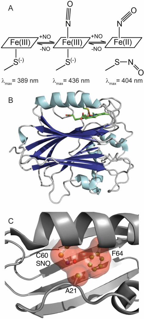
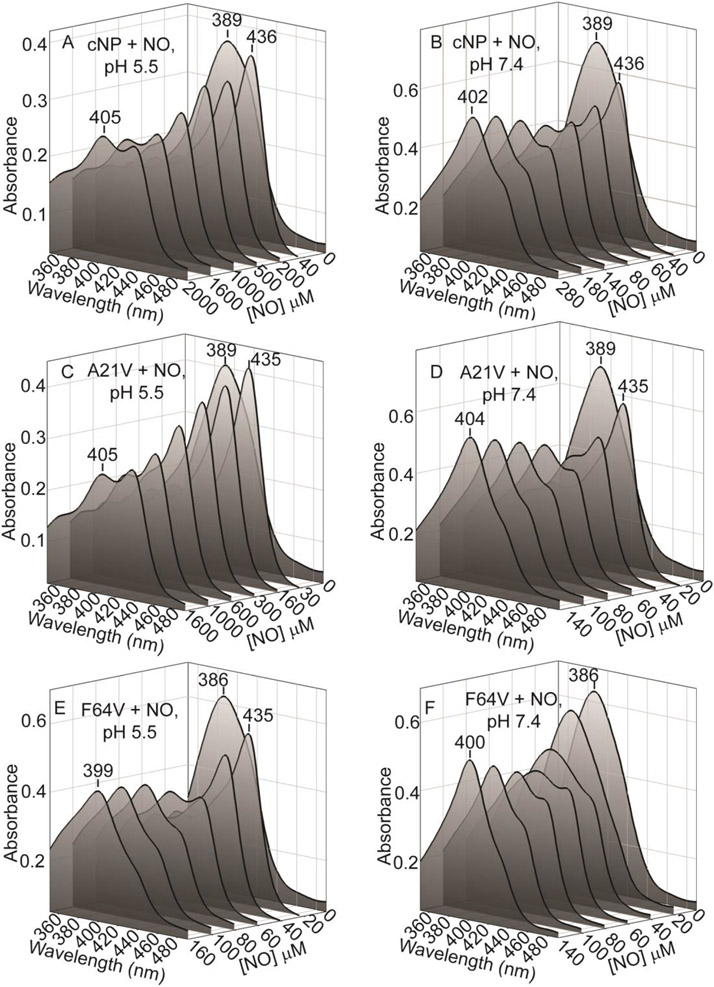
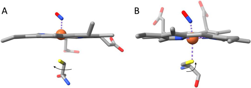
Nitrophorins are heme proteins used by blood feeding insects to deliver nitric oxide (NO) to a victim, leading to vasodilation and antiplatelet activity. Cimex lectularius (bedbug) nitrophorin (cNP) accomplishes this with a cysteine ligated ferric (Fe(III)) heme. In the acidic environment of the insect's salivary glands, NO binds tightly to cNP. During a blood meal, cNP-NO is delivered to the feeding site where dilution and increased pH lead to NO release. In a previous study, cNP was shown to not only bind heme, but to also nitrosate the proximal cysteine, leading to Cys-NO (SNO) formation. SNO formation requires oxidation of the proximal cysteine, which was proposed to be metal-assisted through accompanying reduction of ferric heme and formation of Fe(II)-NO. Here, we report the 1.6 Å crystal structure of cNP first chemically reduced and then exposed to NO, and show that Fe(II)-NO is formed but SNO is not, supporting a metal-assisted SNO formation mechanism. Crystallographic and spectroscopic studies of mutated cNP show that steric crowding of the proximal site inhibits SNO formation while a sterically relaxed proximal site enhances SNO formation, providing insight into specificity for this poorly understood modification. Experiments examining the pH dependence for NO implicate direct protonation of the proximal cysteine as the underlying mechanism. At lower pH, thiol heme ligation predominates, leading to a smaller trans effect and 60-fold enhanced NO affinity (Kd = 70 nM). Unexpectedly, we find that thiol formation interferes with SNO formation, suggesting cNP-SNO is unlikely to form in the insect salivary glands.

Keywords: Heme protein; Nitric oxide; Nitrophorin; Nitrosylation; S-nitrosocysteine.


Conflict of interest statement

Declaration of Competing Interest The authors declare that they have no known competing financial interests or personal relationships that could have appeared to influence the work reported in this paper.

Figures

Fig. 1.
Fig. 1.
Cartoons illustrating structure and NO binding mechanism for Cimex nitrophorin. (A) NO binding is initially to the ferric protein, yielding a six-coordinate nitrosyl complex. A second NO molecule binds at higher NO concentrations, yielding a five-coordinate ferrous nitrosyl complex and S-nitrosocysteine. All steps are reversible. Soret band absorption maxima are shown for each complex. (B) Overall fold for cNP (PDB entry 1YTF). (C) Contacts among Cys-NO, Ala 21 and Phe 64 in the proximal pocket.
Fig. 2.
Fig. 2.
Structures of ferrous cNP. Shown are the heme, Cys 60 and distal pocket solvent and ligand molecules, with electron density (2Fo-Fc coefficients, 1.5 σ). The propionate group attached to pyrrole ring A has been omitted for clarity. The nitrosyl complex has released the proximal thiol but is unmodified (no SNO). (A) cNP(II)-NO. (B) cNP(II)-CO. (C) cNP(II).
Fig. 3.
Fig. 3.
Electronic absorption spectra of cNP complexes in solution and crystal. Shown are spectra recorded in solution (25 °C, ~15 μM cNP) between 350 and 700 nm (thin lines), and in the crystal (100 K) between 450 and 700 nm (inset, thick lines). Normalized solution spectra are also included in the insets for reference (thin lines, ~40 μM cNP). Peaks labeled in the inset are for the crystal spectra. All spectra are recorded at pH 7.0 unless otherwise noted. Where cNP was chemically reduced, the UV regions of the spectra are obscured by the reducing agent, sodium dithionite. (A) cNP(III)-H2O. (B) Dithionite reduced cNP(II) at pH 7.0 (solid line), pH 8.0 (dotted line), pH 9.0 (dashed line), and at pH 7.0 in the crystal (inset, thick line). (C) cNP(II)-NO (dithionite reduced). (D) cNP(II)-NO/SNO (formed from cNP(III) by addition of two NO equivalents). (E) cNP(II)-CO (dithionite reduced). (F) cNP(III)-NO (formed with one NO equivalent).
Fig. 4.
Fig. 4.
Titration of cNP with NO at pH 5.5 and 7.4. (A) Change in absorption at 437 nm (pH 5.5), plotted against total NO concentration and fitted to a mutually depleting model (see methods), yielding Kd=70±30nM (n = 3). Inset: difference spectra. (B) Change in absorption at 437 nm (pH 7.4), plotted against the free NO concentration and fitted to a simple hyperbola, yielding Kd=4.4±1.7μM (n = 5). Inset: difference spectra.
Fig. 5.
Fig. 5.
NO titration with full spectra for wild type and mutant proteins. (A) cNP wild type, pH 5.5. The cNP(II)-NO/SNO species (405 nm) is ~50% at saturating NO concentrations. (B) cNP wild type, pH 7.4. The cNP(II)-NO/SNO Soret band (405 nm) is fully present at an NO concentration of 300 μM. (C) A21V, pH 5.5. The A21V(II)-NO/SNO species (405 nm) is ~50% at saturating NO concentrations. (D) A21V, pH 7.4. The A21V(II)-NO/SNO Soret band (405 nm) is fully present at an NO concentration of 140 μM. (E) F64V, pH 5.5. The F64V(II)-NO/SNO Soret band (399 nm) is fully present at an NO concentration of 160 μM (F) F64V, pH 7.4. The F64V(II)-NO/SNO Soret band (400 nm) is fully present at an NO concentration of 140 μM. Protein concentrations varied from 4-8 μM.
Figure 6.
Figure 6.
Structures of A21V and F64V mutant cNP proteins, highlighting nitrosyl heme and proximal SNO geometry. (A) A21V, (2Fo-Fc coefficients, 1.0 σ). (B) F64V, (2Fo-Fc coefficients, 1.2 σ). In both structures, the Fe-N-O group has geometry consistent with a ferrous nitrosyl complex (Fe-N-O bond angle ~124° and Fe-N bond length ~1.8 Å). (C) Cross-eyed stereo view of the proximal heme pocket of A21V (magenta) superposed on cNP wild type (green, PDB entry 1Y21). Val 21 is pointing away from the heme face and the Cys 60 SNO moiety is in the trans conformation. (D) Cross-eyed stereo view of the proximal heme pocket of F64V (purple) superposed on cNP wild type (green). Val 64 is in a similar position to that of Phe 64 and the Cys 60 SNO moiety is in the cis conformation, similarly to the wild type complex.
Fig. 7.
Fig. 7.
Structures for A21V and F64V SNO moieties along with schematic representation of S-nitroso cysteine. The S-N-O linkage has a partial double bond-character, leading to a planar SNO geometry and CB-S-N-O bond dihedral (χ3) of either 180° (trans) or 0° (cis). Optimal values for χ1 and χ2 are ~165° and ~−70°, respectively. (A) trans geometry. (B) cis geometry. (C) 2Fo-Fc electron density for A21V (1.2σ). (D) 2Fo-Fc electron density for F64V (1.2σ).
Fig. 8.
Fig. 8.
Comparison of proximal thiol orientation in cNP and NOS nitrosyl complexes. (A) cNP(II)-NO. The proximal cysteine sulfur is oriented such that it cannot rotate about the Cα-Cβ bond to follow heme iron as it moves to the distal pocket. (B) NOS (PDB entry 3HSP, [39]). The proximal cysteine sulfur can rotate toward the heme to retain coordination with heme iron.

References

    1. Walker FA. Nitric oxide interaction with insect nitrophorins and thoughts on the electron configuration of the {FeNO}6 complex. J. Inorg. Biochem 2005, 99:216–236. - PubMed
    1. Montfort WR, Weichsel A, Andersen JF. Nitrophorins and related antihemostatic lipocalins from Rhodnius prolixus and other blood-sucking arthropods. Biochim. Biophys. Acta 2000, 1482:110–118. - PubMed
    1. Andersen JF. Structure and mechanism in salivary proteins from blood-feeding arthropods. Toxicon. 2010, 56:1120–1129. - PMC - PubMed
    1. Maes EM, Roberts SA, Weichsel A, Montfort WR. Ultrahigh Resolution Structures of Nitrophorin 4: Heme Distortion in Ferrous CO and NO Complexes. Biochemistry. 2005, 44:12690–12699. - PubMed
    1. Kondrashov DA, Roberts SA, Weichsel A, Montfort WR. Protein functional cycle viewed at atomic resolution: conformational change and mobility in nitrophorin 4 as a function of pH and NO binding. Biochemistry. 2004, 43:13637–13647. - PubMed

Publication types

LinkOut - more resources