. 2023 Jul;64(7):1009-1016.
doi: 10.2967/jnumed.122.265199. Epub 2023 Jun 8.

Theranostics in Hematooncology

Affiliations

Theranostics in Hematooncology

Andreas K Buck et al. J Nucl Med. 2023 Jul.

Abstract

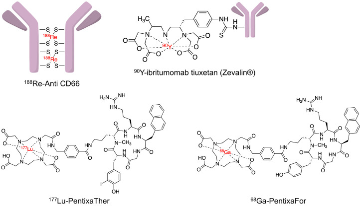
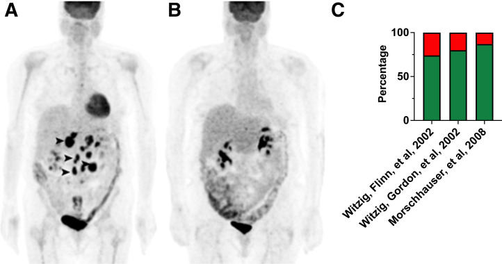
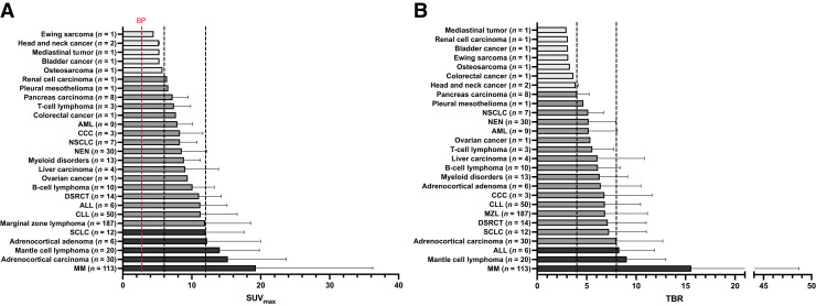
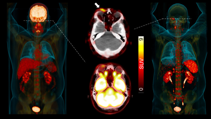
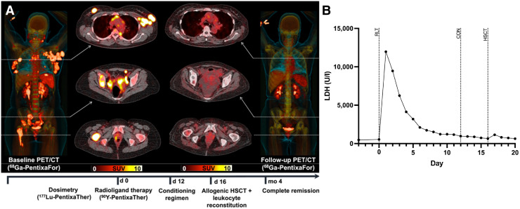
In the early 2000s, major clinical trials provided evidence of a favorable outcome from antibody-mediated radioimmunotherapy for hematologic neoplasms, which then led to Food and Drug Administration approval. For instance, the theranostic armamentarium for the referring hematooncologist now includes 90Y-ibritumomab tiuxetan for refractory low-grade follicular lymphoma or transformed B-cell non-Hodgkin lymphoma, as well as 131I-tositumomab for rituximab-refractory follicular lymphoma. Moreover, the first interim results of the SIERRA phase III trial reported beneficial effects from the use of 131I-anti-CD45 antibodies (Iomab-B) in refractory or relapsed acute myeloid leukemia. During the last decade, the concept of theranostics in hematooncology has been further expanded by C-X-C motif chemokine receptor 4-directed molecular imaging. Beyond improved detection rates of putative sites of disease, C-X-C motif chemokine receptor 4-directed PET/CT also selects candidates for radioligand therapy using β-emitting radioisotopes targeting the identical chemokine receptor on the lymphoma cell surface. Such image-piloted therapeutic strategies provided robust antilymphoma efficacy, along with desired eradication of the bone marrow niche, such as in patients with T- or B-cell lymphoma. As an integral part of the treatment plan, such radioligand therapy-mediated myeloablation also allows one to line up patients for stem cell transplantation, which leads to successful engraftment during the further treatment course. In this continuing education article, we provide an overview of the current advent of theranostics in hematooncology and highlight emerging clinical applications.

Keywords: C-X-C motif chemokine receptor 4; CXCR4; hematooncology; lymphoma; radioimmunotherapy; theranostics.


Figures

FIGURE 1.
FIGURE 1.
Structures of established (188Re-CD66 antibodies, 90Y-ibritumomab tiuxetan) and novel (68Ga-pentixafor, 90Y-pentixather) theranostic agents applied in hematooncology.
FIGURE 2.
FIGURE 2.
Patient with NHL treated with 90Y-ibritumomab tiuxetan. (A) Pretherapeutic maximum-intensity projection derived from 18F-FDG PET revealed multiple lymphoma manifestations in abdomen (arrowheads). (B) PR with inactive disease was achieved as visualized on 18F-FDG PET at 3-mo follow-up. (Modified from (67).) (C) Response rates of major clinical trials using 90Y-ibritumomab tiuxetan (6,22,25) (green indicates disease control depending on study’s definition; red indicates uncontrolled disease).
FIGURE 3.
FIGURE 3.
SUVmax (A) and target-to-background ratios (B) for 690 patients scanned with 68Ga-pentixafor PET/CT for assessment of in vivo CXCR4 extent. Black dashed lines show SUVmax of 6 and 12 and target-to-background ratio of 4 and 8, respectively. ALL = acute lymphoblastoid leukemia; BP = blood pool; CCC = cholangiocarcinoma; CLL = chronic lymphocytic leukemia; DSRCT = desmoplastic small round cell tumor; NEN = neuroendocrine neoplasm; NSCLC = non–small cell lung carcinoma; SCLC = small cell lung carcinoma; adrenocortical adenoma = aldosterone-producing adrenocortical adenoma. (Modified from (40).)
FIGURE 4.
FIGURE 4.
CXCR4-targeting 68Ga-pentixafor PET/CT for dissecting true relapse and autoimmune-mediated side effects in MM patient scheduled for B-cell maturation antigen–targeting CAR T-cell therapy (idecabtagene vicleucel). (A) Before CAR T-cell therapy, 18F-FDG showed osseous lesions (arrows). (B) On restaging 3 mo after CAR T-cell therapy, myeloma clearance in skeleton was observed on maximum-intensity projections of 18F-FDG and 68Ga-pentixafor PET. Only 18F-FDG, however, revealed uptake in pulmonary system; no such radiotracer accumulation was observed on CXCR4-directed imaging. Single-cell RNA sequencing on lung specimen demonstrated upregulation of Th17.1-positive T cells, which are associated with autoimmune diseases such as sarcoidosis. (C) Six months after CAR T-cell therapy, both imaging modalities showed novel manifestations (red box) suggestive of relapse. 68Ga-pentixafor PET–guided biopsy was conducted, and single-cell RNA sequencing then revealed malignant plasma cells along with increased CXCR4 expression (leftmost panel in C). CART = CAR T-cell therapy. (Modified from (15).)
FIGURE 5.
FIGURE 5.
MZL patient with additional periorbital disease site (white arrow) identified on 68Ga-pentixafor PET/CT. On 18F-FDG maximum-intensity projection (MIP, left) and transaxial PET/CT (middle, bottom), periorbital manifestation was masked by normal biodistribution in brain. On CXCR4-targeted 68Ga-pentixafor (MIP, right; transaxial PET/CT, middle top), this additional site of disease can be identified because of missing brain accumulation. (Modified from (53).)
FIGURE 6.
FIGURE 6.
Patient with MZL and scheduled for rituximab-bendamustine. (A and B) Maximum-intensity projection (A) of 68Ga-pentixafor PET revealed multiple lymphoma manifestations, in particular in axilla as seen on transaxial PET/CT (B). (C and D) After treatment, complete remission was achieved on follow-up imaging, indicating that CXCR4-targeted PET/CT may be useful to monitor treatment response. (Modified from (53).)
FIGURE 7.
FIGURE 7.
Patient with relapsed T-cell lymphoma treated with CXCR4-directed RLT and achieving complete remission. (A) Maximum-intensity projection and transaxial 68Ga-pentixafor PET/CT at baseline showed extensive disease in skeleton and lymph nodes. Four months after treatment, CR was noted on follow-up 68Ga-pentixafor PET/CT. (B) Lactate dehydrogenase as surrogate marker of antilymphoma efficacy peaked directly after RLT and then rapidly declined till conditioning regimen and HSCT, thereby suggesting direct antilymphoma effect caused by CXCR4 RLT. CON = conditioning regimen; LDH = lactate dehydrogenase. (Modified from (7).)
FIGURE 8.
FIGURE 8.
Synergistic effects of radioimmunotherapy and CXCR4-targeted RLT in patient with heavily pretreated diffuse large B-cell lymphoma. (A) Pretherapeutic scintigraphy 24 h after 177Lu-pentixafor injection revealed multiple disease sites, allowing for calculations of absorbed doses. (B) Baseline maximum-intensity projection and transaxial 68Ga-pentixafor PET/CT showed multiple CXCR4-expressing mediastinal and abdominal lesions. (C) 90Y-ibritumomab tiuxetan combined with 90Y-pentixather was initiated. On 18F-FDG PET/CT 4 mo later, sites of disease were smaller, indicating PR. (Modified from (63).)

References

    1. Sartor O, de Bono J, Chi KN, et al. . Lutetium-177-PSMA-617 for metastatic castration-resistant prostate cancer. N Engl J Med. 2021;385:1091–1103. - PMC - PubMed
    1. Hofman MS, Emmett L, Sandhu S, et al. . [177Lu]Lu-PSMA-617 versus cabazitaxel in patients with metastatic castration-resistant prostate cancer (TheraP): a randomised, open-label, phase 2 trial. Lancet. 2021;397:797–804. - PubMed
    1. Strosberg JR, Caplin ME, Kunz PL, et al. . 177Lu-Dotatate plus long-acting octreotide versus high-dose long-acting octreotide in patients with midgut neuroendocrine tumours (NETTER-1): final overall survival and long-term safety results from an open-label, randomised, controlled, phase 3 trial. Lancet Oncol. 2021;22:1752–1763. - PubMed
    1. Strosberg J, El-Haddad G, Wolin E, et al. . Phase 3 trial of 177Lu-Dotatate for midgut neuroendocrine tumors. N Engl J Med. 2017;376:125–135. - PMC - PubMed
    1. Kaminski MS, Tuck M, Estes J, et al. . 131I-tositumomab therapy as initial treatment for follicular lymphoma. N Engl J Med. 2005;352:441–449. - PubMed

Publication types

MeSH terms