. 2023 Jun 8;9(1):36.
doi: 10.1038/s41522-023-00401-8.

Combining SNAPs with antibiotics shows enhanced synergistic efficacy against S. aureus and P. aeruginosa biofilms

Affiliations

Combining SNAPs with antibiotics shows enhanced synergistic efficacy against S. aureus and P. aeruginosa biofilms

Ramón Garcia Maset et al. NPJ Biofilms Microbiomes. .

Erratum in

Abstract

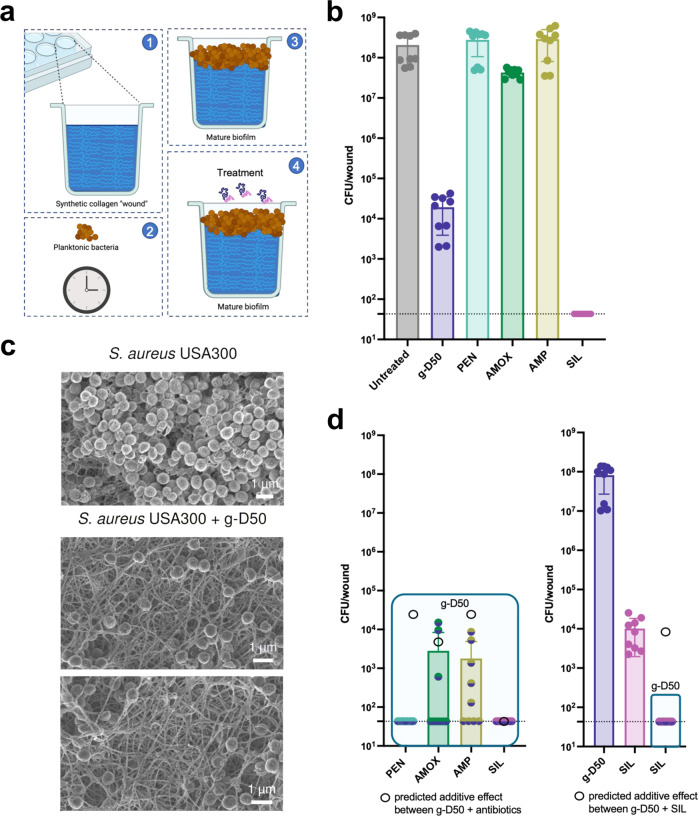
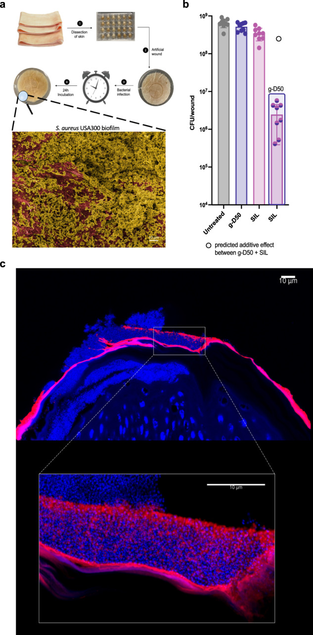
Biofilm infections are associated with a high mortality risk for patients. Antibiotics perform poorly against biofilm communities, so high doses and prolonged treatments are often used in clinical settings. We investigated the pairwise interactions of two synthetic nano-engineered antimicrobial polymers (SNAPs). The g-D50 copolymer was synergistic with penicillin and silver sulfadiazine against planktonic Staphylococcus aureus USA300 in synthetic wound fluid. Furthermore, the combination of g-D50 and silver sulfadiazine showed a potent synergistic antibiofilm activity against S. aureus USA300 using in vitro and ex vivo wound biofilm models. The a-T50 copolymer was synergistic with colistin against planktonic Pseudomonas aeruginosa in synthetic cystic fibrosis medium, and this pair showed a potent synergistic antibiofilm activity against P. aeruginosa in an ex vivo cystic fibrosis lung model. SNAPs thus have the potential for increased antibiofilm performance in combination with certain antibiotics to shorten prolonged treatments and reduce dosages against biofilm infection.


Conflict of interest statement

The authors declare no competing interests.

Figures

Fig. 1
Fig. 1. Synthesis and characterization of g-D50 and a-T50 copolymers.
a General procedure was used for the RAFT polymerisations of the diblock (g-D50) and triblock (a-T50) copolymers. The final degree of polymerization (DP = 50) was kept constant. For ease of reference, the diblock guanidinium copolymer was called g-D50 and the ammonium triblock a-T50. b SEC traces in DMF of the Boc-protected a-T50. Dots represent the first block, dashes the first block extension, and solid lines the second block extension. The polydispersity (Ð) of a-T50 is 1.15, and the molecular weight (Mn-SEC) is 7500 g mol−1. c 1H NMR spectra in DMSO-d6 of the consecutive block extensions to obtain the triblock copolymer a-T50. d SEC traces in DMF of the Boc-protected g-D50. The polydispersity (Ð) of g-D50 is 1.19, and the molecular weight (Mn-SEC) is 10400 g mol−1. e 1H NMR spectra in DMSO-d6 of the consecutive block extensions to obtain g-D50. f The Boc removal was confirmed by 1H NMR (the red box highlights the disappearance of the signal of the Boc groups). g HPLC chromatograms of the copolymers a-T50 and g-D50. h UV-vis transmittance at 633 nm for copolymers a-T50 and g-D50.
Fig. 2
Fig. 2. Checkerboard assay: pairwise interaction of copolymers and antibiotics.
a MIC and FIC values of g-D50 and different antibiotics determined against S. aureus USA300 in caMHB. b MIC and FICs of a-T50 and different antibiotics determined against P. aeruginosa PA14 in caMHB (PEN = penicillin, AMP = ampicillin, AMO = amoxicillin, SIL = silver sulfadiazine, VAN = vancomycin, ERY = erythromycin, CIP = ciprofloxacin, POL = polymyxin B, COL = colistin, TOB = tobramycin, PIP = piperacillin, TET = tetracycline). Three independent experiments (on three different days) were performed. The averages of the three independent experiments were used to calculate the FICs. c FIC values of the combination of g-D50 with PEN, AMOX, AMP and SIL against S. aureus USA300 in SWF and caMHB media. d FIC values of the combination of a-T50 with COL and POL against P. aeruginosa PA14 in SFCM and caMHB media. Source data are provided as Supplementary data file 1.
Fig. 3
Fig. 3. 3D synergy landscapes.
ac 3D synergy plot of g-D50 and silver sulfadiazine against S. aureus USA300 in caMHB using the Bliss, Loewe and ZIP models, respectively. df 3D synergy plot of a-T50 and colistin against P. aeruginosa PA14 in caMHB using the Bliss, Loewe, and ZIP models, respectively. The synergy profile was calculated using SynergyFinder. The colour gradient from blue to magenta shows the shift of drug interactions from antagonism to synergy score. Source data are provided as Supplementary data file 1.
Fig. 4
Fig. 4. Biofilm prevention assay against S. aureus USA300.
a Schematic describing the biofilm prevention assay in the soft tissue collagen wound model. b Effect of single treatments preventing the biofilm formation of S. aureus USA300 in the soft tissue wound model. CFU counts (circles) after 48 h of treatment with g-D50, penicillin (PEN), amoxicillin (AMOX), ampicillin (AMP), silver sulfadiazine (SIL) at 2x MIC against S. aureus USA300 in the soft tissue wound model. A complete killing was not observed for the individual treatments. A Dunnett’s test was performed to compare the CFU of the individual treatments with the CFU of the untreated controls. g-D50 (2x) and SIL (2x) were significantly different from the respective untreated controls (all p < 0.001***, Dunnett’s test). The data were collected from three independent experiments (conducted on different days), using three wounds per treatment. The error bar represents the standard deviation between the replicates. Source data are provided as Supplementary data file 1. c CFU counts after 48 h of treatment of g-D50 in combination with PEN, AMOX, AMP and SIL, respectively, at a final concentration of 2x MIC against S. aureus USA300 in the soft tissue wound model. The combinations of g-D50 and the respective antimicrobial compounds produced total killing and statistical tests were not performed. The horizontal dashed line represents the limit of detection by plating. d SEM images of mature S. aureus USA300 biofilm in the soft tissue collagen model. Images were processed with GIMP1-2 for false colouring. The collagen matrix image was obtained from an uninfected wound control, while the image with biofilm was obtained from an infected wound after 24 h. Blue was used to highlight the collagen matrix and yellow was used to highlight S. aureus USA300 biofilm.
Fig. 5
Fig. 5. Biofilm disruption assay against S. aureus USA300.
a Schematic representation of the biofilm formation and the following antibiofilm treatment in the collagen wound model. b CFU counts (circles) after 24 h of individual treatment of penicillin (PEN), amoxicillin (AMOX), ampicillin (AMP), silver sulfadiazine (SIL) and g-D50 at 10x MIC against mature S. aureus USA300 biofilms in the soft tissue wound model. The data were collected from three independent experiments (conducted on different days), each using three wounds per treatment. A Dunnett’s test was performed to compare the CFU of the combination treatments with the CFU of the untreated controls. Only g-D50 (10x MIC) was significantly different from the untreated controls (p < 0.001***, Dunnett’s test). SIL (10x MIC) caused complete biofilm eradication and was not included in the statistical analysis. The error bar represents the standard deviation between the replicates. c SEM images of (top) mature S. aureus USA300 biofilm in the soft tissue collagen model, and (bottom) treated with g-D50 at 10x MIC. d CFU counts (circles) after 24 h of combinatorial treatment of g-D50 (10x MIC) with PEN, AMOX, AMP and SIL at 10x MIC in mature S. aureus USA300 biofilms in the soft tissue wound model. The data were collected from three independent experiments (conducted on different days), each using three wounds per treatment. T-tests were performed to compare the predicted additive effect (empty black dots) with the CFU counts obtained by the drug combinations. The combination of g-D50 (10x MIC) with AMOX (10x MIC) showed no significant difference in comparison with the additive prediction (t8 = −0.634, p = 0.5438, t-test). The combination of g-D50 (10x MIC) with AMP (10x MIC) showed lower CFU counts in comparison with the additive prediction (t8 = −16.995, p < 0.001***, t-test), indicating a synergistic effect. The combination of g-D50 (10x MIC) with PEN (10x MIC) and SIL (10x MIC), respectively, caused a complete biofilm eradication, and no statistical tests were performed. In the case of g-D50 (10x MIC) in combination with SIL (10x MIC), a synergistic effect could not be assumed since the individual SIL (10x MIC) treatment caused complete biofilm eradication. The horizontal dashed line represents the limit of detection by plating. Individual treatments of silver sulfadiazine (SIL) and g-D50 at 5x MIC, and the combinatorial treatment of both compounds at a final concentration of 5x MIC in mature S. aureus USA300 biofilms in the soft tissue wound model. The combination of g-D50 with SIL (5x MIC) produced complete biofilm eradication and no statistical test was performed. Source data are provided as Supplementary data file 1.
Fig. 6
Fig. 6. Confocal images of S. aureus USA300 biofilm in the soft tissue wound model.
a Top surface view of S. aureus USA300 biofilm in the soft tissue wound collagen model stained with DAPI (blue), visualized by using confocal microscopy after 48 h of infection. b Top surface view of S. aureus USA300 biofilm in the soft tissue wound collagen model treated with Cy5-g-D50 (magenta) and stained with DAPI, visualized by using confocal microscopy after 48 h of infection. c Cross-section view of S. aureus USA300 biofilm in the soft tissue wound collagen model treated with Cy5-g-D50 and stained with DAPI, visualized by using confocal microscopy after 48 h of infection.
Fig. 7
Fig. 7. S. aureus USA300 biofilm in a porcine skin ex vivo model.
a Schematic diagram of the porcine skin wound model. Representative electron micrographs of the untreated S. aureus USA300 biofilm into the porcine wound model. The image was processed with GIMP1-2 for false colouring. Red was used to highlight the porcine skin and dark yellow was used to highlight the bacteria and biofilm matrix. b Biofilm disruption in the porcine ex vivo wound model. S. aureus USA300 biofilms were formed for 24 h and subsequently treated with g-D50 and SIL individually at 5x MIC and with the combination of the two drugs at 5x MIC final concentration for 24 h. The data were collected from three independent experiments (conducted on different days), each using three wounds per treatment. A Dunnett’s test was performed to compare the CFU of the individual and the combination treatment with the CFU of the untreated controls, and a significant difference between g-D50 in combination with SIL at 5x MIC and the untreated controls (p < 0.001***, Dunnett’s test) was found. The individual treatments of g-D50 (5x MIC) and SIL (5x MIC) were not significantly different from the untreated controls (p = 0.3857 and p = 0.0698, respectively). The additive effect of the combination of g-D50 (5x MIC) and silver sulfadiazine (5x MIC) was predicted by the sum of the CFU log reductions obtained for each single treatment (indicated with an empty black circle). A t-test was performed to compare the additive effect with the CFU counts obtained by the drug combination. The combination of g-D50 (5x MIC) with SIL (5x MIC) showed lower CFU counts in comparison with the additive prediction (t8 = −396.25, p < 0.001***, t-test), indicating a synergistic effect against biofilms. The error bar represents the standard deviation between the replicates. Source data are provided as Supplementary data file 1. c Representative fluorescent images of the cross-section of the porcine skin wound biofilm (S. aureus USA300 biofilm) treated with Cy5-g-D50 (128 μg mL−1) in red. DAPI was used to stained nucleic acid (bacterial and mammalian cells) shown in blue. The bottom image is a high magnification of the S. aureus USA300 cross-section of the porcine skin model.
Fig. 8
Fig. 8. P. aeruginosa PA14 biofilm in a pig lung ex vivo model.
a Schematic diagram of lung dissection for the EVPL model and representative electron micrograph of P. aeruginosa PA14 biofilm into pig lung pieces. The image was processed with GIMP1-2 for false colouring. Red was used to highlight the pig lung surface and green was used to highlight the bacteria and biofilm matrix. b Biofilm disruption in the EVPL model. P. aeruginosa PA14 biofilms were grown for 48 h and subsequently treated with a-T50 and COL individually at 32x MIC and with the combination of the two drugs at 32x MIC final concentration for 24 h. The data were collected from three independent experiments (conducted on 3 different days with 3 different lungs), including three lung pieces per treatment in each experiment. A Dunnett’s test was performed to compare the CFU of the individual treatments with the CFU of the controls. a-T50 (32x MIC) was not significantly different from the untreated control. COL (32x MIC) was significantly different to the untreated control (p = 0.00552**, Dunnett’s test). The combination of colistin and a-T50 showed the greatest difference with respect to the untreated control (p < 0.001***, Dunnett’s test). The additive effect of the combination of a-T50 (32x MIC) and colistin (32x MIC) was predicted by the sum of the CFU log reduction obtained for each single treatment (indicated with an empty black circle). Then, a t-test was performed to compare the additive effect with the CFU counts obtained by the drug combination. The combination of a-T50 (32x MIC) with COL (32x MIC) showed lower CFU counts in comparison with the additive prediction (t8 = −2321.9, p < 0.001***, t-test), indicating a synergistic effect against biofilm. The error bar represents the standard deviation between the replicates. Source data are provided as Supplementary data file 1.

References

    1. Holmes AH, et al. Understanding the mechanisms and drivers of antimicrobial resistance. Lancet. 2016;387:176–187. doi: 10.1016/S0140-6736(15)00473-0. - DOI - PubMed
    1. Hall-Stoodley L, Costerton JW, Stoodley P. Bacterial biofilms: from the natural environment to infectious diseases. Nat. Rev. Microbiol. 2004;2:95–108. doi: 10.1038/nrmicro821. - DOI - PubMed
    1. Ciofu O, Moser C, Jensen PO, Hoiby N. Tolerance and resistance of microbial biofilms. Nat. Rev. Microbiol. 2022;20:621–635. doi: 10.1038/s41579-022-00682-4. - DOI - PubMed
    1. Karygianni L, Ren Z, Koo H, Thurnheer T. Biofilm matrixome: extracellular components in structured microbial communities. Trends Microbiol. 2020;28:668–681. doi: 10.1016/j.tim.2020.03.016. - DOI - PubMed
    1. Jamal M, et al. Bacterial biofilm and associated infections. J. Chin. Med. Assoc. 2018;81:7–11. doi: 10.1016/j.jcma.2017.07.012. - DOI - PubMed

Publication types