. 2023 Sep;39(9):1785-1793.
doi: 10.1007/s10554-023-02888-w. Epub 2023 Jun 8.

Native T1 high region and left ventricular ejection fraction recovery in patients with dilated cardiomyopathy

Affiliations

Native T1 high region and left ventricular ejection fraction recovery in patients with dilated cardiomyopathy

Mayu Yazaki et al. Int J Cardiovasc Imaging. 2023 Sep.

Abstract

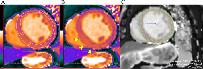
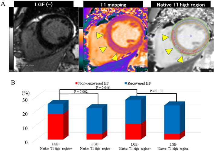
Native T1 mapping is used to assess myocardial tissue characteristics without gadolinium contrast agents. The focal T1 high-intensity region can indicate myocardial alterations. This study aimed to identify the association between the native T1 mapping including the native T1 high region and left ventricular ejection fraction (LVEF) recovery in patients with dilated cardiomyopathy (DCM). Patients with newly diagnosed DCM (LVEF of < 45%) who underwent cardiac magnetic resonance imaging with native T1 mapping were included in the analysis. Native T1 high region was defined as a signal intensity of > 5 SD in the remote myocardium. Recovered EF was defined as a follow-up LVEF of ≥ 45% and an LVEF increase of ≥ 10% after 2 years from baseline. Seventy-one patients met the inclusion criteria for this study. Forty-four patients (61.9%) achieved recovered EF. Logistic regression analysis showed that the native T1 value (OR: 0.98; 95% CI: 0.96-0.99; P = 0.014) and the native T1 high region (OR: 0.17; 95% CI: 0.05-0.55; P = 0.002), but not late gadolinium enhancement, were independent predictors of recovered EF. Compared with native T1 value alone, combined native T1 high region and native T1 value improved the area under the curve from 0.703 to 0.788 for predicting recovered EF. Myocardial damage, which was quantified using native T1 mapping and the native T1 high region were independently associated with recovered EF in patients with newly diagnosed DCM.

Keywords: Cardiac magnetic resonance; Dilated cardiomyopathy; Left ventricular ejection fraction recovery; T1 mapping.


Conflict of interest statement

The authors declare no competing interests.

Figures

Fig. 1
Fig. 1
Evaluation of native T1 values and native T1 high region. A Native T1 values were determined by drawing the region-of-interest in each segment of the six regions in the mid-left ventricle. B The native T1 high region (arrowheads). C A region-of-interest of 0.5−1 cm2 was manually drawn in the visually normal myocardium. The native T1 high region ratio was defined as a signal intensity of > 5 SD in the remote myocardium and was expressed as a percentage of the left ventricular area
Fig. 2
Fig. 2
Patient selection.  CMR cardiac magnetic resonance, EF ejection fraction
Fig. 3
Fig. 3
Native T1 high region and LGE.  A Representative case. The patient presented with the native T1 high regions (arrowheads) without LGE. B Native T1 high region and LGE proportions. EF ejection fraction, LGE late gadolinium enhancement
Fig. 4
Fig. 4
Kaplan–Meier curves of hospitalisation due to heart failure. Based on the KaplanMeier curves, patients with the non-recovered EF group had a significantly higher risk of hospitalisation due to heart failure than those with the recovered EF group. EF ejection fraction
Fig. 5
Fig. 5
Receiver-operating characteristic curve for predicting LVEF recovery. The receiver-operating characteristic curves were used to compare the discriminative power of predicting recovered left ventricular ejection fraction between the native T1 value and the native T1 high region

References

    1. Elliott P, Andersson B, Arbustini E, Bilinska Z, Cecchi F, Charron P, et al. Classification of the cardiomyopathies: a position statement from the european society of cardiology working group on myocardial and pericardial diseases. Eur Heart J. 2008;29(2):270–276. doi: 10.1093/eurheartj/ehm342. - DOI - PubMed
    1. Merlo M, Cannatá A, Vitagliano A, Zambon E, Lardieri G, Sinagra G. Clinical management of dilated cardiomyopathy: current knowledge and future perspectives. Expert Rev Cardiovasc Ther. 2016;14(2):137–140. doi: 10.1586/14779072.2016.1125292. - DOI - PubMed
    1. Wilcox JE, Fang JC, Margulies KB, Mann DL. Heart failure with recovered left ventricular ejection Fraction: JACC scientific expert panel. J Am Coll Cardiol. 2020;76(6):719–734. doi: 10.1016/j.jacc.2020.05.075. - DOI - PubMed
    1. Lupón J, Díez-López C, de Antonio M, Domingo M, Zamora E, Moliner P, et al. Recovered heart failure with reduced ejection fraction and outcomes: a prospective study. Eur J Heart Fail. 2017;19(12):1615–1623. doi: 10.1002/ejhf.824. - DOI - PubMed
    1. Gulati A, Jabbour A, Ismail TF, Guha K, Khwaja J, Raza S, et al. Association of fibrosis with mortality and sudden cardiac death in patients with nonischemic dilated cardiomyopathy. JAMA. 2013;309(9):896–908. doi: 10.1001/jama.2013.1363. - DOI - PubMed

LinkOut - more resources