. 2023 Aug;30(8):1119-1131.
doi: 10.1038/s41594-023-01006-7. Epub 2023 Jun 8.

Structure of nascent 5S RNPs at the crossroad between ribosome assembly and MDM2-p53 pathways

Affiliations

Structure of nascent 5S RNPs at the crossroad between ribosome assembly and MDM2-p53 pathways

Nestor Miguel Castillo Duque de Estrada et al. Nat Struct Mol Biol. 2023 Aug.

Abstract

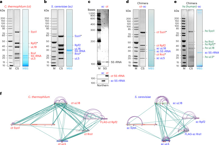
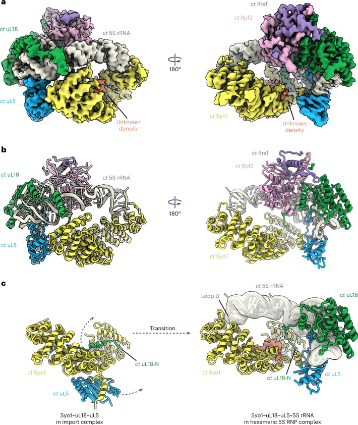
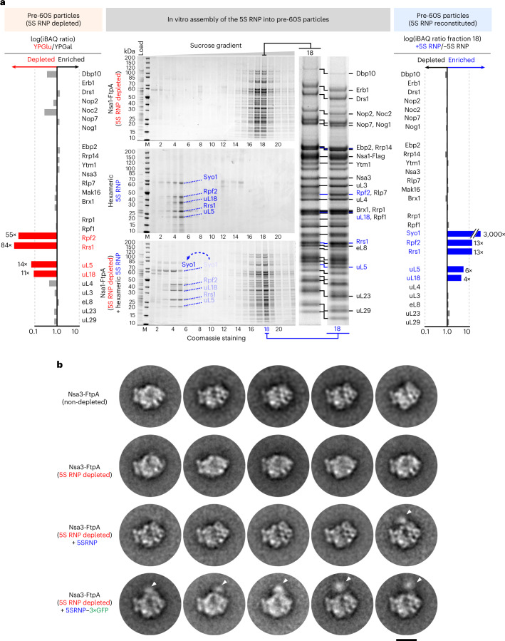
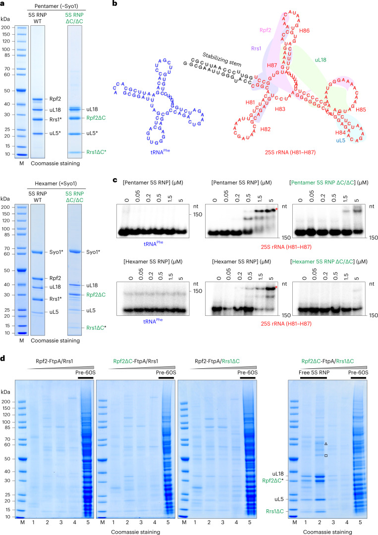
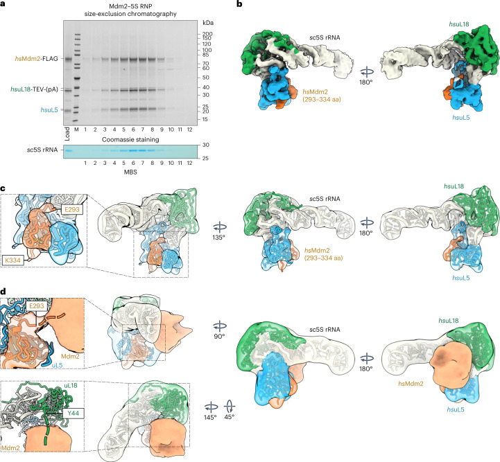
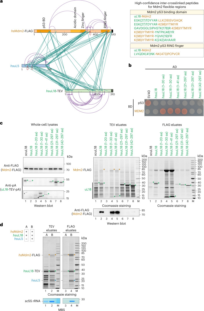
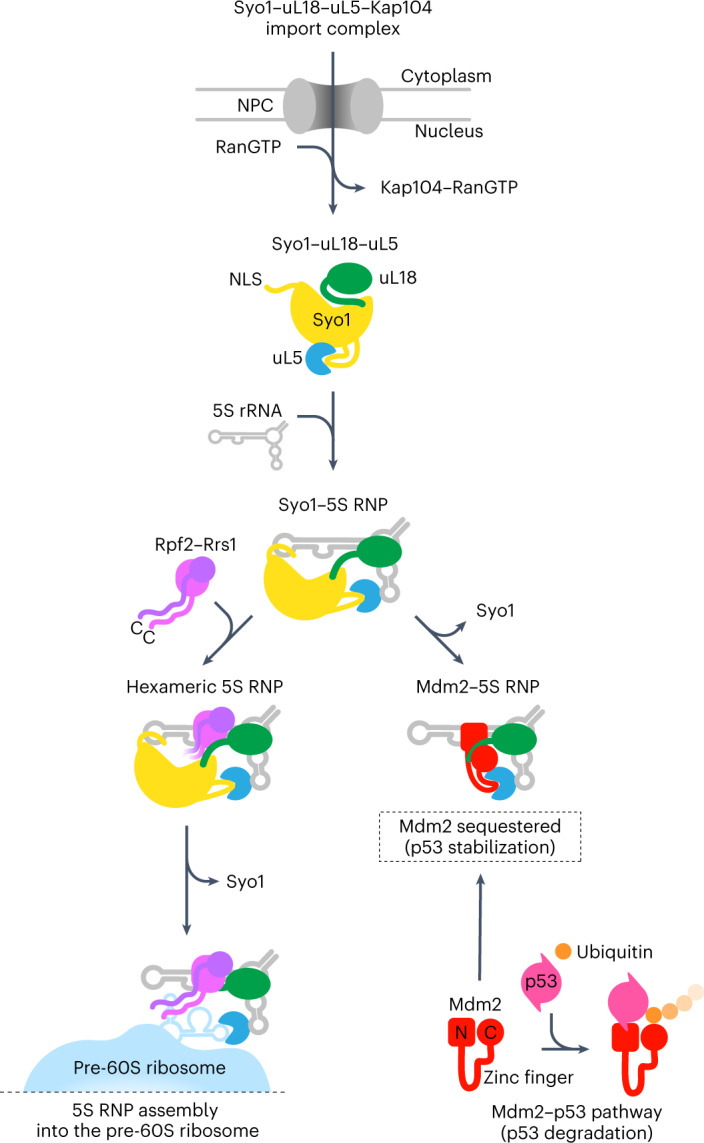
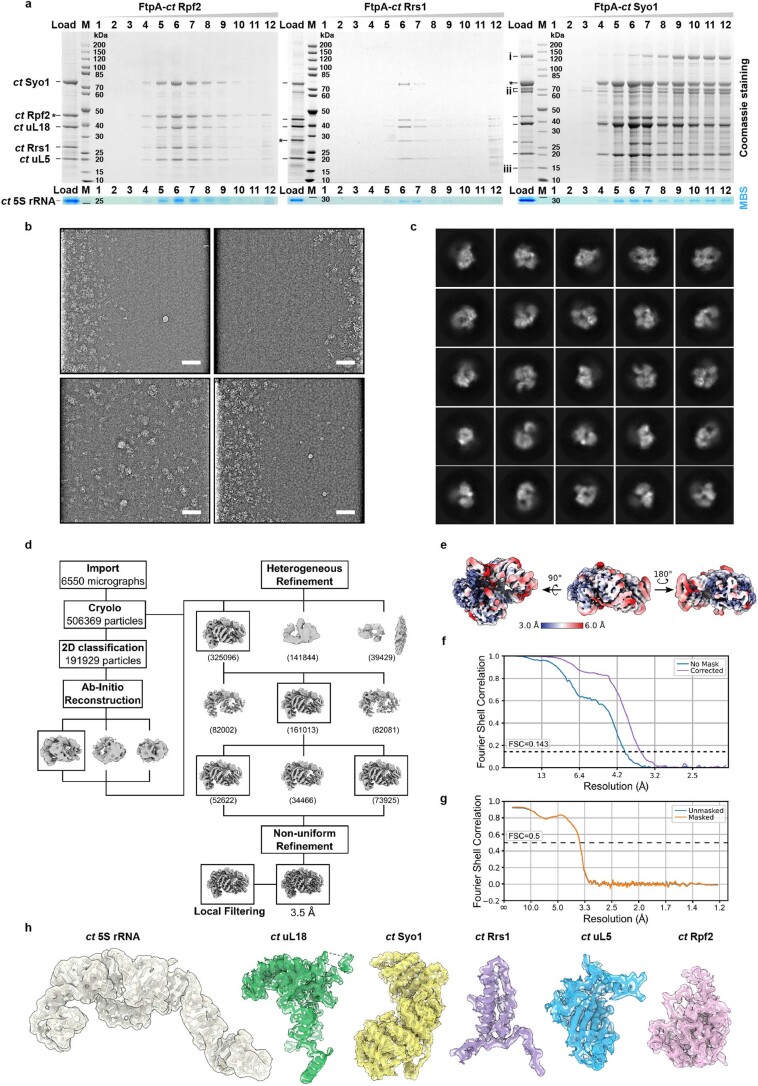
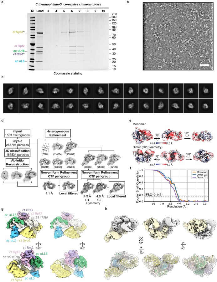
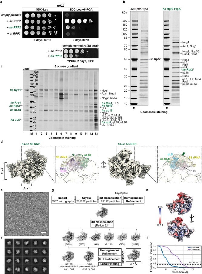
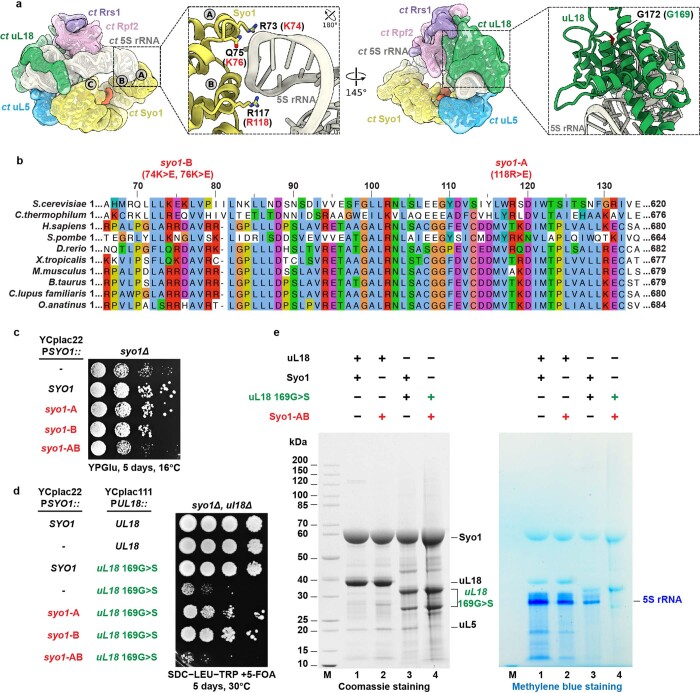
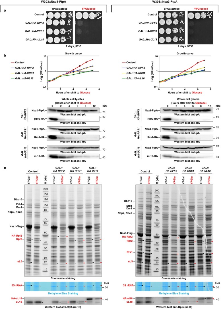
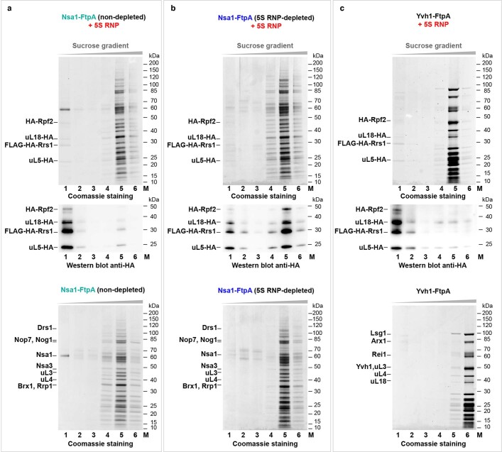
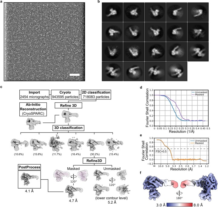
The 5S ribonucleoprotein (RNP) is assembled from its three components (5S rRNA, Rpl5/uL18 and Rpl11/uL5) before being incorporated into the pre-60S subunit. However, when ribosome synthesis is disturbed, a free 5S RNP can enter the MDM2-p53 pathway to regulate cell cycle and apoptotic signaling. Here we reconstitute and determine the cryo-electron microscopy structure of the conserved hexameric 5S RNP with fungal or human factors. This reveals how the nascent 5S rRNA associates with the initial nuclear import complex Syo1-uL18-uL5 and, upon further recruitment of the nucleolar factors Rpf2 and Rrs1, develops into the 5S RNP precursor that can assemble into the pre-ribosome. In addition, we elucidate the structure of another 5S RNP intermediate, carrying the human ubiquitin ligase Mdm2, which unravels how this enzyme can be sequestered from its target substrate p53. Our data provide molecular insight into how the 5S RNP can mediate between ribosome biogenesis and cell proliferation.


Conflict of interest statement

The authors declare no competing interests.

Figures

Fig. 1
Fig. 1. Isolation of hexameric 5S RNP complexes from Chaetomium thermophilum and Saccharomyces cerevisiae and reconstitution of thermophile–yeast and human–yeast 5S RNP chimeras.
a, Affinity purification of C. thermophilum (ct) 5S RNP via ctRpf2. b, Split-tag affinity purification of S. cerevisiae (sc) 5S RNP via scSyo1–scRrs1 pair. c, SYBR Green II staining (SG) to detect the total RNA and northern blot analysis (Northern) of ct and sc 5S rRNA extracted from the isolated 5S RNP complexes and probed with sc-specific and ct-specific 5S rRNA oligonucleotide probes. d, Split-tag affinity purification of the thermophile–yeast 5S RNP (ctsc chimera) via ctSyo1–ctRrs1 pair. e, Split-tag affinity purification of the human–yeast 5S RNP (hssc chimera) via hsuL5–hsRpf2 pair. The final eluates from a, b, d and e were analyzed by SDS–PAGE and Coomassie staining (CS). Labeled bands were identified by mass spectrometry or by methylene blue staining (MBS) to reveal the 5S rRNA. One caveat of SDS–PAGE/MBS staining is that the structured 5S RNA may not be fully denatured by SDS, causing different running behavior. To correctly analyze the 5S RNA, we also performed denaturing urea PAGE of the 5S RNP samples from sc and ct (see Fig. 1c). A protein molecular-weight marker standard (M) is shown on the left for the SDS–PAGE gels (a,b,d,e). An RNA molecular-weight standard (indicated in bases) is shown for the urea PAGE gel (c). Asterisks indicate the baits used for each affinity purification step. All purifications were done at least twice with a similar outcome. f, XL-MS of the affinity-purified 5S RNP from C. thermophilum (left) and S. cerevisiae (right) using DSS-H12. The protein primary structures of Syo1, uL18, uL5, Rpf2 and Rrs1 are shown, and specific regions are indicated in darker colors. Intermolecular crosslinks are shown in green, and intramolecular crosslinks are shown in purple. The xiNET tool was used for visualization. Source data
Fig. 2
Fig. 2. Cryo-EM structure of the conserved 5S RNP.
a,b, Cryo-EM map (a) and model (b) of the C. thermophilum 5S RNP (for purification, see Fig. 1a). The subunits of the 5S RNP are shown in different colors and labeled. c, Rearrangement of Syo1, uL5 and uL18-N before (left; PDB ID 5AFF) and after incorporation into the 5S RNP complex.
Fig. 3
Fig. 3. In vitro assembly of the yeast hexameric 5S RNP into early nucleolar pre-60S particles depleted of endogenous 5S RNP.
a, Left: SQ-MS analysis of the pre-60S particles depleted for the 5S RNP and used for the in vitro assembly (affinity purified via Nsa1-FtpA, derived from the GAL-HA-RPF2 yeast strain) compared with the same 5S RNP-non-depleted particles (Extended Data Fig. 5 and Source Data Excel file). Middle: in vitro assembly of the reconstituted yeast hexameric 5S RNP into the pre-60S particles depleted of 5S RNP. Samples were subjected to sucrose gradient centrifugation, fractioned and analyzed by SDS–PAGE. Labeled bands were identified by mass spectrometry. Right: SQ-MS analysis of pre-60S particles after binding of the 5S RNP compared with the non-reconstituted particles (fraction 18 of the sucrose gradients) (Source Data Excel file). Because Syo1 is absent in the Nsa1-FtpA sample, small amounts of Syo1 detected in the reconstituted sample (+5S RNP) may explain the high enrichment factor, which accordingly should be interpreted with caution. The iBAQ values were normalized to Erb1 in both SQ-MS analyses (left and right). The in vitro binding assay was repeated at least ten times with consistent results. b, Negative-stain EM of the pre-60S particles before and after 5S RNP depletion and after 5S RNP reconstitution. The assay was performed with 5S RNP-depleted pre-60S particles (affinity purified via Nsa3-FtpA from the GAL-HA-RPF2 strain) and 5S RNP containing uL18 untagged or tagged with three GFP moieties (3×GFP). The portions of the sucrose gradient with high molecular weight were analyzed by negative-stain EM, from which 2D class averages showed pre-60S alterations in the 5S RNP region. Specifically, pre-60S particles reconstituted with the 5S RNP–3×GFP exhibited an extra density (indicated by white arrows) that corresponds to the central protuberance of the pre-60S. Scale bar, 20 nm. For the entire 2D classes dataset, see Supplementary Information.
Fig. 4
Fig. 4. Rpf2–Rrs1 C-terminal extensions are required for 5S RNP assembly into pre-60S ribosomes.
a, SDS–PAGE analysis of the yeast 5S RNP complexes, Syo1 lacking (top) or Syo1 containing (bottom), with either wild-type (WT) or truncated (ΔC/ΔC) Rpf2/Rrs1 factors, used for the EMSA. Asterisks indicate bait proteins used for the split-tag affinity purification. The purifications of these 5S RNP complexes were done more than three times with consistent results. b, Folding prediction, calculated by RNAfold, of the yeast 25S rRNA fragment H81–H87 (red, right) and tRNAPhe (blue, left) used for the EMSA with the 5S RNP complexes. The colored areas illustrate the contacts of the 5S RNP proteins to the 25S rRNA helixes (labeled from H81 to H87) after binding to pre-60S ribosome (PDB ID 3JCT). c, EMSA radiographs showing the specific band shift (asterisks) of the radiolabeled 25S rRNA fragment upon binding to increasing amounts of the indicated 5S RNP complexes. nt, nucleotides. All of the EMSA assays were done twice with a similar outcome. d, SDS–PAGE analysis of sucrose gradient fractions from in vivo 5S RNP assembly into pre-60S ribosomes in yeast cells. 5S RNP incorporation into pre-60S particles was monitored upon affinity purification using Rpf2 as bait, from yeast wild-type, rpf2∆C and rrs1∆C single mutants, and rpf2∆C rrs1∆C double mutants. The typical protein pattern of pre-60S ribosomes is visible in the fractions with high molecular weight (lane 5), whereas the free 5S RNP (unbound) is visible in the fractions with low molecular weight (lanes 1 and 2). The bands corresponding to the 5S RNP factors were identified by mass spectrometry and are labeled. Co-precipitation of Fpr3 (triangle) and Fpr4 (square) with the free 5S RNP was also detected. This in vivo binding experiment was done at least twice with a similar outcome. Source data
Fig. 5
Fig. 5. Reconstitution and cryo-EM structure of the Mdm2–5S RNP complex.
a, Split-tag affinity purification of the reconstituted Mdm2–5S RNP complex (hsMdm2–hsuL18–hsuL5–sc5S rRNA) using hsuL18-TEV-ProtA as first bait and hsMdm2-FLAG as second bait, followed by size-exclusion chromatography. The final eluate (Load) and fractions 1–12 from the gel-filtration column were analyzed by SDS–PAGE. Labeled bands were identified by mass spectrometry. The gel was also stained with MBS to reveal the 5S rRNA. The purification of the Mdm2–5S RNP complex was performed more than five times with similar outcomes. b,c, Cryo-EM map (b) and fitted model (c) of the Mdm2–5S RNP complex (PDB IDs 4XXB for Mdm2, 6ZM7 for hsuL5 and hsuL18, and 3JCT for sc5S rRNA). aa, amino acids. The components of the complex are shown in different colors and labeled. d, Model and Gaussian filtered map of the Mdm2–5S RNP complex refined without mask and at lower contour levels revealing additional Mdm2 electron density. The appearing flexible density (unresolved parts of Mdm2) contacts the N-terminal residue E293 of the Mdm2 zinc finger domain, as well as the uL18 N-terminal residue Y44 (expansions, left). The connections of the Mdm2 zinc finger and the unresolved N terminus of uL18 are indicated with dashed lines. Source data
Fig. 6
Fig. 6. The human uL18 N-terminal sequence specifically binds to the Mdm2 N domain.
a, Left: XL-MS of the purified Mdm2–5S RNP complex, using DSS-H12. All inter-crosslinks and self-crosslinks are depicted. The xiNET tool was used for visualization of the crosslinks and the primary structure of the proteins. The domain organization of Mdm2 is also displayed. Right: manually curated list of the high-confidence inter-crosslinked peptides for the flexible regions of Mdm2 unresolved by cryo-EM. b, Yeast two-hybrid interaction between the indicated hsuL18 constructs and hsMdm2. AD, activation domain; BD, binding domain. The yeast two-hybrid assay was performed twice with a similar outcome. c, Sequence-specific binding of hsuL18 to hsMdm2, analyzed by co-expression and pull-down assays in yeast cells. GAL-induced co-expression of hsuL18 N-terminal constructs (fused at the C terminus to TEV-ProtA) and hsMdm2-FLAG, followed by IgG Sepharose chromatography and TEV cleavage (TEV eluates). Total lysates (left) were analyzed by western blotting for hsuL18-TEV-ProtA and Mdm2-FLAG using anti-ProtA and anti-FLAG antibodies, respectively. The TEV eluates were further affinity purified on FLAG beads to enrich for Mdm2-FLAG. Both TEV (middle) and FLAG (right) elutes were analyzed by SDS–PAGE. Mdm2 and uL18 bands are indicated by orange and green asterisks, respectively. The FLAG-labeled Mdm2 bands in lanes 2, 4 and 5 (orange asterisks) of the Coomassie-stained gel (TEV eluates) were also verified by mass spectrometry. This co-immunoprecipitation assay was performed twice with similar outcomes. d, Cooperative binding of hsuL5 and hsuL18 to Mdm2, analyzed by co-expression and pull-down assays in yeast cells. Sample A corresponds to the co-expression of hsuL18 and hsMdm2, whereas in sample B, hsuL5 was added to the in vivo co-expression system. Tandem affinity purifications from the cell lysates were performed by pulling down hsuL18-TEV-ProtA (TEV eluates) in the first step and hsMdm2-FLAG (FLAG eluates) in the second step. Eluates were analyzed by SDS–PAGE and Coomassie staining or methylene blue staining. Mdm2, uL18 and uL5 bands are indicated by orange, green and blue asterisks, respectively. This co-immunoprecipitation assay was performed twice with similar outcomes. Source data
Fig. 7
Fig. 7. Model of the formation of 5S RNP that assembles into nascent 60S subunits or signals the MDM2–p53 pathway.
After Syo1-mediated nuclear import of uL5–uL18, the 5S RNP assembles upon binding to the nascent 5S rRNA. Under normal circumstances (for example, cell proliferation), the 5S RNP is targeted to the ribosome biogenesis pathway by the binding of Rpf2–Rrs1, thereby forming the conserved hexameric 5S RNP that eventually integrates into pre-60S particles. Under nucleolar stress (for example, impaired ribosome biogenesis), the 5S RNP instead accumulates in a free pool that can sequester the ubiquitin ligase Mdm2, which in consequence stabilizes p53 levels that can eventually cause cell cycle arrest and apoptosis (see Discussion). NPC, nuclear pore complex.
Extended Data Fig. 1
Extended Data Fig. 1. Purification and cryo-EM analysis of the hexameric 5S RNP complex isolated directly from Chaetomium thermophilum.
a, Affinity purification of FLAG-TEV-ProtA (FtpA)-tagged ctRpf2 (left), ctRrs1 (middle) and ctSyo1 (right) from C. thermophilum. The final FLAG eluates (Load) were separated by sucrose gradient centrifugation, and fractions were analyzed by SDS-PAGE. Bait proteins are labeled with asterisks. M: Molecular weight marker. The hexameric Syo1–uL18–uL5–Rpf2–Rrs1-5S rRNA pulled down by these different baits is typically recovered in fractions 6-7. A simpler Syo1–uL18–uL5-5S rRNA complex is also detected in fractions 4-5 of the FtpA-ctSyo1 purification. Presence of 5S rRNA was identified by methylene blue staining (MBS). The labeled proteins were identified by mass spectrometry: ctSyo1 (CTHT_0033460), ctRpf2 (CTHT_0061110), ctRrs1 (CTHT_0057260), ctuL18 (CTHT_0063640), and ctuL5 (CTHT_0072970); additional proteins in the FtpA-ctSyo1 purification were identified as (i) CTHT_0061210, (ii) CTHT_ 0056370 (Fpr4 homologue) and (iii) CTHT_0025560. These purifications have been done at least 3 times with similar outcome. b,c, Cryo-EM representative micrographs of the ct 5S RNP low-pass filtered to 20 Å, displayed with inverted contrast (out of 6550 micrographs used for processing) (b) and selected 2D classification averages (c). Scale bar: 50 nm. d, Cryo-EM processing scheme. The overall resolution of the final 3D reconstruction was 3.5 Å. e, Final 3D reconstruction of the ct 5S RNP filtered and colored according to local resolution. f, Fourier shell correlation (FSC) curves of the final structure showing unmasked (No mask) and masked (Corrected) FSC curves. g, Model-to-map FSC curve. h, Segmented cryo-EM densities and models of the individual subunits of the ct 5S RNP. Source data
Extended Data Fig. 2
Extended Data Fig. 2. Purification, cryo-EM analysis and structure of the ct–sc 5S RNP chimera.
a, Split-tag affinity purification of the ct–sc 5S RNP chimera from yeast cells via ProtA-TEV-ctSyo1 and FLAG-ctRrs1 under conditions of ctRpf2 co-expression. The final FLAG eluate (Load) was separated by sucrose gradient centrifugation, and fractions were analyzed by SDS-PAGE. Bait proteins are labeled with asterisks. M: Molecular weight marker. Labeled bands were identified by MS. This purification has been done at least 5 times with similar results. b, c, Cryo-EM representative micrograph of the ct–sc 5S RNP chimera low-pass filtered to 20 Å, displayed with inverted contrast (out of 1583 micrographs used for processing) (b) and selected 2D classification averages (c). d, Cryo-EM processing scheme. The overall resolution of the final 3D reconstructions was 4.1 Å for the monomeric 5S RNP and 4.3 Å/4.0 Å for the dimeric 5S RNP without/with application of C2 symmetry. e, Final 3D reconstructions filtered and colored according to local resolutions. f, FSC curves of the final 3D reconstructions (masked). g, h, Cryo-EM densities and fitted models of the ct–sc 5S RNP assembled with C. thermophilum and S. cerevisiae factors for the monomeric (g) and dimeric (h) forms. 5S RNP factors are labeled in (g).
Extended Data Fig. 3
Extended Data Fig. 3. Cryo-EM analysis of the human–yeast 5S RNP chimera within yeast pre-60S particles.
a, Growth analysis of the nonviable yeast rpf2Δ strain complemented by hsRpf2 but not ctRpf2 on 5-FOA plates (upper panel) and YPGlu-plates (lower panel). To test for complementation, yeast rpf2Δ or rrs1Δ strains carrying RPF2 or RRS1 in a URA3-containing plasmid were transformed with plasmids carrying scRPF2, hsRPF2, ctRPF2, scRRS1, hsRRS1 or ctRRS1, and grown on selective SDC-Leu or SDC-Leu + 5-FOA plates for 3 or 5 days at 30 °C. b, Affinity purification of yeast Rpf2-FtpA or human Rpf2-FtpA from the complemented yeast rpf2Δ strain (growth analysis is shown in panel a), yielding pre-60S particles with a similar composition. Bait proteins are labeled with asterisks. Purifications have been done twice with similar results. c, Sucrose gradient centrifugation of the final eluate from the hs–sc 5S RNP chimera obtained from yeast co-overexpressing hsuL18, hsRrs1 and hsSyo1 and baits hsuL5 and hsRpf2 (labeled with asterisks). The free 5S RNP was detected in fraction 3 and pre-60S particles containing the human 5S RNP factors in fraction 13. Labeled bands were identified by mass spectrometry. The minor band above hsuL18 is hsuL18 with small amounts of scuL18. The two intense bands in fraction 6 (□) were human keratin contaminants. This purification has been done twice with similar results. d, Cryo-EM structure of the yeast pre-60S particle carrying hsRpf2, hsRrs1, hsuL18, and hsuL5 (panel C, fraction 13), which, together with the sc 5S rRNA, are shown in different colors. e, f, Cryo-EM micrograph of human 5S RNP assembled into yeast pre-60S, low-pass filtered to 20 Å and displayed with inverted contrast (out of 3937 micrographs used for processing) (e) and selected 2D classification averages (f). Scale bar: 50 nm. g, Cryo-EM processing scheme. The overall resolution was 3.7 Å. h, Final 3D reconstruction filtered and colored according to local resolution. i, Fourier shell correlation curves of showing unmasked (No mask) and masked (Corrected) FSC curves.
Extended Data Fig. 4
Extended Data Fig. 4. Structure-based mutations in Syo1 mapping at the contact site to 5S rRNA helix IV.
a, Overview and zoomed-in view of the ct 5S RNP cryo-EM structure showing contact of Syo1 N-terminal end to the 5S rRNA tip in the region of helix IV. Sites A and B correspond to regions in the Syo1 N-terminal domain with direct contact to the 5S rRNA. The residues of sites A and B for ctSyo1 are labeled in black and the corresponding residues in yeast Syo1 in red. Site C corresponds to a region in C-terminal domain of Syo1 that is also in close proximity to the 5S rRNA. Highlighted is also residue G169 of S. cerevisiae uL18 (G172 in C. thermophilum), which upon mutation yields a slow-growing yeast mutant (see also panel D). b, Multiple sequence alignment of the Syo1 N-terminal region, indicating the positively charged amino acids at the 5S rRNA contact sites: K74 and K76 (termed syo1-B) and R118 (termed syo1-A). These sites were mutated to glutamic acids. c, Dot–spot growth analysis of the syo1∆ strain transformed with plasmid-borne SYO1, syo1-B (74K>E, 76K>E), syo1-A (118R>E), and the combination syo1-AB (74K>E, 76K>E, 118R>E). Growth analyses have been performed twice with a similar outcome. d, Synthetic lethality relationship between syo1-AB and uL18 169G>S mutant alleles. The yeast syo1∆ uL18∆ double-disruption strain (carrying wild type uL18 on pRS316-URA3) was transformed with plasmids carrying either uL18 or uL18 169G>S in combination with plasmids carrying either empty (-), SYO1 or the indicated syo1 mutant alleles. This genetic experiment has been repeated twice with a similar outcome. e, Split-tag affinity purification of overexpressed Syo1 or Syo1-AB mutant (both ProtA-TEV-tagged) in combination with uL18 or uL18 169G>S (both FLAG-tagged). The final eluate was analyzed by SDS-PAGE and Coomassie staining to reveal Syo1 and uL18 (left panel), and methylene blue staining to visualize the 5S rRNA (right panel). The uL18 169G>S mutant protein is partially degraded, yielding two smaller breakdown products verified by mass spectrometry. This co-immunoprecipitation assay has been performed twice with a similar outcome. Source data
Extended Data Fig. 5
Extended Data Fig. 5. In vivo depletion of the yeast 5S RNP from pre-60S particles by GAL promoter-driven repression of either GAL::HA-RPF2, GAL::HA-RRS1 or GAL::HA-uL18.
a, b, Yeast strains (W303) carrying genomically integrated FLAG-TEV-ProtA tag fused at the C-terminus of either NSA1 (left) or NSA3 (right), each under control of the endogenous promoter. Dot–spot (a) and liquid culture (b) growth analysis of the indicated yeast strains on galactose (inducing) and glucose (non-inducing) containing medium. Samples were collected at the indicated hours and whole lysates were analyzed by western blot (lower panel) to follow the depletion of the indicated gene constructs. The growth curves show the mean ± STD of 3 replicas per culture. c, SDS-PAGE analysis of pre-60S particles isolated via tandem affinity purification based FtpA-tagged bait constructs (left: Nsa1-FtpA; right, Nsa3-FtpA) of the corresponding yeast strains, grown after 6 hours in either glucose or galactose containing medium. Black arrows point to the 5S RNP factors, and red arrows to the verified depletion (analyzed by mass spectrometry) of these factors. Labeled bands were identified by mass spectrometry. Staining with methylene blue revealed the 5S rRNA, and uL18 was detected by Western blot. Total YPGal and YPGlu eluate samples marked with asterisks were analyzed by SQ-MS. (Source Dataset 2; see also Fig. 3a, left panel). These depletion experiments have been done at least twice for all the groups with consistent results, and the Rpf2-depletion was further used as routine for the production of pre-60S particles depleted of 5S RNP. Source data
Extended Data Fig. 6
Extended Data Fig. 6. Yeast 5S RNP in vitro binding reconstitution into pre-60S particles using epitope-labeled subunits.
ac, The yeast hexameric 5S RNP was incubated with Nsa1-FtpA pre-60S particles, either non-depleted (a) or 5S RNP-depleted via GAL::HA-RPF2 (b), and as a further control, with Yvh1-FtpA-derived pre-60S particles (c). After sucrose gradient centrifugation of the assay mixtures, all fractions (fractions 1–6) were analyzed by SDS-PAGE (upper panel), and western blotting using the anti-HA antibody (middle panel). The bands corresponding to HA-Rpf2, uL18-HA, FLAG-HA-Rrs1, and uL5-HA are indicated. The different Nsa1-FtpA and Yvh1-FtpA pre-60S particles without addition of 5S RNP were also analyzed by sucrose gradient centrifugation (lower panels). M: Molecular weight marker. This in vitro binding assay has been done at least twice with a similar outcome.
Extended Data Fig. 7
Extended Data Fig. 7. Cryo-EM analysis of the Mdm2-5S RNP complex.
a,b, Representative micrograph of the Mdm2-5S RNP assembly low-pass filtered to 20 Å, displayed with inverted contrast (out of 2454 micrographs used for processing) (a) and selected 2D classification averages (b). Scale bar: 50 nm. c, Cryo-EM processing scheme of the Mdm2-5S RNP. The overall resolution of the final 3D reconstruction was 4.1 Å. d, FSC curves of the final 3D reconstructions showing masked and unmasked FSC curves. e, Model-to-map FSC curves. f, Final 3D reconstruction filtered and colored according to local resolution.

References

    1. Woolford JLJr, Baserga SJ. Ribosome biogenesis in the yeast Saccharomyces cerevisiae. Genetics. 2013;195:643–681. doi: 10.1534/genetics.113.153197. - DOI - PMC - PubMed
    1. Bohnsack KE, Bohnsack MT. Uncovering the assembly pathway of human ribosomes and its emerging links to disease. EMBO J. 2019;38:e100278. doi: 10.15252/embj.2018100278. - DOI - PMC - PubMed
    1. Ban N, et al. A new system for naming ribosomal proteins. Curr. Opin. Struct. Biol. 2014;24:165–169. doi: 10.1016/j.sbi.2014.01.002. - DOI - PMC - PubMed
    1. Sloan KE, Bohnsack MT, Watkins NJ. The 5S RNP couples p53 homeostasis to ribosome biogenesis and nucleolar stress. Cell Rep. 2013;5:237–247. doi: 10.1016/j.celrep.2013.08.049. - DOI - PMC - PubMed
    1. Ciganda M, Williams N. Eukaryotic 5S rRNA biogenesis. Wiley Interdiscip. Rev. RNA. 2011;2:523–533. doi: 10.1002/wrna.74. - DOI - PMC - PubMed

Publication types

MeSH terms