. 2023 Sep;7(9):1097-1112.
doi: 10.1038/s41551-023-01050-0. Epub 2023 Jun 8.

Resolving sepsis-induced immunoparalysis via trained immunity by targeting interleukin-4 to myeloid cells

Affiliations

Resolving sepsis-induced immunoparalysis via trained immunity by targeting interleukin-4 to myeloid cells

David P Schrijver et al. Nat Biomed Eng. 2023 Sep.

Abstract

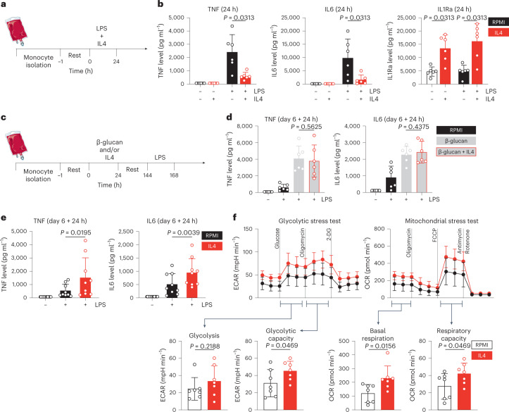
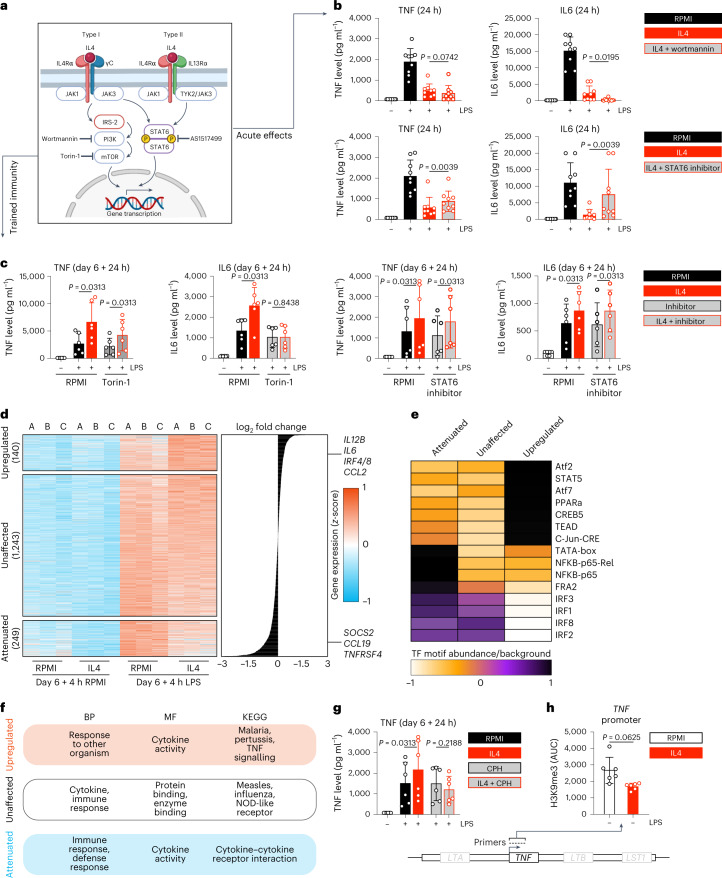
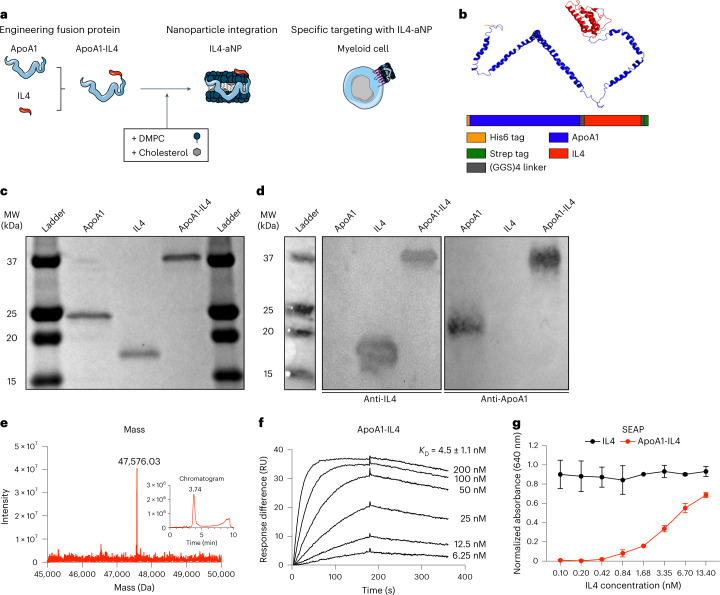
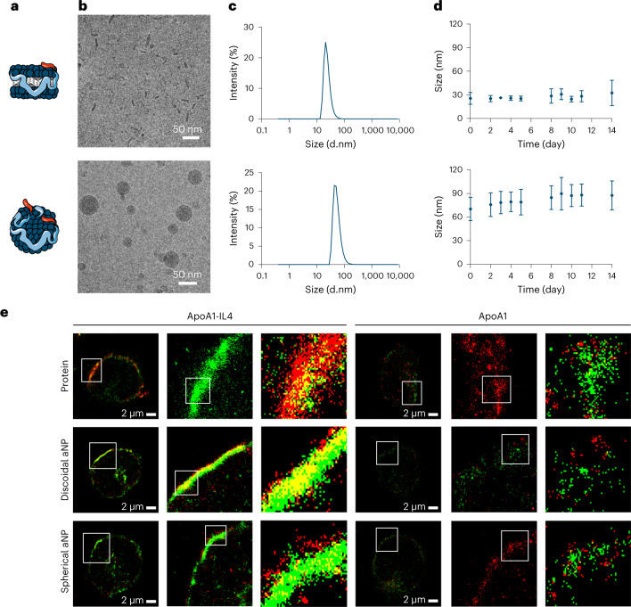
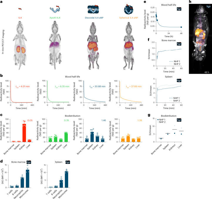
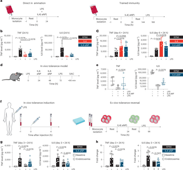
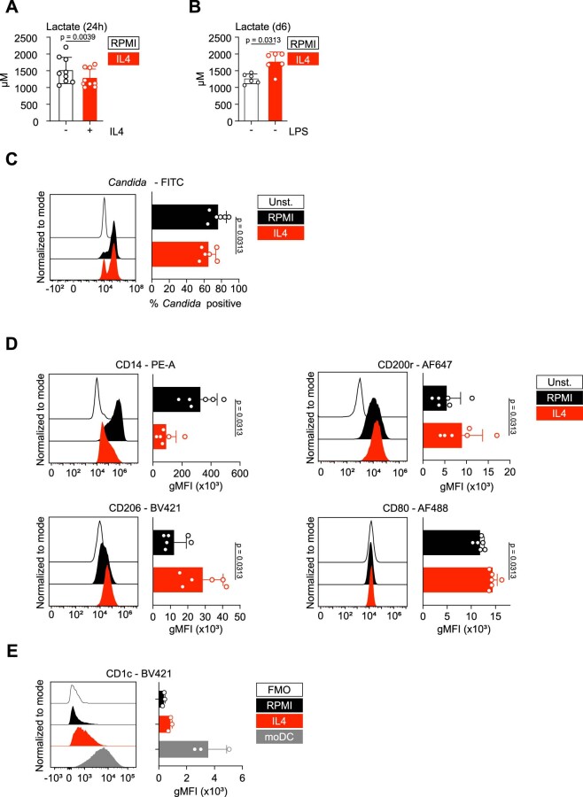
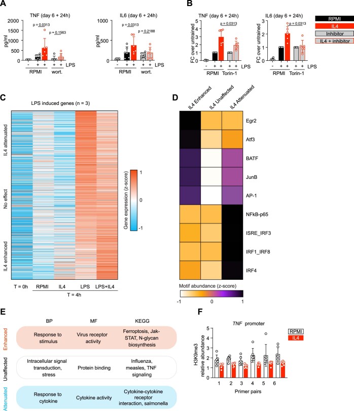
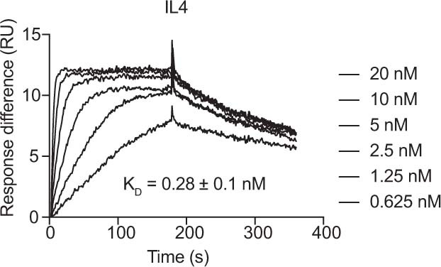
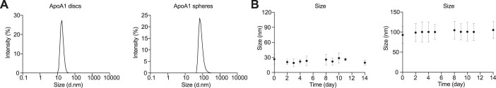
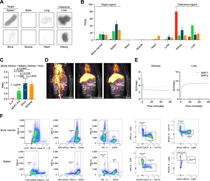
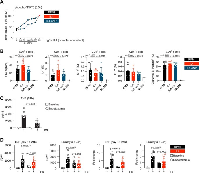
Immunoparalysis is a compensatory and persistent anti-inflammatory response to trauma, sepsis or another serious insult, which increases the risk of opportunistic infections, morbidity and mortality. Here, we show that in cultured primary human monocytes, interleukin-4 (IL4) inhibits acute inflammation, while simultaneously inducing a long-lasting innate immune memory named trained immunity. To take advantage of this paradoxical IL4 feature in vivo, we developed a fusion protein of apolipoprotein A1 (apoA1) and IL4, which integrates into a lipid nanoparticle. In mice and non-human primates, an intravenously injected apoA1-IL4-embedding nanoparticle targets myeloid-cell-rich haematopoietic organs, in particular, the spleen and bone marrow. We subsequently demonstrate that IL4 nanotherapy resolved immunoparalysis in mice with lipopolysaccharide-induced hyperinflammation, as well as in ex vivo human sepsis models and in experimental endotoxemia. Our findings support the translational development of nanoparticle formulations of apoA1-IL4 for the treatment of patients with sepsis at risk of immunoparalysis-induced complications.


Conflict of interest statement

W.J.M.M., L.A.B.J. and M.G.N. are scientific co-founders of and have equity in Trained Therapeutix Discovery. W.J.M.M. is CSO of Trained Therapeutix Discovery. W.J.M.M. and M.G.N. are scientific co-founders of and have equity in BioTrip.

Figures

Fig. 1
Fig. 1. IL4 inhibits acute inflammation, yet induces trained immunity.
a, Schematic of in vitro direct inflammation experiments. b, TNF, IL6 and IL1Ra levels after 24 h stimulation of human primary monocytes. c, Schematic of in vitro trained immunity experiments. d, TNF and IL6 levels after re-stimulation of β-glucan-trained cells. e, TNF and IL6 levels after re-stimulation of IL4-trained cells. f, Seahorse analysis of glycolytic (left) and mitochondrial (right) metabolism in IL4-trained cells. Data are presented as mean ± s.d. OCR, oxygen consumption rate; 2-DG, 2-deoxy-D-glucose.
Fig. 2
Fig. 2. Immune and epigenetic mechanisms mediating IL4-induced trained immunity.
a, Schematic overview of previously described premier IL4 signalling pathways. Generated using Biorender. b, TNF and IL6 levels after 24 h stimulation of monocytes while blocking key IL4 signalling routes. c, TNF and IL6 levels after re-stimulation of cells that were trained with IL4 while blocking key IL4 signalling routes. d, Heat map of the transcriptome of IL4-trained cells, before and after re-stimulation. e, TF motif enrichment analysis in IL4-trained immunity (heat map indicates z-scores). f, Pathway enrichment analyses of the IL4-trained immunity transcriptome. g, TNF and IL6 levels after re-stimulation of cells that were trained with IL4 in the presence of a SET7 methyltransferase inhibitor. CPH, cyproheptadine. h, ChIP-qPCR AUC analysis of TNF in IL4-trained cells. Data in bar graphs are presented as mean ± s.d.
Fig. 3
Fig. 3. Engineering an apoA1–IL4 fusion protein.
a, Schematic overview of apoA1-based fusion protein technology. b, Schematic of apoA1–IL4 fusion protein structure. c,d SDS–PAGE (c) and western blot (d) of recombinantly expressed proteins. Antibodies specific for endogenous IL4 and apoA1. e, Chromatogram and Q-ToF-MS spectrum of apoA1–IL4. f, Kinetics of apoA1–IL4 binding to IL4Rα using SPR. g, Activation of HEK-Blue cells expressing IL4Rα and IL13Rα1 by apoA1–IL4. Data are presented as mean ± s.d. DMPC, 1,2-dimyristoyl-sn-glycero-3-phosphocholine; GGS, glycine–glycine–serine; RU, resonance units.
Fig. 4
Fig. 4. Integrating apoA1–IL4 in nanoparticles.
a,b, Schematic representation (a) and cryo-TEM images (b) of discoidal (upper panel) and spherical IL4-aNPs (lower panel). c,d, IL4-aNP size distribution (c) and stability (d) of IL4-aNPs over time as determined by DLS. IL4-aNP size is reported as the number mean. e, Super-resolution fluorescence microscopy (dSTORM) images of human monocytes incubated with either fluorescently labelled apoA1(-IL4) or (IL4-)aNPs (red) and stained with anti-IL4Rα antibody (green). The co-localization between the proteins and IL4Rα can be appreciated in yellow. White ROIs are magnified in subsequent images on the right. Data are presented as mean ± s.d.
Fig. 5
Fig. 5. In vivo pharmacokinetics, biodistribution and safety profile after intravenous injection.
a, PET-CT render at 24 h after injecting 89Zr-labelled constructs. b, 89Zr-labelled construct blood half-life (n = 5, as fitted with a two-phase decay function). ID, injected dose. c, Ex vivo gamma counting of tissues 24 h after 89Zr-labelled construct injection (n = 5), number represents ratio target to clearance organs. d, Cell type-specific biodistribution of DiO-labelled discoid IL4-aNPs in spleen and bone marrow, as measured by flow cytometry. e, 89Zr-IL4-aNP blood half-life in non-human primates. f, Organ SUV mean over time in 89Zr-IL4-aNPs-injected non-human primates (n = 2). g, Organ-specific SUV mean 48 h after 89Zr-IL4-aNPs injection in non-human primates (n = 2). h, PET-MRI scan of non-human primate 48 h after 89Zr-IL4-aNPs injection. Data are presented as mean ± s.d. where appropriate. DIO, 3,3′-dioctadecyloxacarbocyanine perchlorate; MFI, mean fluorescence intensity; NHP, non-human primate.
Fig. 6
Fig. 6. Immunological in vitro, in vivo and ex vivo therapeutic evaluation of IL4-aNPs.
a, Schematic overview of the direct inflammation and trained immunity experiments in vitro. b. TNF and IL6 levels after 24 h stimulation of monocytes in the presence of IL4-aNPs. c, TNF and IL6 levels after re-stimulation of IL4(-aNP)-trained cells. d, Schematic overview of murine in vivo tolerance model, including IL4-nanotherapy. e, Serum TNF and IL6 levels following LPS re-challenge of mice treated with IL4m-aNPs. The Mann–Whitney U test was used for statistical comparisons. f, Schematic overview of human experimental endotoxaemia model, including ex vivo tolerance reversal. g, TNF and IL6 levels after ex vivo re-stimulation of human in vivo LPS-tolerized cells. h, TNF and IL6 fold increase after ex vivo re-stimulation of human in vivo LPS-tolerized cells. Data are presented as mean ± s.d.
Extended Data Fig. 1
Extended Data Fig. 1. IL4 inhibits acute inflammation, yet induces trained immunity.
(a) Supernatant lactate levels after 24-hour stimulation with IL4. (b) Accumulated supernatant lactate levels on day 6 after IL4 training. (c) Flow cytometric measurement of FITC-labelled Candida albicans phagocytosis on day 6 after IL4-training. (d) Flow cytometric measurement of surface markers commonly associated with IL4 activation of monocytes/macrophages, on day 6 after IL4-training. (e) Flow cytometric measurement of the dendritic cell marker CD1c on IL4-trained macrophages and monocyte-derived dendritic cells (moDC). Data are presented as mean (flow cytometry: geometric mean fluorescence intensity) ± SD.
Extended Data Fig. 2
Extended Data Fig. 2. Immune and epigenetic mechanisms mediating IL4-induced trained immunity.
(a) TNF/IL6 production after re-stimulation of cells that were trained with IL4 whilst blocking key IL4 signaling routes (b) Fold increase of TNF/IL6 after re-stimulation of cells that were trained with IL4 whilst blocking key IL4 signaling routes. (c) Heatmap of the transcriptome of monocytes directly after isolation, and after stimulation with RPMI, IL4, LPS, or LPS+IL4. (d) Transcription factor motif enrichment analysis of acute IL4 effects on LPS-stimulated monocytes. (e) Pathway enrichment analyses of IL4’s effect on acute LPS stimulation. (f) ChIP-qPCR analysis of TNF in IL4-trained cells. Data are presented as mean ± SD.
Extended Data Fig. 3
Extended Data Fig. 3. Engineering an apoA1–IL4 fusion protein.
Kinetics of IL4 binding to IL4Rα using SPR.
Extended Data Fig. 4
Extended Data Fig. 4. Integrating in aNP technology.
(a) DLS evaluation of conventional aNPs particle size. (b) DLS stability of conventional aNPs over time.
Extended Data Fig. 5
Extended Data Fig. 5. In vivo pharmacokinetics, biodistribution and safety profile.
(a) Organ autoradiography 24 h after 89Zr-IL4-aNPs injection in mice. (b) Gamma count in vital organs 24 h after 89Zr-IL4-aNPs injection in mice (n = 5). (c) Uptake ratios by target organs over clearance organs in mice 2-way ANOVA with Turkey post-hoc analysis. (d) Dynamic PET/MRI scans of non-human primates at 1, 30, and 60 minutes after 89Zr-IL4-aNPs injection. (e) Organ-specific SUVmean in non-human primates over 1 hour following 89Zr-IL4-aNPs administration. (f) Gating strategy for bone marrow (top) and spleen (bottom) in experiments to measure cell type-specificity of DiO-labelled discoidal IL4-aNPs. Data are presented as mean ± SD where appropriate.
Extended Data Fig. 6
Extended Data Fig. 6. Therapeutic evaluation of IL4-aNPs.
(a) Phosphorylation of STAT6, measured by flow cytometry after 30 minutes stimulation with RPMI, IL4, or different concentrations of IL4-aNPs. The data are expressed relative to the signal elicited by bare IL4. (d) T cell polarization assay by flow cytometry, after 7 days of mixed leukocyte reaction with IL4(-aNP)-trained macrophages or control. (b) TNF production following LPS-stimulation of monocytes isolated before and 4 hours after in vivo endotoxin challenge in humans. (c) TNF and IL6 levels after ex vivo re-stimulation of human in vivo LPS-tolerized cells (left) and the same data expressed as fold changes relative to the tolerant endotoxemia samples (right).

Similar articles

Cited by

References

    1. Singer M, et al. The third international consensus definitions for sepsis and septic shock (sepsis-3) JAMA. 2016;315:801–810. doi: 10.1001/jama.2016.0287. - DOI - PMC - PubMed
    1. van der Poll T, van de Veerdonk FL, Scicluna BP, Netea MG. The immunopathology of sepsis and potential therapeutic targets. Nat. Rev. Immunol. 2017;17:407–420. doi: 10.1038/nri.2017.36. - DOI - PubMed
    1. Bruse, N., Leijte, G. P., Pickkers, P. & Kox, M. in Expert Review of Clinical Immunology, Vol. 15, 251–263 (Taylor and Francis, 2019). - PubMed
    1. van der Poll T, Shankar-Hari M, Wiersinga WJ. The immunology of sepsis. Immunity. 2021;54:2450–2464. doi: 10.1016/j.immuni.2021.10.012. - DOI - PubMed
    1. Frazier WJ, Hall MW. Immunoparalysis and adverse outcomes from critical illness. Pediatr. Clin. North Am. 2008;55:647–668. doi: 10.1016/j.pcl.2008.02.009. - DOI - PMC - PubMed

Publication types