. 2023 Jun 8;6(1):591.
doi: 10.1038/s42003-023-04945-5.

The functional anatomy of elephant trunk whiskers

Affiliations

The functional anatomy of elephant trunk whiskers

Nora Deiringer et al. Commun Biol. .

Abstract

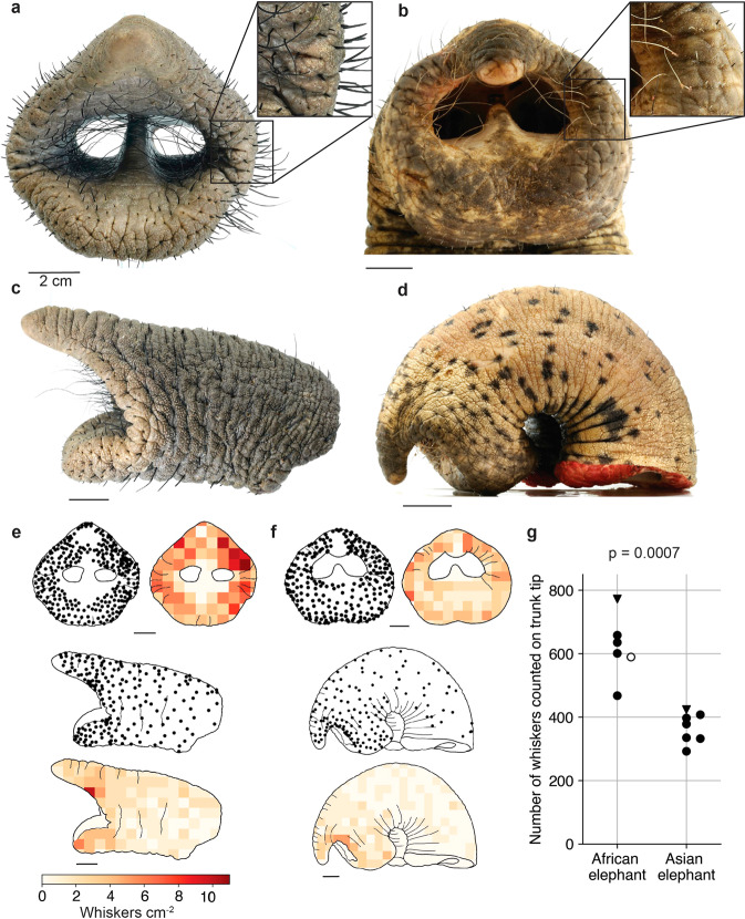
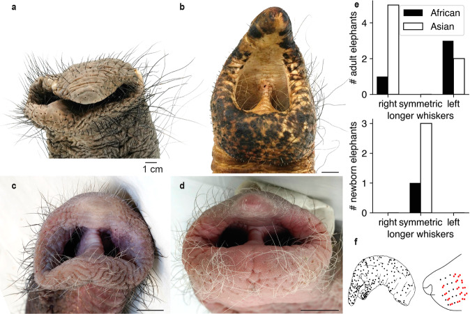
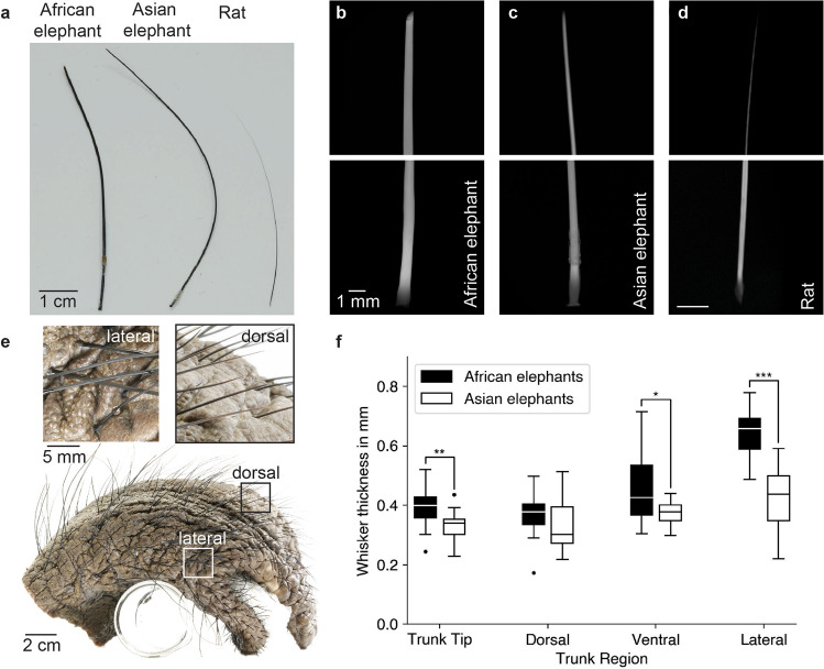
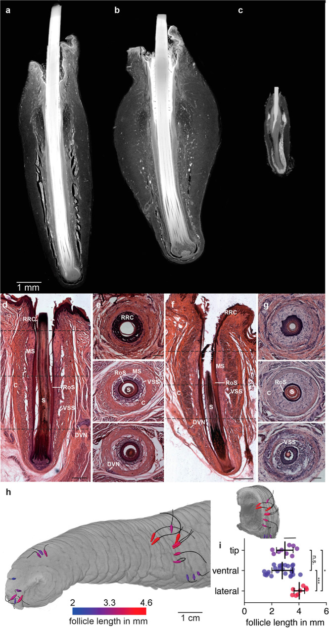
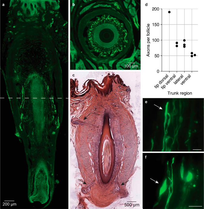
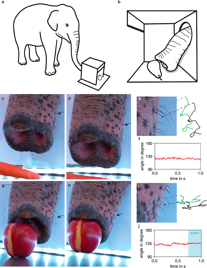
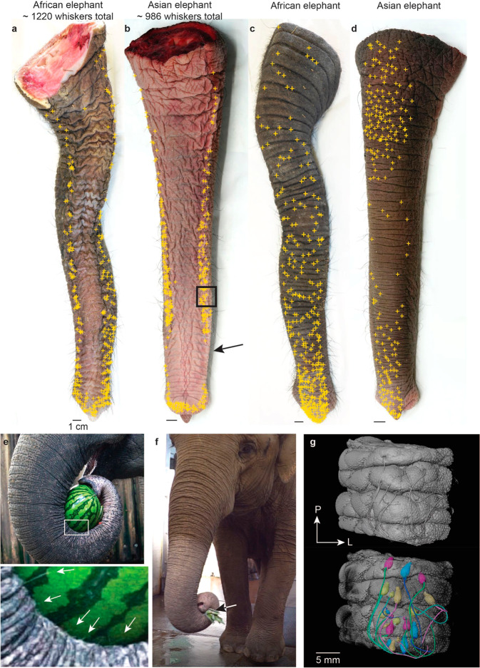
Behavior and innervation suggest a high tactile sensitivity of elephant trunks. To clarify the tactile trunk periphery we studied whiskers with the following findings. Whisker density is high at the trunk tip and African savanna elephants have more trunk tip whiskers than Asian elephants. Adult elephants show striking lateralized whisker abrasion caused by lateralized trunk behavior. Elephant whiskers are thick and show little tapering. Whisker follicles are large, lack a ring sinus and their organization varies across the trunk. Follicles are innervated by ~90 axons from multiple nerves. Because elephants don't whisk, trunk movements determine whisker contacts. Whisker-arrays on the ventral trunk-ridge contact objects balanced on the ventral trunk. Trunk whiskers differ from the mobile, thin and tapered facial whiskers that sample peri-rostrum space symmetrically in many mammals. We suggest their distinctive features-being thick, non-tapered, lateralized and arranged in specific high-density arrays-evolved along with the manipulative capacities of the trunk.


Conflict of interest statement

The authors declare no competing interests.

Figures

Fig. 1
Fig. 1. Trunk tip whisker number and density differ between African and Asian elephants.
a Frontal view of an adult African elephant cow trunk tip (Linda, see Table 1). Whiskers protrude from skinfolds (inset). b Frontal view of an Asian elephant trunk tip (Unknown Asian, see Table 1). Whiskers protrude from skinfolds (inset). c Side view of an African elephant trunk tip. d Side view of an Asian elephant trunk tip. e Upper left, dot display of whisker positions of an African elephant trunk tip from a frontal view. Upper right, whisker density (whiskers per cm2) of an African elephant trunk tip from a frontal view. Middle, dot display of whisker positions of an African elephant trunk tip from a side view. Lower, whisker density of an African elephant trunk tip from a side view. f Whisker positions and whisker density on an Asian elephant trunk tip, conventions as in e. g Number of whiskers counted on the trunk tip of six African and seven Asian elephants. Dots (adults); triangles (newborns). One count was partially extrapolated due to an incomplete sample (empty dot). p = 0.0007, Welch’s t-test (Hedges’ g = 3.59).
Fig. 2
Fig. 2. Whisker lengths varies with usage/ abrasion and is lateralized in adult elephants.
a Frontal view of an adult African elephant trunk tip. Note the asymmetric whisker length. b As in a but for an adult Asian elephant trunk tip. Note the asymmetry. c Frontal view of a newborn African elephant trunk tip. Note the symmetric whisker length. d As in c but for a newborn Asian elephant trunk tip. Note the symmetry. e Number of African (black) and Asian (white) elephant trunk tips with symmetrical whisker lengths or longer whiskers on the right or left trunk side. Upper, adult elephants. Lower, newborn elephants. f Left, dot display of whisker positions on the side of an African elephant trunk tip. Right, dot display of whisker positions in a rat whisker pad. Single whiskers are marked in black, double whiskers in red as reported by Maier & Brecht (2018). No double whiskers were observed in elephants.
Fig. 3
Fig. 3. Elephant trunk whiskers are thick and show little tapering.
a Photograph of an African elephant lateral trunk whisker (left), Asian elephant lateral trunk whisker (middle) and rat δ- whisker (right). b Upper, volume rendering of a microCT scan showing the distal part of an iodide-stained African elephant whisker from the lateral trunk tip. Lower, proximal part of the whisker. Note the similar thickness distally and proximally. c Asian elephant whisker sampled on the lateral trunk tip, conventions and scaling as in b. Note the similar thickness distally and proximally. d A rat whisker (δ), conventions as in b. Note the tapering. e Side view of an African elephant trunk tip. Insets show the difference in whisker thickness between lateral and dorsal trunk tip regions. f Whisker thickness in different regions of the trunk tip of African and Asian elephants. While in Asian elephants, whiskers of different trunk tip regions do not differ in thickness, in African elephants, lateral whiskers are thicker than whiskers from other regions of the trunk tip. Thickness was measured from two adult Asian and two adult African elephants. A comparison of whisker thickness was done using a Welch’s t-test (*P < 0.05, **P < 0.01, ***P < 0.001). Central line, median; box limits, upper and lower quartiles; whiskers, 1.5x interquartile range; dots, outliers.
Fig. 4
Fig. 4. Organization of elephant trunk whisker follicles.
Virtual longitudinal microCT section of a a dorsal African elephant trunk follicle-sinus-complex (FSC). The follicle was stained in 1% iodine for 48 h. The surrounding tissue was cropped, and whisker contrast was selectively adjusted in some areas to maintain visibility and avoid saturation. b a lateral Asian elephant trunk FSC, conventions as in a. c a rat δ whisker FSC. The FSC was stained in 1% iodine for 24 h. d Micrograph of a hematoxylin-eosin stained longitudinal section of a lateral adult African elephant trunk FSC (scale bar = 1 mm). e Micrograph of hematoxylin-eosin stained transversal sections of a lateral adult African elephant trunk FSC. Approximate planes of the sections are indicated in d with dotted lines (scale bar = 500 µm). f As d but for a lateral adult Asian elephant trunk FSC (scale bar = 1 mm). Note that the whisker partially broke during cryosectioning and embedding. g As e but for a lateral newborn Asian elephant trunk FSC (scale bar = 100 µm). h Volume rendering of an Asian newborn elephant trunk microCT scan with single FSCs and their respective whiskers segmented on the trunk tip and a proximal trunk piece. FSCs are color-coded depending on their lengths. Left, view of the whole scan. Right, Volume rendering of the proximal trunk piece. i Follicle length in the lateral and ventral trunk and the trunk tip of an Asian newborn elephant (Hoa’s Baby, see Table 1). One-way ANOVA, P < 0.001, post hoc Scheffé test for pairwise comparison (*P < 0.05, ***P < 0.001). Error bars indicate the standard deviation. RRC Rete ridge collar, MS Mesenchymal sheath, C Vibrissal capsule, S Vibrissal shaft, RoS Root sheath, VSS Vascular sinus spaces, DVN Deep vibrissal nerve.
Fig. 5
Fig. 5. Innervation of elephant trunk follicle-sinus-complexes.
a Longitudinal section of a newborn Asian elephant trunk whisker FSC stained for Neurofilament H using immunohistochemistry. The dashed line indicates the level of the transversal section shown in b. Axon bundles enter the follicle in multiple nerves. Accordingly, we observed the axons penetrating the capsule in this and adjacent sections and ascending through the follicle while distributing evenly around the hair shaft. b Transversal section of a newborn Asian elephant trunk whisker FSC stained for Neurofilament H using immunohistochemistry. The approximate level of the section is indicated by the dashed line in a. c Longitudinal section of a hematoxylin-eosin stained adult Asian elephant trunk whisker FSC. Arrows indicate multiple nerves penetrating the follicle at different levels. In addition to their distinct appearance in hematoxylin-eosin staining, we identified nerves by Neurofilament H antibody labeling of alternating serial sections. d Number of axons per follicle counted for follicles of different trunk areas. The number indicated is a cumulative axon count corresponding to the sum of all axons counted in the various nerves innervating the respective follicle. Data refers to one newborn Asian elephant (Hoa’s baby, see Table 1). e Micrograph of a lanceolate ending (indicated by an arrow) at the upper third level of a newborn Asian elephant trunk FSC stained for Neurofilament H using immunohistochemistry (scale bar = 20 µm). f Micrograph of lanceolate-like droplet-shaped nerve ending (indicated by an arrow) at the midlevel of a newborn Asian elephant trunk FSC, conventions as in e.
Fig. 6
Fig. 6. Elephants do not whisk (even during haptic pinching and vacuuming).
a Schematics of the experimental setup used for investigating whisker movement during haptically controlled retrieval of fruit from a wooden box. b View from the side of the box during the grasping task pictured in a. c Still of a grasping video clip, in which the female Asian elephant Anchali pinches a carrot; the frame shown is the first one we analyzed in the clip. Also see Supplementary Movie 1. The arrow points to the tracked whisker. d The final frame we analyzed in the same video clip. e Left, close-up view of the tracked whisker with the whisker base marked by a green dot and the tip of the whisker marked by a black dot. We measured the angle between the vertical of the trunk and the chosen whisker (marked in black). Right, trajectories of the base (in green) and tip (in black) during the tracking time of 1 s. The trajectories look identical indicating a lack of relative whisker movement (whisking). f Angle between the whisker and the vertical over the tracking time. Measurements were taken as indicated in e. Little or no whisker movement is observed during the pinching behavior. g Still of a video clip, in which Anchali vacuums an apple. Conventions as in c. Also see Supplementary Movie 1. h The final frame we analyzed in the same video clip. i As in e, but for the vacuuming behavior. j Angle between the whisker and the vertical over the tracking time. The angle is shown in e. The onset and time of suction is marked in blue and was inferred from the audio trace of the video. See also Supplementary Movie 1.
Fig. 7
Fig. 7. Ventral trunk ridge whisker arrays and their behavioral contact patterns.
a Photograph of the ventral side of a newborn African elephant trunk with whisker positions marked in yellow. All trunks investigated show two distinct whisker bands on the ventral trunk. In African elephants, whisker bands are more marked than in Asian elephants and extend over the whole trunk length. On top, we provide the total number of whiskers counted on the entire trunk of this newborn elephant. b Photograph of the ventral side of an Asian newborn elephant trunk with whisker positions marked in yellow. In Asian elephants, whiskers are missing in the ‘clamp- zone’ (black arrow). On top, we provide the total trunk whisker count. c Photograph of the dorsal side of a newborn African elephant trunk with whisker positions marked in yellow. d Photograph of the dorsal side of a newborn Asian elephant trunk with whisker positions marked in yellow. e Upper, photograph of an Asian elephant balancing a watermelon. Lower, high magnification view of the same picture, with white arrows pointing to ventral ridge whiskers that are visibly in contact with the melon during balancing. f Photograph of an Asian elephant clamping a pineapple; ventral ridge whiskers are distinctly missing in the trunk region posterior to the tip, where Asian elephants clamp objects. g Upper, volume rendering of a microCT scan of an iodine-stained trunk piece with ventral ridge whisker arrays from a newborn Asian elephant (black box in b). Lower, volume rendering of segmented whisker follicles. Note the ventral position and orientation of all whiskers. P posterior, L lateral.

References

    1. Dehnhardt G, Friese C, Sachser N. Sensitivity of the trunk of Asian elephants for texture differences of actively touched objects. Z. Saugetierkd. 1997;62:37–39.
    1. Purkart L, et al. Trigeminal ganglion and sensory nerves suggest tactile specialization of elephants. Curr. Biol. 2022;32:904–910. doi: 10.1016/j.cub.2021.12.051. - DOI - PubMed
    1. Boas, J. E. V., & Paulli, S. The Elephant’s Head: Studies in the Comparative Anatomy of the Organs of the Head of the Indian Elephant and Other Mammals. The Facial Muscles and the Proboscis. 1st Part. (G. Fischer, 1908).
    1. Fischer MS, Trautmann U. Fetuses of African elephants (Loxodonta africana) in photographs. Elephant. 1987;2:6. doi: 10.22237/elephant/1521732098. - DOI
    1. Cuvier, G., and Dumeril, A. M. Lecons D’anatomie Comparée: 2. Societe Typographique Belge (Crochard, 1838).

Publication types