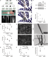
TAPT1-at the crossroads of extracellular matrix and signaling in Osteogenesis imperfecta
- PMID: 37292039
- PMCID: PMC10331569
- DOI: 10.15252/emmm.202317528
TAPT1-at the crossroads of extracellular matrix and signaling in Osteogenesis imperfecta
Abstract
Osteogenesis imperfecta (OI) is a hereditary skeletal disorder primarily affecting collagen type I structure and function, causing bone fragility and occasionally versatile extraskeletal symptoms. This study expands the spectrum of OI-causing TAPT1 mutations and links extracellular matrix changes to signaling regulation.
© 2023 The Authors. Published under the terms of the CC BY 4.0 license.
Conflict of interest statement
The authors declare that they have no conflict of interest.
Figures

Similar articles
-
[Genetic basis for skeletal disease. Osteogenesis imperfecta and genetic abnormalities].Clin Calcium. 2010 Aug;20(8):1190-5. Clin Calcium. 2010. PMID: 20675929 Review. Japanese.
-
Osteogenesis Imperfecta: Mechanisms and Signaling Pathways Connecting Classical and Rare OI Types.Endocr Rev. 2022 Jan 12;43(1):61-90. doi: 10.1210/endrev/bnab017. Endocr Rev. 2022. PMID: 34007986 Free PMC article. Review.
-
Osteogenesis imperfecta and therapeutics.Matrix Biol. 2018 Oct;71-72:294-312. doi: 10.1016/j.matbio.2018.03.010. Epub 2018 Mar 11. Matrix Biol. 2018. PMID: 29540309 Free PMC article. Review.
-
Osteogenesis imperfecta and its molecular diagnosis by determination of mutations of type I collagen genes.Pediatr Endocrinol Rev. 2006 Sep;4(1):40-6. Pediatr Endocrinol Rev. 2006. PMID: 17021582 Review.
-
Molecular diagnosis in children with fractures but no extraskeletal signs of osteogenesis imperfecta.Osteoporos Int. 2017 Jul;28(7):2095-2101. doi: 10.1007/s00198-017-4031-2. Epub 2017 Apr 4. Osteoporos Int. 2017. PMID: 28378289
Cited by
-
TENT5A-associated osteogenesis imperfecta: long-term follow-up and molecular insights.JBMR Plus. 2025 May 11;9(7):ziaf083. doi: 10.1093/jbmrpl/ziaf083. eCollection 2025 Jul. JBMR Plus. 2025. PMID: 40575455 Free PMC article.
-
ANTXR1 deficiency promotes fibroblast senescence: implications for GAPO syndrome as a progeroid disorder.Sci Rep. 2024 Apr 23;14(1):9321. doi: 10.1038/s41598-024-59901-y. Sci Rep. 2024. PMID: 38653789 Free PMC article.
-
Genome-wide association study for bone quality of ducks during the laying period.Poult Sci. 2024 May;103(5):103575. doi: 10.1016/j.psj.2024.103575. Epub 2024 Feb 25. Poult Sci. 2024. PMID: 38447311 Free PMC article.
References
-
- Bodine PVN, Zhao W, Kharode YP, Bex FJ, Lambert AJ, Goad MB, Gaur T, Stein GS, Lian JB, Komm BS (2004) The Wnt antagonist secreted frizzled‐related protein‐1 is a negative regulator of trabecular bone formation in adult mice. Mol Endocrinol 18: 1222–1237 - PubMed
-
- Bodine PVN, Stauffer B, Ponce‐de‐Leon H, Bhat RA, Mangine A, Seestaller‐Wehr LM, Moran RA, Billiard J, Fukayama S, Komm BS et al (2009) A small molecule inhibitor of the Wnt antagonist secreted frizzled‐related protein‐1 stimulates bone formation. Bone 44: 1063–1068 - PubMed
-
- Häusler KD, Horwood NJ, Chuman Y, Fisher JL, Ellis J, Martin TJ, Rubin JS, Gillespie MT (2004) Secreted frizzled‐related Protein‐1 inhibits RANKL‐dependent osteoclast formation. J Bone Miner Res 19: 1873–1881 - PubMed
-
- He J, Sheng T, Stelter AA, Li C, Zhang X, Sinha M, Luxon BA, Xie J (2006) Suppressing Wnt signaling by the hedgehog pathway through sFRP‐1. J Biol Chem 281: 35598–35602 - PubMed
Publication types
MeSH terms
Substances
Grants and funding
LinkOut - more resources
Full Text Sources
Medical
Molecular Biology Databases

