. 2023 Mar 20;4(6):386-398.
doi: 10.1039/d2cb00158f. eCollection 2023 Jun 7.

Using light scattering to assess how phospholipid-protein interactions affect complex I functionality in liposomes

Affiliations

Using light scattering to assess how phospholipid-protein interactions affect complex I functionality in liposomes

Jana Eisermann et al. RSC Chem Biol. .

Abstract

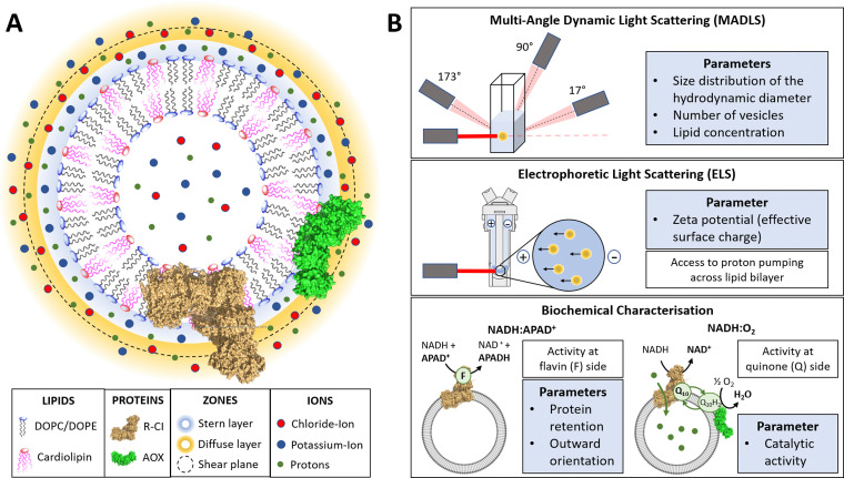
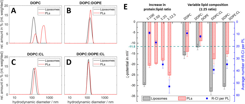
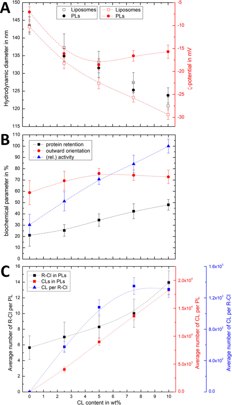
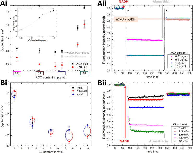
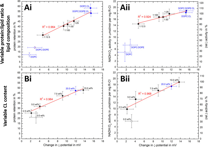
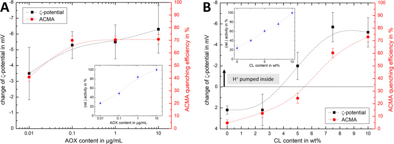
Complex I is an essential membrane protein in respiration, oxidising NADH and reducing ubiquinone to contribute to the proton-motive force that powers ATP synthesis. Liposomes provide an attractive platform to investigate complex I in a phospholipid membrane with the native hydrophobic ubiquinone substrate and proton transport across the membrane, but without convoluting contributions from other proteins present in the native mitochondrial inner membrane. Here, we use dynamic and electrophoretic light scattering techniques (DLS and ELS) to show how physical parameters, in particular the zeta potential (ζ-potential), correlate strongly with the biochemical functionality of complex I-containing proteoliposomes. We find that cardiolipin plays a crucial role in the reconstitution and functioning of complex I and that, as a highly charged lipid, it acts as a sensitive reporter on the biochemical competence of proteoliposomes in ELS measurements. We show that the change in ζ-potential between liposomes and proteoliposomes correlates linearly with protein retention and catalytic oxidoreduction activity of complex I. These correlations are dependent on the presence of cardiolipin, but are otherwise independent of the liposome lipid composition. Moreover, changes in the ζ-potential are sensitive to the proton motive force established upon proton pumping by complex I, thereby constituting a complementary technique to established biochemical assays. ELS measurements may thus serve as a more widely useful tool to investigate membrane proteins in lipid systems, especially those that contain charged lipids.


Conflict of interest statement

There are no conflicts to declare.

Figures

Fig. 1
Fig. 1. Introduction to PLs and characterisation parameters. (A) Schematic representation of PLs containing R-CI (PDB 5LDW) as well as AOX (PDB 3VV9) which was added for all activity assays focusing on the Q10 site to re-oxidise the quinone pool. The lipid bilayer contains the synthetic lipids DOPC, DOPE and 18 : 1 CL in a defined ratio. Immediately surrounding the liposome is a layer of tightly associated ions, opposite in charge to the surface of the liposome (here potassium ions). Surrounding this Stern layer is a second layer (diffuse layer) of loosely associated ions. The point at which the second layer of ions moves with the liposome as a single entity is termed the slipping plane. This plane defines the ζ-potential. (B) Summary of the main physico-chemical and biochemical characterisation techniques.
Fig. 2
Fig. 2. DLS and ELS characterisation of liposomes and PLs with variable lipid composition and R-CI content. (A–D) Intensity-weighted size distributions for liposomes and PLs for lipid systems: DOPC (A), DOPC : DOPE 8.9 : 1.1 (B), DOPC : CL 8.9 : 1.1 (C), DOPC : DOPE : CL 8 : 1 : 1 (D, optimised mixture). The mean diameter relating to the first peak in the polydisperse size distribution of the PLs (A) was used to calculate their ζ-potential in E. (E) ζ-potentials of liposomes and PLs with increasing protein : lipid ratios from 1 : 100 to 1 : 12.5 (left) and simplified lipid compositions (right), including the average number of reconstituted R-CI per PL (blue data points). The green dashed line is the ζ-potential of as-isolated bovine R-CI (10 μg mL−1) in aqueous buffer with detergent (no lipids). DOPC : DOPE : CL PLs with protein : lipid ratios of 1 : 25 for the left and right data sets were obtained from different R-CI batches and hence show some variation.
Fig. 3
Fig. 3. Effect of CL on hydrodynamic diameter, ζ-potential and biochemical competence of PLs, and average number of CL per PL and R-CI. (A) Change in hydrodynamic diameter (black) and ζ-potential (red) with increasing CL content in PLs (1 : 50 protein : lipid ratio) and liposomes. (B) Biochemical parameters (protein retention, outward orientation, activity) against CL content. The activity, determined for a fixed amount of outward-facing R-CI of 1.5 μg mL−1, is given as a relative value compared to the optimised system (DOPC : DOPE : CL 8 : 1 : 1). Data points represent mean values of two batches of PLs prepared with increasing CL content. (C) Effect of increasing CL content on the average number of R-CI in PLs with protein : lipid ratio 1 : 50 (black), with number of CL units in PLs (red) and the average number of CLs per reconstituted R-CI (blue). Dashed lines serve as a guide to the observed trends.
Fig. 4
Fig. 4. Influence of AOX and CL content on the build-up PMF in PLs. (Ai): changes in the ζ-potential with increasing amounts of AOX for PLs with 10 wt% CL (1.5 μg mL−1 outward-facing R-CI) before and after injection of NADH (see also Fig. S5, ESI†). Controls with 20 μg mL−1 alamethicin (+ala) or 1 μM of piericidin A (+pier A) are shown in grey. NADH:O2 activity as a function of AOX content is given in the inset. (Aii) Proton pumping in R-CI PLs with varying AOX content monitored using ACMA fluorescence. All PLs (1.0 μg mL−1 outward-facing R-CI) were treated with 0.1 μM valinomycin. Proton pumping was initiated with 500 μM of NADH and addition of 10 μg mL−1 of alamethicin led to the collapse of the PMF. (Bi) Comparison of ζ-potential for PLs (1.5 μg mL−1 outward-facing R-CI) with varying CL content under the influence of AOX (1 μg mL−1). NADH induced the formation of a PMF, subsequently collapsed with the addition of valinomycin. (Bii) Proton pumping in R-CI PLs (+1 μg mL−1 AOX) with increasing CL-content monitored using ACMA fluorescence. Other conditions were as in (Aii).
Fig. 5
Fig. 5. Correlation of ζ-potential changes with biochemical parameters. Plots of Δζ from PLs with (A) increase in protein : lipid ratio (fixed 10 wt% Cl content) and variable lipid composition and (B) increased in CL content (fixed 1 : 50 protein : lipid ratio) against protein retention (i) and catalytic activity (ii). The relative activity values use (A) protein : lipid ratio 1 : 25 or (B) the 10 wt% CL data as reference point. Data points for Ai/Aii are taken from Fig. 2E and Table 1, whereas Bi/Bii are the mean values for two measured PLs sets (see Fig. S8, ESI for individual data sets). Data points linked to lipid mixtures without CL are present as hollow spheres or circles. The primary data for the 20 wt% CL sample can be found in Table S9 (ESI†).
Fig. 6
Fig. 6. Comparison of ELS and ACMA measurements. Comparison of the change in ζ-potential for AOX-R-CI PLs before and after initiating proton pumping via NADH addition with the ACMA quenching efficiency, with (A) showing variable AOX content with fixed 10 wt% CL (linked to Fig. 4Ai and Aii) and (B) showing variable CL content (linked to Fig. 4B). Dashed lines in all plots serve as guide for the observed trends. Insets show the change in relative activity as determined with the NADH:O2 assay. The y-axis for the ζ-potential change was flipped (from positive to negative values) to allow easier comparison.

Similar articles

Cited by

References

    1. Amati A. M. Graf S. Deutschmann S. Dolder N. von Ballmoos C. Biochem. Soc. Trans. 2020;48:1473–1492. doi: 10.1042/BST20190966. - DOI - PubMed
    1. Stillwell W., in An Introduction to Biological Membranes, ed. W. Stillwell, Elsevier, 2016, pp. 3–15
    1. Shen H. H. Lithgow T. Martin L. L. Int. J. Mol. Sci. 2013;14:1589–1607. doi: 10.3390/ijms14011589. - DOI - PMC - PubMed
    1. Beales P. A. Khan S. Muench S. P. Jeuken L. J. C. Biochem. Soc. Trans. 2017;45:15–26. doi: 10.1042/BST20160019. - DOI - PMC - PubMed
    1. Rigaud J.-L. and Lévy D., Membrane Protein Reconstitution, Elsevier, 2003, vol. 372, pp. 65–86 - PubMed